. 2014 Aug 15;196(16):2934-43.
doi: 10.1128/JB.01675-14. Epub 2014 Jun 9.

Transcription of the lysine-2,3-aminomutase gene in the kam locus of Bacillus thuringiensis subsp. kurstaki HD73 is controlled by both σ54 and σK factors

Affiliations

Transcription of the lysine-2,3-aminomutase gene in the kam locus of Bacillus thuringiensis subsp. kurstaki HD73 is controlled by both σ54 and σK factors

Zhe Zhang et al. J Bacteriol. .

Abstract

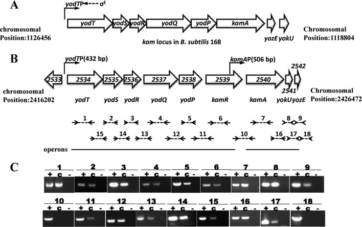
Lysine 2,3-aminomutase (KAM; EC 5.4.3.2) catalyzes the interconversion of l-lysine and l-β-lysine. The transcription and regulation of the kam locus, including lysine-2,3-aminomutase-encoding genes, in Bacillus thuringiensis were analyzed in this study. Reverse transcription-PCR (RT-PCR) analysis revealed that this locus forms two operons: yodT (yodT-yodS-yodR-yodQ-yodP-kamR) and kamA (kamA-yokU-yozE). The transcriptional start sites (TSSs) of the kamA gene were determined using 5' rapid amplification of cDNA ends (RACE). A typical -12/-24 σ(54) binding site was identified in the promoter PkamA, which is located upstream of the kamA gene TSS. A β-galactosidase assay showed that PkamA, which directs the transcription of the kamA operon, is controlled by the σ(54) factor and is activated through the σ(54)-dependent transcriptional regulator KamR. The kamA operon is also controlled by σ(K) and regulated by the GerE protein in the late stage of sporulation. kamR and kamA mutants were prepared by homologous recombination to examine the role of the kam locus. The results showed that the sporulation rate in B. thuringiensis HD(ΔkamR) was slightly decreased compared to that in HD73, whereas that in HD(ΔkamA) was similar to that in HD73. This means that other genes regulated by KamR are important for sporulation.


Figures

FIG 1
FIG 1
Gene organization of the kam locus in B. subtilis 168 and B. thuringiensis HD73. (A) Gene organization of the kam locus in B. subtilis 168. The tentative assigned functions are as follows: yodT, Nε-acetyl-β-lysine transaminase; yodS and yodR, 3-keto-6-acetamidohexanoate cleavage enzyme; yodQ, 4-acetamidobutyryl-CoA deacetylase; yodP, β-lysine acetyltransferase; and kamA, lysine 2,3-aminomutase. (B) The kam locus in B. thuringiensis strain HD73. The white arrows represent open reading frames (ORFs). The small arrows denote the lengths of the promoters present upstream of the yodT and kamA genes. The dashed lines with small black arrows that are annotated with numbers correspond to the RT-PCR amplicons (see lanes in panel C). The full lines below the ORFs indicate operons. (C) RT-PCR analysis of the kam locus in B. thuringiensis strain HD73. The RNA samples were prepared at T7 of bacterial growth in SSM. The RT-PCRs labeled with “c” were performed with 500 ng of RNA. The positive controls are labeled “+” (PCR with 100 ng of genomic DNA). The negative controls are labeled “−” (RT-PCR with 500 ng of RNA with heat-inactivated reverse transcriptase). The numbers represent different RT-PCR amplicons: numbers 1 to 9 represent yodT, yodS, yodR, yodQ, yodP, kamR, kamA, yokU, and yozE, respectively; numbers 10 to 18 represent kamR-kamA, yodP-kamR, yodQ-yodP, yodR-yodQ, yodS-yodR, yodT-yodS, kamA-yokU, yokU-yozE, and yozE-HD_2543, respectively.
FIG 2
FIG 2
The transcriptional start sites (TSSs) of the kamA gene. The −12/−24 consensus sequences, the GG-N10-GC σ54 factor binding sites, and σK factor-dependent −10/−35 sequences are in bold font; the TSSs are indicated numerically (+1) and marked in bold font. The codon underlined is the transcription start site of kamA.
FIG 3
FIG 3
Analysis of the transcription of PkamA. (A) Analysis of the activity of PkamA (T0 to T10). The promoter-directed β-galactosidase synthesis of these strains was determined at the indicated times after culturing the cells in SSM at 30°C. Tn is n hours after the end of the exponential phase. Each value represents the mean of results of at least three independent replicates. Error bars show standard deviations. (B) Analysis of the activity of PkamA in HD(ΔsigK). The promoter-directed β-galactosidase synthesis of these strains was determined at the indicated times after culturing the cells in SSM at 30°C. Tn is n hours after the end of the exponential phase. Each value represents the mean of results of at least three independent replicates. Error bars show standard deviations. (C) Analysis of the activity of PkamA in HD(ΔgerE). The promoter-directed β-galactosidase synthesis of these strains was determined at the indicated times after culturing the cells in SSM at 30°C. Tn is n hours after the end of the exponential phase. Each value represents the mean of results of at least three independent replicates. Error bars show standard deviations. (D) Electrophoretic mobility shift assay (EMSA) for detecting protein-nucleic acid interactions, using labeled PkamA and increasing concentrations of recombinant SigK-His. Lanes 1 to lane 8 contained 0, 15, 30, 45, 60, 75, 90, and 105 ng/μl of SigK-His, respectively. F, free DNA; B, bound DNA. (E) EMSA for detecting protein-nucleic acid interactions, using labeled PkamA and increasing concentrations of recombinant GerE-GST. Lanes 1 to lane 7 contained 0, 10.5, 21, 42, 63, 84, and 126 ng/μl of GerE-GST, respectively.
FIG 4
FIG 4
Analysis of the activity of PyodT. The promoter-directed β-galactosidase synthesis of these strains was determined at the indicated times after culturing the cells in SSM at 30°C. Tn is n hours after the end of the exponential phase. Each value represents the mean of results of at least three independent replicates. Error bars show standard deviations.
FIG 5
FIG 5
Comparisons of sporulation frequency between the HD(ΔkamR) mutant, HD(ΔkamA) mutant, and wild-type strain, HD73. The graph depicts sporulation frequency. Error bars show standard deviations.
FIG 6
FIG 6
The l-lysine degradation pathway in HD73. l-Lysine is converted to β-lysine by a lysine-2,3-aminomutase (KamA) that is subsequently acetylated to Nε-acetyl-β-lysine by the action of an acetyltransferase (YodP); Nε-acetyl-l-β-lysine is deaminated to 3-keto-6-acetamidohexanoate by an Nε-acetyl-β-lysine transaminase (YodT) in the presence of 2-ketoglutarate. The conversion of 3-keto-6-acetamidohexanoate and acetyl-CoA to 4-acetamidobutyryl-CoA and acetoacetate is catalyzed by a 3-keto-6-acetamidohexanoate cleavage enzyme (YodS and YodR); 4-acetamidobutyryl-CoA deacetylase (YodQ) converts 4-acetamidobutyryl-CoA to 4-aminobutyrate. 4-Aminobutyrate is readily converted via succinic semialdehyde to succinate, which is called GABA shunt. The succinate product will enter the tricarboxylic acid (TCA) cycle. The encoding genes are annotated in parentheses, and the dashed arrows marked the genes controlled by σ54.

References

    1. Merrick M. 1993. In a class of its own—the RNA polymerase sigma factor σ54 (σN). Mol. Microbiol. 10:903–909. 10.1111/j.1365-2958.1993.tb00961.x - DOI - PubMed
    1. Wigneshweraraj S, Burrows P, Bordes P, Schumacher J, Rappas M, Finn R, Cannon W, Zhang X, Buck M. 2005. The second paradigm for activation of transcription. Prog. Nucleic Acids Res. Mol. Biol. 79:339–369. 10.1016/S0079-6603(04)79007-8 - DOI - PubMed
    1. Buck M, Gallegos M-T, Studholme DJ, Guo Y, Gralla JD. 2000. The bacterial enhancer-dependent σ54 (σN) transcription factor. J. Bacteriol. 182:4129–4136. 10.1128/JB.182.15.4129-4136.2000 - DOI - PMC - PubMed
    1. Lonetto M, Gribskov M, Gross C. 1992. The sigma 70 family: sequence conservation and evolutionary relationships. J. Bacteriol. 174:3843–3849 - PMC - PubMed
    1. Wigneshweraraj S, Bose D, Burrows PC, Joly N, Schumacher J, Rappas M, Pape T, Zhang X, Stockley P, Severinov K. 2008. Modus operandi of the bacterial RNA polymerase containing the σ54 promoter-specificity factor. Mol. Microbiol. 68:538–546. 10.1111/j.1365-2958.2008.06181.x - DOI - PubMed

Publication types

MeSH terms

LinkOut - more resources