

A pilot proof-of-concept study of a modified device for one-step endoscopic ultrasound-guided biliary drainage in a new experimental biliary dilatation animal model
- PMID: 24914346
- PMCID: PMC4024795
- DOI: 10.3748/wjg.v20.i19.5859
A pilot proof-of-concept study of a modified device for one-step endoscopic ultrasound-guided biliary drainage in a new experimental biliary dilatation animal model
Abstract
Aim: To evaluate the technical feasibility of a modified tapered metal tip and low profile introducer for one-step endoscopic ultrasound (EUS)-guided biliary drainage (EUS-BD) in a new experimental biliary dilatation porcine model.
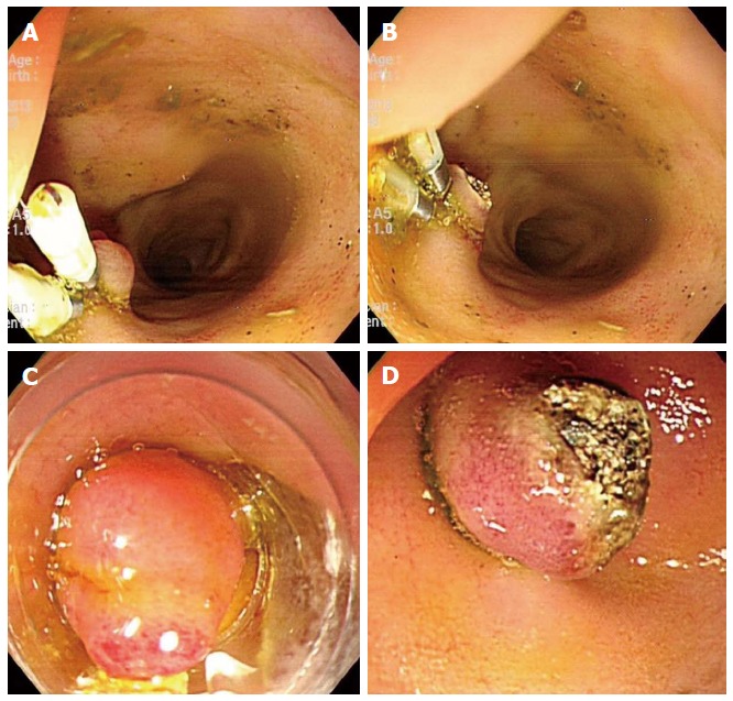
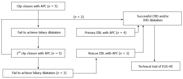
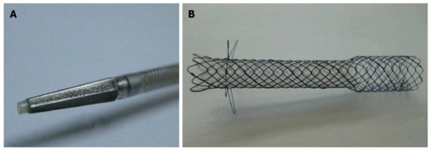
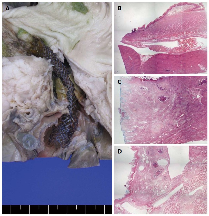
Methods: A novel dedicated device for one-step EUS-guided biliary drainage system (DEUS) introducer has size 3F tapered catheter with size 4F metal tip for simple puncture of the intestinal wall and liver parenchyma without graded dilation. A self-expandable metal stent, consisting of both uncovered and nitinol-covered portions, was preloaded into DEUS introducer. After establishment of a biliary dilatation model using endoscopic hemoclips or band ligation with argon plasma coagulation in 9 mini-pigs, EUS-BD using a DEUS was performed following 19-G needle puncture without the use of fistula dilation devices.
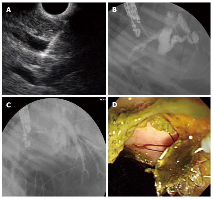
Results: One-step EUS-BD was technically successful in seven pigs [7/9 (77.8%) as intention to treat] without the aid of devices for fistula dilation from the high body of stomach or far distal esophagus to the intrahepatic (n = 2) or common hepatic (n = 5) duct. Primary technical failure occurred in two cases that did not show adequate biliary dilatation. In seven pigs with a successful bile duct dilatation, the technical success rate was 100% (7/7 as per protocol). Median procedure time from confirmation of the dilated bile duct to successful placement of a metallic stent was 10 min (IQR; 8.9-18.1). There were no immediate procedure-related complications.
Conclusion: Modified tapered metal tip and low profile introducer may be technically feasible for one-step EUS-BD in experimental porcine model.
Keywords: Biliary dilation; Biliary drainage; Complications; Endoscopic ultrasound; Feasibility.
Figures
References
-
- Beissert M, Wittenberg G, Sandstede J, Beer M, Tschammler A, Burghardt W, Jahns R, Hahn D. Metallic stents and plastic endoprostheses in percutaneous treatment of biliary obstruction. Z Gastroenterol. 2002;40:503–510. - PubMed
-
- Doctor N, Dick R, Rai R, Dafnios N, Salamat A, Whiteway H, Dooley J, Davidson BR. Results of percutaneous plastic stents for malignant distal biliary obstruction following failed endoscopic stent insertion and comparison with current literature on expandable metallic stents. Eur J Gastroenterol Hepatol. 1999;11:775–780. - PubMed
-
- Kühn JP, Busemann A, Lerch MM, Heidecke CD, Hosten N, Puls R. Percutaneous biliary drainage in patients with nondilated intrahepatic bile ducts compared with patients with dilated intrahepatic bile ducts. AJR Am J Roentgenol. 2010;195:851–857. - PubMed
-
- Weber A, Gaa J, Rosca B, Born P, Neu B, Schmid RM, Prinz C. Complications of percutaneous transhepatic biliary drainage in patients with dilated and nondilated intrahepatic bile ducts. Eur J Radiol. 2009;72:412–417. - PubMed
-
- Park do H. Endoscopic ultrasonography-guided hepaticogastrostomy. Gastrointest Endosc Clin N Am. 2012;22:271–280, ix. - PubMed
Publication types
MeSH terms
Substances
LinkOut - more resources
Full Text Sources
Other Literature Sources
Medical
