. 2014 Jun 7;20(21):6523-33.
doi: 10.3748/wjg.v20.i21.6523.

IGFBPrP1 induces liver fibrosis by inducing hepatic stellate cell activation and hepatocyte apoptosis via Smad2/3 signaling

Affiliations

IGFBPrP1 induces liver fibrosis by inducing hepatic stellate cell activation and hepatocyte apoptosis via Smad2/3 signaling

Yun Zhang et al. World J Gastroenterol. .

Abstract

Aim: To investigate the role and mechanism of insulin-like growth factor binding protein-related protein 1 (IGFBPrP1) in the development of liver fibrosis.

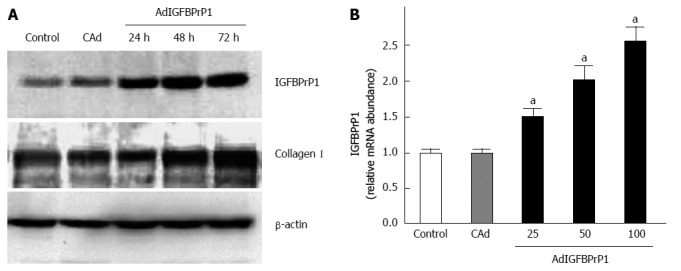
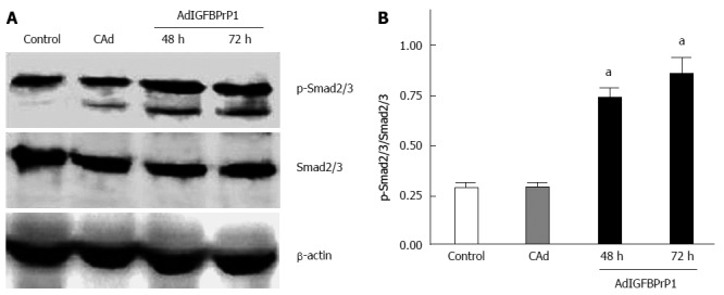
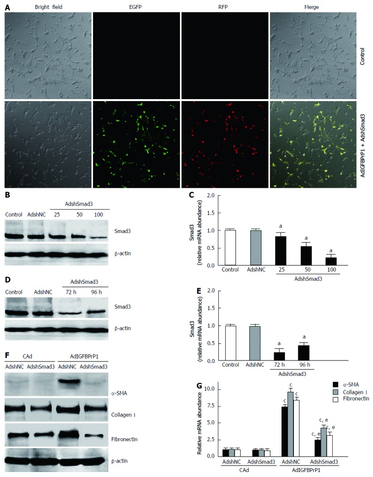
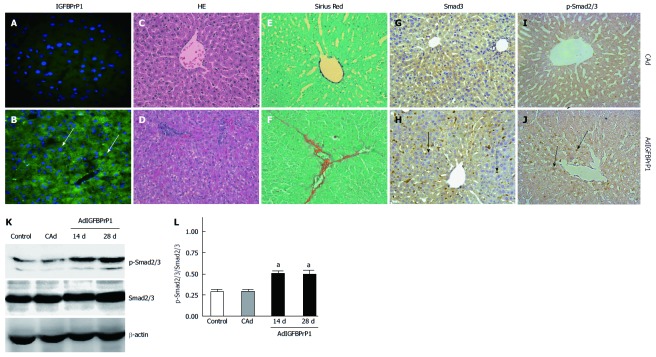
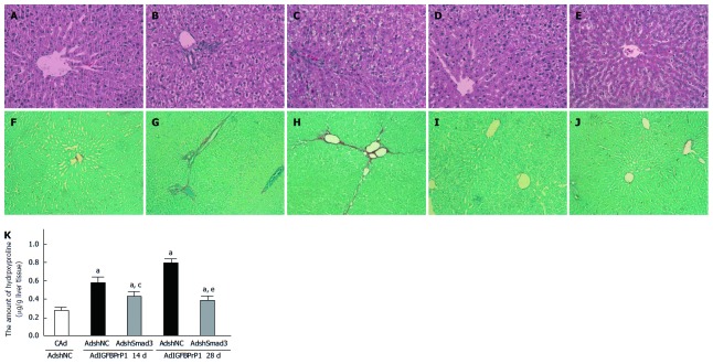
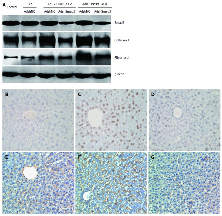
Methods: An in vitro model using hepatic stellate cell (HSC)-T6 cells and an in vivo model of rat liver overexpressing IGFBPrP1 were established using an IGFBPrP1-expressing recombinant adenovirus. The expression of IGFBPrP1 was examined by immunofluorescence, and the expression of collagen I and fibronectin was measured by real-time reverse transcription-polymerase chain reaction and Western blot analysis. The expression of Smad2/3 and p-Smad2/3 was examined by Western blot and immunohistochemistry. A shSmad3-expressing recombinant adenovirus (AdshSmad3) was designed and used to knockdown the Smad3 gene in HSC-T6 cells and rat liver fibrosis transfected with IGFBPrP1. The expression of collagen I, fibronectin, and α-smooth muscle actin (α-SMA) was determined by Western blot analysis and immunohistochemistry. Hepatocyte apoptosis was assessed using TUNEL assay.

Results: IGFBPrP1 overexpression induced collagen deposition and up-regulated the expression of α-SMA and p-Smad2/3, and AdshSmad3 inhibited IGFBPrP1-stimulated p-Smad2/3 activation and the expression of α-SMA, collagen I and fibronectin in HSC-T6 cells. Similarly, increased hepatocyte apoptosis (38.56% ± 3.42% vs 0.24% ± 0.03%, P < 0.05), α-SMA positive stained cells (29.84% ± 1.36% vs 5.83% ± 1.47%, P < 0.05), and increased numbers of Smad3 (35.88% ± 2.15% vs 10.24% ± 1.31%, P < 0.05) and p-Smad2/3 positive cells (28.87% ± 2.73% vs 8.23% ± 0.98%, P < 0.05) were detected in the livers of IGFBPrP1-overexpressing rats compared with the control group. Moreover, AdshSmad3 reduced IGFBPrP1-stimulated Smad3 expression and attenuated α-SMA expression (29.84% ± 1.36% vs 8.23% ± 1.28%, P < 0.05), hepatocyte apoptosis (38.56% ± 3.42% vs 6.75% ± 0.52%, P < 0.05), and both collagen I and fibronectin deposition in the livers of AdIGFBPrP1-treated rats.

Conclusion: IGFBPrP1 induces liver fibrosis by mediating the activation of hepatic stellate cells and hepatocyte apoptosis in a Smad3-dependent mechanism.

Keywords: Hepatic stellate cells; Hepatocyte apoptosis; Insulin-like growth factor binding protein-related protein 1; Liver fibrosis; Smad pathway.


Figures

Figure 1
Figure 1
Insulin-like growth factor binding protein-related protein 1 overexpression induces extracellular matrix production in hepatic stellate cell-T6 cells. Hepatic stellate cell-T6 cells were infected with cDNA (cAd) or adenovirus vector carrying insulin-like growth factor binding protein-related protein 1 (IGFBPrP1) (AdIGFBPrP1) for 24, 48 and 72 h at a multiplicity of infection of 100. Cell lysates were prepared from each culture. A: IGFBPrP1 and collagen I protein expression was detected by Western blot; B: IGFBPrP1 mRNA level was analyzed by real-time polymerase chain reaction. Data are expressed as mean ± SD (n = 4 per group). aP < 0.05 vs the levels in the control group.
Figure 2
Figure 2
Insulin-like growth factor binding protein-related protein 1 overexpression stimulates Smad2/3 phosphorylation in hepatic stellate cell-T6 cells. A: Total and phosphorylated Smad2/3 protein expression in hepatic stellate cell-T6 (HSC-T6) cells was examined by Western blot 48 and 72 h after cDNA (cAd) or adenovirus vector carrying insulin-like growth factor binding protein-related protein 1 (IGFBPrP1) (AdIGFBPrP1) transfection (multiplicity of infection = 100); B: P-Smad2/3 content normalized with total Smad3 content in cAd or AdIGFBPrP1-treated HSC-T6 cells. Data are expressed as mean ± SD (n = 4 per group). aP < 0.05 vs the levels in the control group.
Figure 3
Figure 3
Insulin-like growth factor binding protein-related protein 1-induced extracellular matrix expression is mediated through the Smad pathway in hepatic stellate cell-T6 cells. Hepatic stellate cell-T6 (HSC-T6) cells were co-infected with adenovirus vectors containing shSmad3 (AdshSmad3) or shNC (AdshNC) and adenovirus vector carrying insulin-like growth factor binding protein-related protein 1 (IGFBPrP1) (AdIGFBPrP1). A: Expression of enhanced green fluorescent protein (EGFP) and red fluorescent protein (RFP) in HSC-T6 cells was visualized by confocal microscopy after treatment with AdshSmad3 (magnification ×200); B, C: Smad3 protein (B) and mRNA (C) expression in HSC-T6 cells was detected by Western blot and real-time polymerase chain reaction (RT-PCR) after treatment with different multiplicity of infection (MOI) of AdshSmad3, respectively; D, E: Smad3 protein (D) and mRNA (E) expression was detected by Western blot and real-time RT-PCR 72 h or 96 h after AdshSmad3 treatment (MOI = 100), respectively; F, G: Protein (F) and mRNA (G) expression of α-smooth muscle actin (α-SMA) and extracellular matrix in HSC-T6 cells was analyzed by Western blot and real-time RT-PCR 72 h after AdshSmad3 treatment (MOI = 100), respectively. Data are expressed as mean ± SD (n = 4 per group). aP < 0.05 vs the levels in the control group; cP < 0.05 vs the levels in cAd + AdshNC; eP < 0.05 vs the levels in AdIGFBPrP1 + AdshNC.
Figure 4
Figure 4
Smad2/3 expression in insulin-like growth factor binding protein-related protein 1-induced rat hepatic fibrosis. Rats were injected with cDNA (cAd) or adenovirus vector carrying insulin-like growth factor binding protein-related protein 1 (IGFBPrP1) (AdIGFBPrP1) and sacrificed 28 d after injection. A-J: Protein expression of IGFBPrP1 in hepatocytes and hepatic stellate cells (HSCs) (arrows) in the livers of cAd-treated (A) and AdIGFBPrP1-treated (B) rats was evaluated by immunofluorescence; Histopathology (C, D) and collagen content (E, F) in the livers of cAd-treated and AdIGFBPrP1-treated rats were demonstrated by HE and Sirius Red staining; expression of total Smad2/3 (G, H) and phosphorylated Smad2/3 (I, J) in hepatocytes and HSCs (arrows) in the livers of cAd-treated and AdIGFBPrP1-treated rats was examined by immunohistochemistry (magnification × 400); K: Protein expression of total and phosphorylated Smad2/3 in the livers was examined by Western blot analysis; L: P-Smad2/3 content normalized with Smad3 content in the livers of the control, cAd or IGFBPrP1 group. Data are expressed as mean ± SD (n = 10 per group). aP < 0.05 vs the levels in the control group.
Figure 5
Figure 5
Adenoviral vectors containing shSmad3 ameliorated insulin-like growth factor binding protein-related protein 1-induced liver fibrosis. Rats were sacrificed 14 and 28 d after injection with adenovirus vectors containing shSmad3 (AdshSmad3) or shNC (AdshNC) in the presence of cDNA (cAd) or adenovirus vector carrying insulin-like growth factor binding protein-related protein 1 (IGFBPrP1) (AdIGFBPrP1). Histopathology (A-E) and collagen content (F-J) in the livers were demonstrated by HE and Sirius Red staining (A, F: cAd + AdshNC; B, G: AdIGFBPrP1 + AdshNC 14 d; C, H: AdIGFBPrP1 + AdshSmad3 14 d; D, I: AdIGFBPrP1 + AdshNC 28 d; E, J: AdIGFBPrP1 + AdshSmad3 28 d; magnification × 200); K: The amount of collagen in rat livers was determined using the hydroxyproline assay. Data are expressed as mean ± SD (n = 10 per group). aP < 0.05 vs the levels in the cAd + AdshNC group; cP < 0.05 the levels in the AdIGFBPrP1 + AdshSmad3 14 d group vs those in the AdIGFBPrP1 + AdshNC 14 d group; eP < 0.05 the levels in the AdIGFBPrP1 + AdshSmad3 28 d group vs those in the AdIGFBPrP1 + AdshNC 28 d group.
Figure 6
Figure 6
Adenoviral vector containing shSmad3 inhibits fibrogenic expression in insulin-like growth factor binding protein-related protein 1-treated rats. A: Expression of Smad3, collagen I and fibronectin in the livers were analyzed 28 d after treatment by Western blot; B-D: Hepatocyte apoptosis was examined 28 d after treatment by TUNEL assay; B: cDNA (cAd) + adenovirus vector containing shNC (AdshNC); C: Adenovirus vector carrying insulin-like growth factor binding protein-related protein 1 (IGFBPrP1) (AdIGFBPrP1) + AdshNC; D: AdIGFBPrP1 + adenovirus vector containing shSmad3 (AdshSmad3); E-G: α-smooth muscle actin (α-SMA) expression was examined 28 d after treatment by immunohistochemistry; E: cAd + AdshNC; F: AdIGFBPrP1 + AdshNC; G: AdIGFBPrP1 + AdshSmad3. Data are expressed as mean ± SD (n = 10 per group).

References

    1. Bataller R, Brenner DA. Liver fibrosis. J Clin Invest. 2005;115:209–218. - PMC - PubMed
    1. Leask A, Denton CP, Abraham DJ. Insights into the molecular mechanism of chronic fibrosis: the role of connective tissue growth factor in scleroderma. J Invest Dermatol. 2004;122:1–6. - PubMed
    1. Friedman SL. Mechanisms of hepatic fibrogenesis. Gastroenterology. 2008;134:1655–1669. - PMC - PubMed
    1. Pinzani M. Novel insights into the biology and physiology of the Ito cell. Pharmacol Ther. 1995;66:387–412. - PubMed
    1. Wong L, Yamasaki G, Johnson RJ, Friedman SL. Induction of beta-platelet-derived growth factor receptor in rat hepatic lipocytes during cellular activation in vivo and in culture. J Clin Invest. 1994;94:1563–1569. - PMC - PubMed

Publication types

MeSH terms