

Diagnostic accuracy of MR imaging to identify and characterize focal liver lesions: comparison between gadolinium and superparamagnetic iron oxide contrast media
- PMID: 24914419
- PMCID: PMC4032916
- DOI: 10.3978/j.issn.2223-4292.2014.01.02
Diagnostic accuracy of MR imaging to identify and characterize focal liver lesions: comparison between gadolinium and superparamagnetic iron oxide contrast media
Abstract
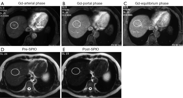
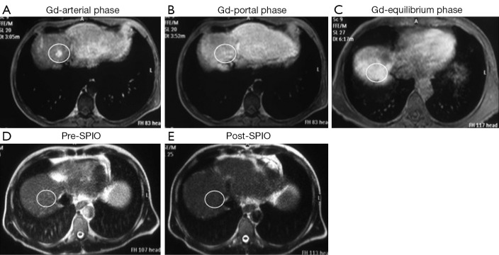
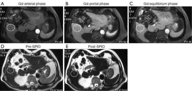
To compare the diagnostic value of gadolinium (Gd) and ultrasmall superparamagnetic iron oxide (SPIO) contrast media for characterization of focal liver lesions (FLL), we retrospectively evaluated the results of magnetic resonance (MR) imaging in 68 patients (40 M, 28 F, age from 22 to 81 yrs) of which 36 with diagnosis of colo-rectal cancer, 26 with hepatic cirrhosis and 6 with incidental imaging detection of FLL. MR (Gyroscan Intera 1.5 T, Philips Medical Systems) study was performed using T1 and T2 fast-field-echo (FFE) and T2 turbo-spin-echo (TSE) sequences in axial and coronal views. Dynamic multi-phases gadolinium Gd-enhanced T1-FFE-Bh images were obtained in arterial, portal and equilibrium phases, followed by SPIO-enhanced T2-FFE scans. A qualitative analysis of pre- and post-contrast MR images to classify FLL as benign or malignant was performed using a 3-point scoring system: 0= benign; 1= suspicious for malignancy; 2= malignant. A total of 118 lesions were evaluated. In particular, histology (n=18), cytology (n=14) or clinical-imaging follow-up data (n=86) demonstrated 4 adenomas, 29 cysts, 3 focal steatosis, 25 hemangiomas, 1 focal vascular abnormality, 5 fibrotic lesions as well as 13 regenerative nodules, 6 dysplastic, 14 hepatocellular carcinomas (HCC), 17 metastasis and 1 cholangiocarcinoma. For MR imaging, diagnostic accuracy, sensitivity, specificity, positive (PPV) and negative (NPV) predictive values of Gd vs. SPIO images were respectively 83% vs. 92%, 79% vs. 74%, 85% vs. 99% (P=0.002), 68% vs. 96% (P=0.005) and 91% vs. 90%, respectively. The results suggest that SPIO-MR provides a diagnostic incremental value, as specificity and PPV, particularly to characterize FLL compared to Gd-MR; thus, we strongly recommend the use of SPIO when liver lesion characterization is requested and Gd images are uncertain.
Keywords: MR imaging; contrast media; focal liver lesions (FLL); gadolinium; superparamagnetic iron oxide (SPIO).
Figures
References
-
- Gervais DA, Goldberg SN, Brown DB, et al. Society of Interventional Radiology position statement on percutaneous radiofrequency ablation for the treatment of liver tumors. J Vasc Interv Radiol 2009;20:3-8 - PubMed
-
- Helmberger T.Interventional procedures for hepatic metastases. Chirurg 2010;81:542-50 - PubMed
-
- Poon RT, Fan ST. Hepatectomy for hepatocellular carcinoma: patient selection and postoperative outcome. Liver Transpl 2004;10:S39-45 - PubMed
-
- Schwartz M.Liver transplantation in patients with hepatocellular carcinoma. Liver Transpl 2004;10:S81-5 - PubMed
-
- Dai Y, Chen MH, Yin SS, et al. Focal liver lesions: can SonoVue-enhanced ultrasound be used to differentiate malignant from benign lesions? Invest Radiol 2007;42:596-603 - PubMed
LinkOut - more resources
Full Text Sources