. 2014 Jun 10;9(6):e99080.
doi: 10.1371/journal.pone.0099080. eCollection 2014.

Age-dependent transcriptome and proteome following transection of neonatal spinal cord of Monodelphis domestica (South American grey short-tailed opossum)

Affiliations

Age-dependent transcriptome and proteome following transection of neonatal spinal cord of Monodelphis domestica (South American grey short-tailed opossum)

Norman R Saunders et al. PLoS One. .

Abstract

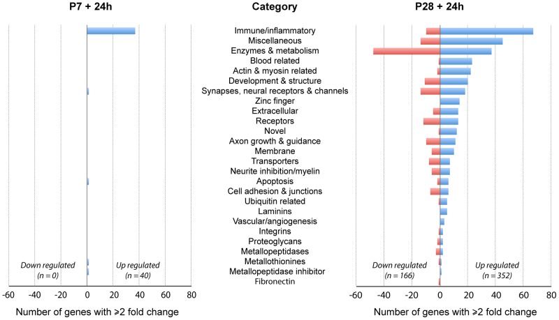
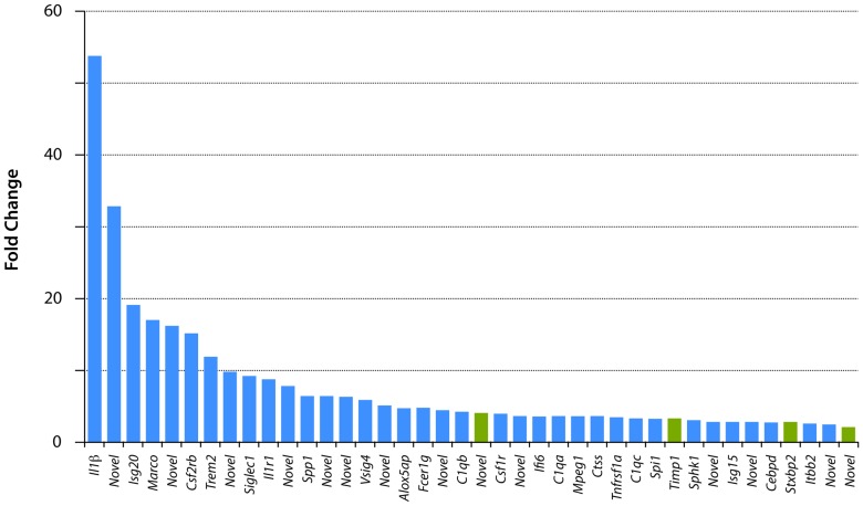
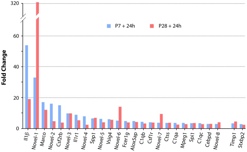
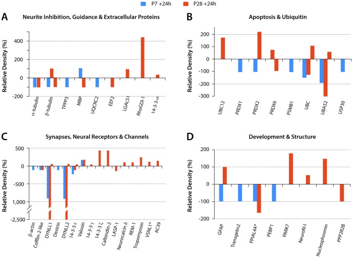
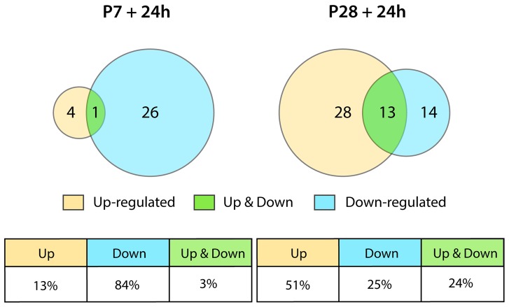
This study describes a combined transcriptome and proteome analysis of Monodelphis domestica response to spinal cord injury at two different postnatal ages. Previously we showed that complete transection at postnatal day 7 (P7) is followed by profuse axon growth across the lesion with near-normal locomotion and swimming when adult. In contrast, at P28 there is no axon growth across the lesion, the animals exhibit weight-bearing locomotion, but cannot use hind limbs when swimming. Here we examined changes in gene and protein expression in the segment of spinal cord rostral to the lesion at 24 h after transection at P7 and at P28. Following injury at P7 only forty genes changed (all increased expression); most were immune/inflammatory genes. Following injury at P28 many more genes changed their expression and the magnitude of change for some genes was strikingly greater. Again many were associated with the immune/inflammation response. In functional groups known to be inhibitory to regeneration in adult cords the expression changes were generally muted, in some cases opposite to that required to account for neurite inhibition. For example myelin basic protein expression was reduced following injury at P28 both at the gene and protein levels. Only four genes from families with extracellular matrix functions thought to influence neurite outgrowth in adult injured cords showed substantial changes in expression following injury at P28: Olfactomedin 4 (Olfm4, 480 fold compared to controls), matrix metallopeptidase (Mmp1, 104 fold), papilin (Papln, 152 fold) and integrin α4 (Itga4, 57 fold). These data provide a resource for investigation of a priori hypotheses in future studies of mechanisms of spinal cord regeneration in immature animals compared to lack of regeneration at more mature stages.


Conflict of interest statement

Competing Interests: The authors have declared that no competing interests exist.

Figures

Figure 1
Figure 1. Monodelphis domestica spinal cords injured at P7 or P28.
Longitudinal sections (hematoxylin & eosin staining) of spinal cords injured at P7 or P28 shown immediately after complete spinal transection at T10 (A, B) or 24 hours later (C, D). Note obvious bleeding into the injury site at P28 (B), which was more pronounced than at P7 (A) One day after transection (+24 h) the gap between severed ends of the cord was larger in P28 injured animals (D) than in P7 injured animals (C). Rostral end is to the left, caudal to the right, dorsal is uppermost. Scale bar is 500 µm.
Figure 2
Figure 2. Changes in gene expression in spinal cord 24Monodelphis domestica.
Numbers of genes in each functional category that showed ±≥2 fold change compared to uninjured aged matched controls. Note dominance of immune/inflammatory genes particularly at P7. Also note that at P28 many more genes showed expression changes (both up and down). See Tables S1 and S2 for gene descriptions, fold change and p values.
Figure 3
Figure 3. Changes in gene expression 24
Only 40 genes changed their expression levels by ≥2 fold. All were upregulated. See Table S1 for gene descriptions and statistics. There were 12 “novel” genes; search of GO categories showed that these have immune/inflammatory properties. Note that only four of the genes in this figure (green bars) are not in the immune/inflammatory category (blue bars).
Figure 4
Figure 4. Interleukin-1β in Monodelphis spinal cord 24 hours after a complete transection at P7 or P28.
In the segment of the cord rostral to the site of injury Il-1β was detected using cross-reacting antibodies to the human cytokine. Note strong immunopositive signal in the tissue surrounding the cords at P7-injured (A) and P28 (B) but lack of significant staining within the spinal tissue especially at P7 (A). One day following injury at P28 a few immunopositive cells with the general morphology of monocytes were detected, especially in segments of the cord more rostral to the injury (C). Scale bars A, B = 500 µm, C = 100 µm.
Figure 5
Figure 5. Comparison of expression levels of the twenty-six genes that changed expression in spinal cord 24 h following transection at both P7 and P28.
All but two of these genes (Timp1 and Stxbp2) were in the immune/inflammatory category. The magnitude of the expression changes was generally similar at the two ages, but more genes (six: IL1β, MARCO, novel-2, CSF2RB, IL1-R1, novel-4) showed greater upregulation at P7 than at P28 (three: novel-1, novel-6, novel-7). A search of Ensembl, NCBI “Gene” and GO categories showed that all of these novel genes are involved in immune/inflammatory functions (see Tables S1 and S2).
Figure 6
Figure 6. Myelin staining in the developing spinal cord of Monodelphis domestica.
Transverse sections through thoracic spinal cord of P8 (A) and P29 (B) spinal cord stained with Luxol fast blue (LFB). There was no LFB stained myelin at P8 (A) but relatively well- developed myelin was present at P28 (B). However even at P28 the myelination is only beginning to appear (first detected between P21 and P28) and does not reach adult levels until several weeks later (not illustrated). Dorsal is uppermost. Scale bar is 500 µm.
Figure 7
Figure 7. Proteins by functional groups with changed expression levels after spinal transection at P7 or P28.
Estimates of protein expression levels from densitometry measurements. Values are expressed as % change from control values (100%). y axis: Relative Density (%). Proteins grouped by functions as listed in Tables S4 and S5. Abbreviations: TPPP3-Brain specific protein; UQCRC2-Ubiquinol-cytochrome c reductase core protein II; EEF2-Elongation factor 2 isoform 1; LGALS1-galactin1; MBP–Myelin basic protein; PRDX1-peroxiredoxin1; PRDX2–peroxiredoxin 2; PRDX6–peroxiredoxin 6; PSMB1-Proteasome subunit β type 1; UBC–Ubiquitin C; UBA52-Ubiquitin A-52; USP30-Ubiquitin specific peptidase 30 phosphoglycerate kinase; BC12-NEDD8-conjugating enzyme UBC12; DYNLL1- Cytoplasmic dynein light chain 1; DYNLL2-Dynein light chain LC8 type 2; VSLN1-Visinin like-1; AC39-AC39/physophilin; GFAP–glial fibrillary acidic protein; TAGLN2-transgelin 2; PPIAL4A - Peptidylprolyl isomerase A-like PEBP1 - Phosphatidylethanolamine-binding protein 1; PARK7 - Parkinson protein 7; Neurofil-L – Neurofilament-L subunit; PPP2R2B - Protein phosphatase 2 regulatory subunit B. Two proteins, VSNL1 and PPIAL4A (marked with *) are mean values as their expression levels changes were detected in more than one fraction (see Table S4 and S5 for individual changes). Note also that a few of the proteins listed showed both up- and downregulation but only in P28 injury group.
Figure 8
Figure 8. Summary of numbers of proteins that changed expression levels 24
All but five of the proteins identified at P7 as showing a change in expression after injury were downregulated. At P28 half of the proteins were upregulated.

References

    1. Aguayo AJ, David S, Bray GM (1981) Influences of the glial environment on the elongation of axons after injury: transplantation studies in adult rodents. The Journal of experimental biology 95: 231–240. - PubMed
    1. David S, Aguayo AJ (1981) Axonal elongation into peripheral nervous system "bridges" after central nervous system injury in adult rats. Science 214: 931–933. - PubMed
    1. Tello F (1911) La influencia del neurotropismo en la regeneracion de los centros nerviosos. Trab Laborator Invest Biol University Madrid 9: 123–159.
    1. Akbik F, Cafferty WB, Strittmatter SM (2011) Myelin associated inhibitors: A link between injury-induced and experience-dependent plasticity. Experimental neurology. - PMC - PubMed
    1. Buchli AD, Schwab ME (2005) Inhibition of Nogo: a key strategy to increase regeneration, plasticity and functional recovery of the lesioned central nervous system. Annals of medicine 37: 556–567. - PubMed

Publication types

MeSH terms