. 2014 Jun;70(Pt 6):1561-71.
doi: 10.1107/S1399004714005331. Epub 2014 May 23.

Structure of the phosphotransferase domain of the bifunctional aminoglycoside-resistance enzyme AAC(6')-Ie-APH(2'')-Ia

Affiliations

Structure of the phosphotransferase domain of the bifunctional aminoglycoside-resistance enzyme AAC(6')-Ie-APH(2'')-Ia

Clyde A Smith et al. Acta Crystallogr D Biol Crystallogr. 2014 Jun.

Abstract

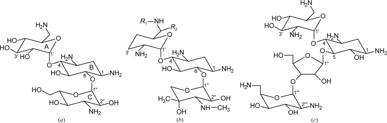
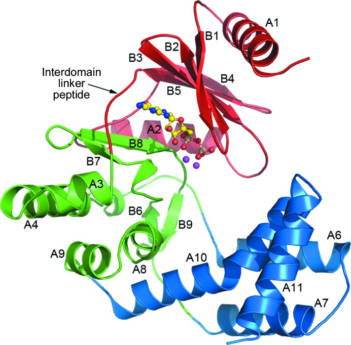
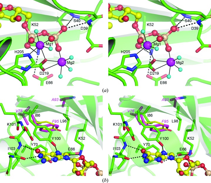
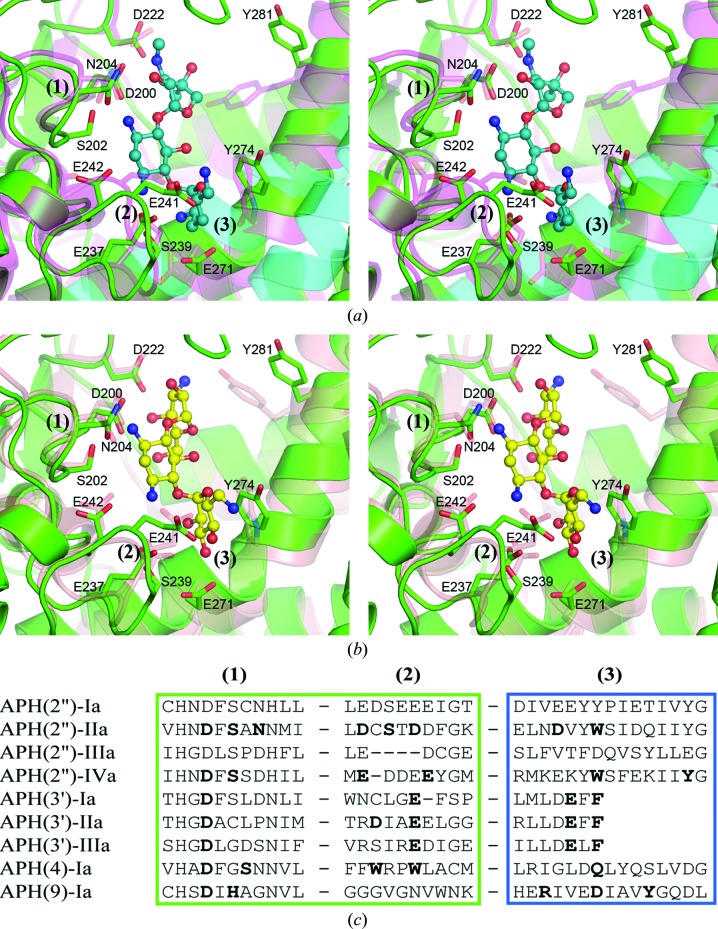
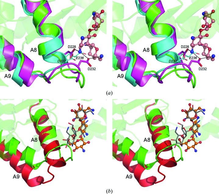
The bifunctional acetyltransferase(6')-Ie-phosphotransferase(2'')-Ia [AAC(6')-Ie-APH(2'')-Ia] is the most important aminoglycoside-resistance enzyme in Gram-positive bacteria, conferring resistance to almost all known aminoglycoside antibiotics in clinical use. Owing to its importance, this enzyme has been the focus of intensive research since its isolation in the mid-1980s but, despite much effort, structural details of AAC(6')-Ie-APH(2'')-Ia have remained elusive. The structure of the Mg2GDP complex of the APH(2'')-Ia domain of the bifunctional enzyme has now been determined at 2.3 Å resolution. The structure of APH(2'')-Ia is reminiscent of the structures of other aminoglycoside phosphotransferases, having a two-domain architecture with the nucleotide-binding site located at the junction of the two domains. Unlike the previously characterized APH(2'')-IIa and APH(2'')-IVa enzymes, which are capable of utilizing both ATP and GTP as the phosphate donors, APH(2'')-Ia uses GTP exclusively in the phosphorylation of the aminoglycoside antibiotics, and in this regard closely resembles the GTP-dependent APH(2'')-IIIa enzyme. In APH(2'')-Ia this GTP selectivity is governed by the presence of a `gatekeeper' residue, Tyr100, the side chain of which projects into the active site and effectively blocks access to the adenine-binding template. Mutation of this tyrosine residue to a less bulky phenylalanine provides better access for ATP to the NTP-binding template and converts APH(2'')-Ia into a dual-specificity enzyme.

Keywords: AAC(6′)-Ie-APH(2′′)-Ia; aminoglycoside-resistance enzymes.


Figures

Figure 1
Figure 1
Aminoglycoside antibiotics. (a) Kanamycin A. (b) Gentamicin C, typically a mixture of types C1 (R 1 = R 2 = CH3), C1a (R 1 = R 2 = H) and C2 (R 1 = CH3, R 2 = H). (c) Neomycin.
Figure 2
Figure 2
Ribbon representation of the APH(2′′)-Ia structure showing the three structural subdivisions: the N-terminal domain (red), the core subdomain (green) and the helical subdomain (blue). The location of the GDP molecule is shown as a yellow ball-and-stick representation and the two associated magnesium ions are represented as magenta spheres. The secondary-structure numbering is also given, and the interdomain linker peptide between the N-terminal domain and the core subdomain is indicated.
Figure 3
Figure 3
The APH(2′′)-Ia nucleotide-binding site. (a) Stereoview of the binding site showing the interactions which anchor the GDP (yellow ball-and-stick representation) in APH(2′′)-Ia (green). The two magnesium ions (Mg1 and Mg2) are shown as magenta spheres, with their coordinating water molecules shown as small cyan spheres. Hydrogen bonds are shown as dashed lines and the magnesium coordinate bonds as thin solid lines. The amino-acid residues involved in nucleotide binding and magnesium coordination are indicated. (b) Stereoview of the secondary hydrophobic binding pocket in APH(2′′)-Ia. The gatekeeper residue Tyr100 is shown in the center of the figure. The residues which make up the secondary hydrophobic pocket are indicated for APH(2′′)-Ia (green sticks) and APH(2′′)-IVa (magenta sticks). The location of the gatekeeper Phe95 in APH(2′′)-IVa is shown bound in the pocket formed by the less bulky residues Val78 and Ala93 (residue labels in magenta). The Tyr100 side chain, were it to swing into the pocket, is shown as thin yellow sticks and the severe steric clashes that this side chain would make with the pocket residues can be seen.
Figure 4
Figure 4
The substrate-binding site in the APH enzymes. (a) Stereoview of the superposition of APH(2′′)-Ia (green) and APH(2′′)-IIa (magenta, partially transparent). The gentamicin bound to APH(2′′)-IIa is shown in a blue/dull green ball-and-stick representation. The residues which interact with gentamicin are shown as partially transparent magenta sticks. The corresponding residues in APH(2′′)-Ia are shown as green sticks and their residue numbers are given. Helices A10 and A11 from APH(2′′)-IIIa are shown as partially transparent cyan coils. (b) Stereoview of the superposition of APH(2′′)-Ia (green) and APH(2′′)-IVa (salmon, partially transparent). The kanamycin bound to APH(2′′)-IVa is shown in a yellow ball-and-stick representation. The residues which interact with kanamycin are shown as partially transparent salmon sticks. The corresponding residues in APH(2′′)-Ia are shown as green sticks. The three substrate-binding motifs (1), (2) and (3) are indicated in both (a) and (b). (c) Partial sequence alignment of the three substrate-binding motifs identified in the APH(2′′) enzymes. The corresponding sequences from three APH(3′) enzymes, APH(4)-Ia and APH(9)-Ia are also aligned. Residues highlighted in bold are known to interact with substrate molecules based upon the known structures of enzyme–substrate complexes.
Figure 5
Figure 5
The structure of the A8–A9 loop in the APHs. (a) Superposition of APH(2′′)-Ia (green, partially transparent), APH(2′′)-IIa (magenta) and APH(2′′)-IIIa (cyan). Only helices A8 and A9 are shown for the latter two enzymes for clarity. Gentamicin from APH(2′′)-IIa (Young et al., 2009 ▶) is shown in a pink ball-and-stick representation. APH(2′′)-IIIa lacks the extended A8–A9 loop that the other three APH(2′′) enzymes possess. (b) Superposition of APH(2′′)-Ia (green, partially transparent) and APH(3′)-IIIa (red). The latter enzyme also lacks the extended A8–A9 loop. Kanamycin (orange ball-and-stick representation) and neomycin (gray sticks) are shown in the location observed in complexes with APH(3′)-IIIa (Fong & Berghuis, 2002 ▶).

Similar articles

Cited by

References

    1. Adams, P. D. et al. (2010). Acta Cryst. D66, 213–221.
    1. Aimes, R. T., Hemmer, W. & Taylor, S. S. (2000). Biochemistry, 39, 8325–8332. - PubMed
    1. Berman, H. M., Westbrook, J., Feng, Z., Gilliland, G., Bhat, T. N., Weissig, H., Shindyalov, I. N. & Bourne, P. E. (2000). Nucleic Acids Res. 28, 235–242. - PMC - PubMed
    1. Bhattacharya, M., Toth, M., Smith, C. A. & Vakulenko, S. B. (2013). Antimicrob. Agents Chemother. 57, 3763–3766. - PMC - PubMed
    1. Boehr, D. D., Daigle, D. M. & Wright, G. D. (2004). Biochemistry, 43, 9846–9855. - PubMed

Publication types

Associated data

LinkOut - more resources