. 2014 Jun;70(Pt 6):1622-30.
doi: 10.1107/S1399004714006439. Epub 2014 May 24.

Structural and functional analysis of the human spliceosomal DEAD-box helicase Prp28

Affiliations

Structural and functional analysis of the human spliceosomal DEAD-box helicase Prp28

Sina Möhlmann et al. Acta Crystallogr D Biol Crystallogr. 2014 Jun.

Abstract

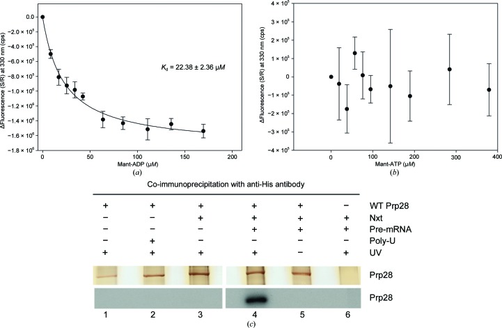
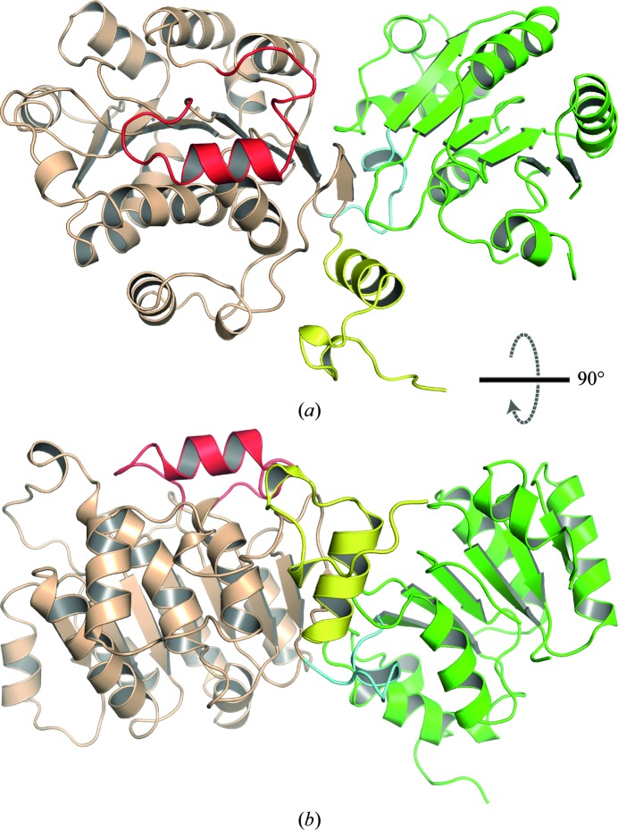
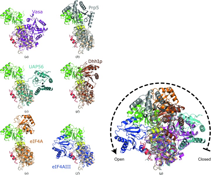
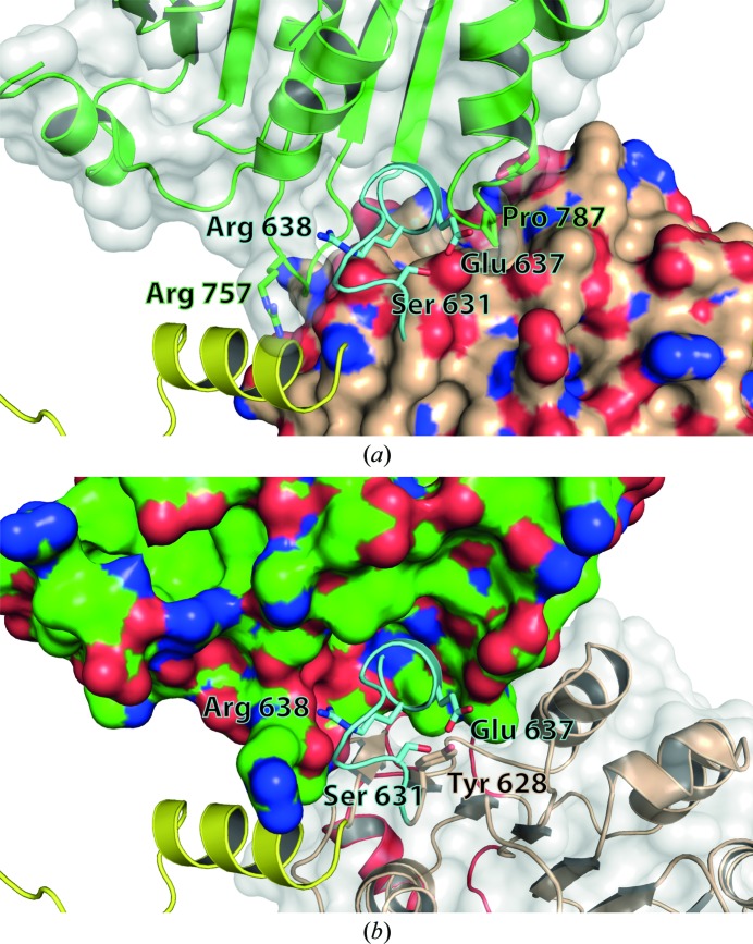
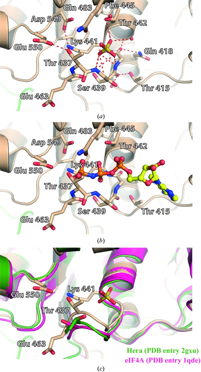
The DEAD-box protein Prp28 is essential for pre-mRNA splicing as it plays a key role in the formation of an active spliceosome. Prp28 participates in the release of the U1 snRNP from the 5'-splice site during association of the U5·U4/U6 tri-snRNP, which is a crucial step in the transition from a pre-catalytic spliceosome to an activated spliceosome. Here, it is demonstrated that the purified helicase domain of human Prp28 (hPrp28ΔN) binds ADP, whereas binding of ATP and ATPase activity could not be detected. ATP binding could not be observed for purified full-length hPrp28 either, but within an assembled spliceosomal complex hPrp28 gains ATP-binding activity. In order to understand the structural basis for the ATP-binding deficiency of isolated hPrp28, the crystal structure of hPrp28ΔN was determined at 2.0 Å resolution. In the crystal the helicase domain adopts a wide-open conformation, as the two RecA-like domains are extraordinarily displaced from the productive ATPase conformation. Binding of ATP is hindered by a closed conformation of the P-loop, which occupies the space required for the γ-phosphate of ATP.

Keywords: DEAD-box proteins; Prp28; helicase domain.


Figures

Figure 1
Figure 1
Adenosine nucleotide binding of hPrp28 determined by (a, b) fluorescence titration using mant-ADP or mant-ATP and (c) UV-induced cross-linking using α-32P-labelled ATP. (a, b) The difference in corrected fluorescence signal is plotted against the ADP/ATP concentration and fitted to the Michaelis–Menten equation. For mant-ADP, a K d of 22.4 ± 2.4 µM was calculated, while in the case of mant-ATP curve fitting was not possible. (c) UV-induced cross-linking of α-32P ATP to purified hPrp28. 30 pmol purified hPrp28 was incubated in the absence of nuclear extract (Nxt; lanes 1–2) without (lane 1) or with (lane 2) poly-U (0.6 µg µl−1) or in the presence of nuclear extract (lanes 3–6) in the absence (lane 3) or presence (lane 4) of MINX pre-mRNA under splicing conditions (see §2) and subjected to UV irradiation. After cross-linking, hPrp28 was immunoprecipitated with anti-His antibody. The precipitates were loaded onto a 10% SDS–PAGE and radioactive hPrp28 (cross-linked to ATP) was detected by autoradiography. Cross-linking of ATP to hPrp28 was also assayed in the absence of UV irradiation (lane 5). As control for immunoprecipitation the reaction was carried out without purified hPrp28 (lane 6).
Figure 2
Figure 2
Two perpendicular views (a, b) of the hPrp28ΔN structure. The N-­terminal extension is coloured yellow, the RecA-1 domain brown and the RecA-2 domain green. Additionally, the insertion in RecA-1 is highlighted in red and the linker connecting RecA-1 and RecA-2 is coloured light blue.
Figure 3
Figure 3
The helicase core of hPrp28ΔN has an open overall conformation as the two RecA domains are displaced with respect to the active conformation represented by Vasa (purple). Superposition of hPrp28ΔN with (a) Vasa in its active closed conformation and other DEAD-box proteins exhibiting an open conformation: (b) Prp5, (c) UAP56, (d) Dhh1p, (e) eIF4A and (f) eIF4AIII. (g) Superposition of (a)–(f).
Figure 4
Figure 4
Interactions of the back side of the RecA-2 domain (green ribbon model) with the RecA-1 domain, the linker between the two RecA domains (blue) and the N-terminal extension (yellow). (a) The surface of RecA-1 is coloured gold and surface-exposed N and O atoms are coloured blue and red, respectively. (b) The surface of RecA-2 is coloured green and surface-exposed N and O atoms are coloured blue and red, respectively.
Figure 5
Figure 5
Conformation of the P-loop. (a) The conformation of the P-loop is stabilized by multiple hydrogen bonds (red dashed lines). The ATP binding pocket is occupied by a sulfate ion. (b) Model of bound ATP based on the Vasa–ATP complex structure. ATP binding is hindered by the P-loop owing to the hydrogen bonds between Thr437, Glu550 and Lys441. (c) The P-loop conformation of hPrp28ΔN is unique, as in the structures of eIF4A with bound sulfate or of the DEAD-box helicase Hera with bound phosphate the hydrogen bond between the Glu of the DEAD motif and the Thr or Ser of the P-loop is broken.

References

    1. Abrahams, J. P. & Leslie, A. G. W. (1996). Acta Cryst. D52, 30–42. - PubMed
    1. Adams, P. D. et al. (2010). Acta Cryst. D66, 213–221.
    1. Battye, T. G. G., Kontogiannis, L., Johnson, O., Powell, H. R. & Leslie, A. G. W. (2011). Acta Cryst. D67, 271–281. - PMC - PubMed
    1. Benz, J., Trachsel, H. & Baumann, U. (1999). Structure, 7, 671–679. - PubMed
    1. Birdsall, B., King, R. W., Wheeler, M. R., Lewis, C. A. Jr, Goode, S. R., Dunlap, R. B. & Roberts, G. C. (1983). Anal. Biochem. 132, 353–361. - PubMed

Publication types

MeSH terms

Associated data

LinkOut - more resources