. 2014 Jun 10;9(6):e99083.
doi: 10.1371/journal.pone.0099083. eCollection 2014.

Laminin α2-mediated focal adhesion kinase activation triggers Alport glomerular pathogenesis

Affiliations

Laminin α2-mediated focal adhesion kinase activation triggers Alport glomerular pathogenesis

Duane Delimont et al. PLoS One. .

Abstract

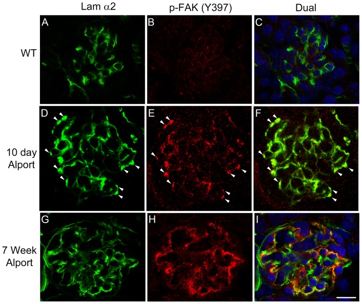
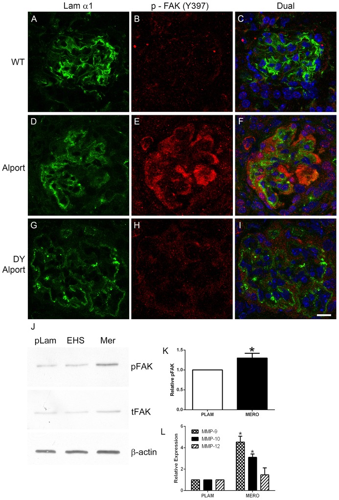
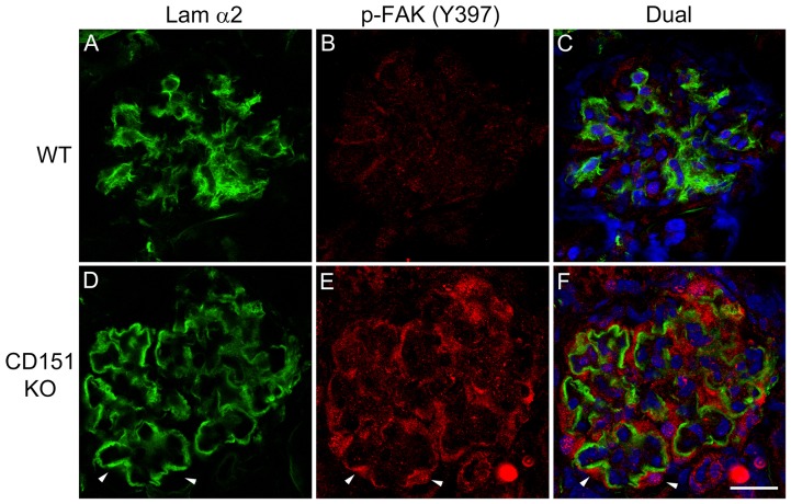
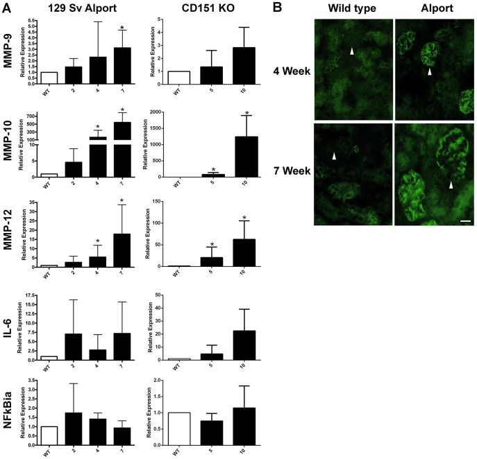
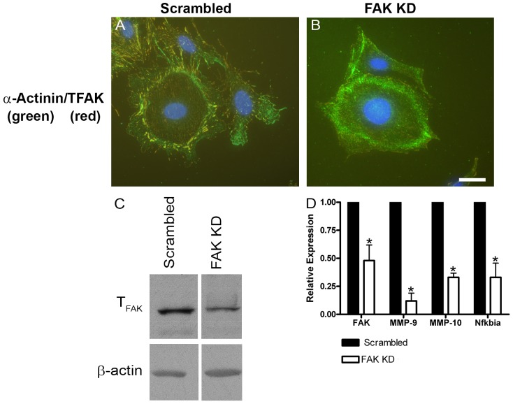
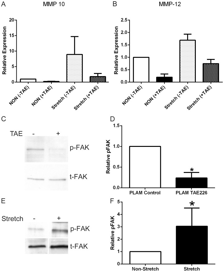
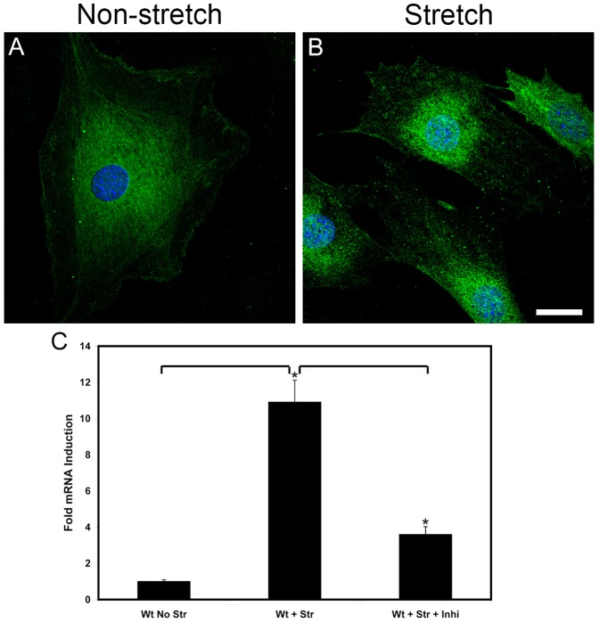
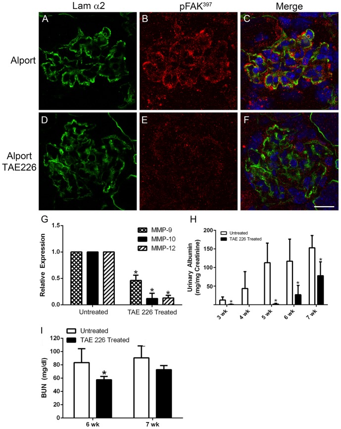
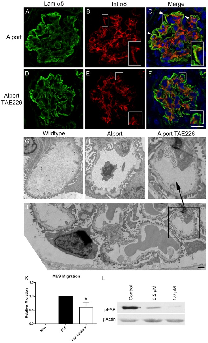
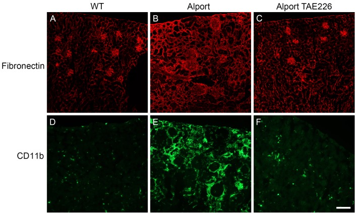
It has been known for some time that laminins containing α1 and α2 chains, which are normally restricted to the mesangial matrix, accumulate in the glomerular basement membranes (GBM) of Alport mice, dogs, and humans. We show that laminins containing the α2 chain, but not those containing the α1 chain activates focal adhesion kinase (FAK) on glomerular podocytes in vitro and in vivo. CD151-null mice, which have weakened podocyte adhesion to the GBM rendering these mice more susceptible to biomechanical strain in the glomerulus, also show progressive accumulation of α2 laminins in the GBM, and podocyte FAK activation. Analysis of glomerular mRNA from both models demonstrates significant induction of MMP-9, MMP-10, MMP-12, MMPs linked to GBM destruction in Alport disease models, as well as the pro-inflammatory cytokine IL-6. SiRNA knockdown of FAK in cultured podocytes significantly reduced expression of MMP-9, MMP-10 and IL-6, but not MMP-12. Treatment of Alport mice with TAE226, a small molecule inhibitor of FAK activation, ameliorated fibrosis and glomerulosclerosis, significantly reduced proteinuria and blood urea nitrogen levels, and partially restored GBM ultrastructure. Glomerular expression of MMP-9, MMP-10 and MMP-12 mRNAs was significantly reduced in TAE226 treated animals. Collectively, this work identifies laminin α2-mediated FAK activation in podocytes as an important early event in Alport glomerular pathogenesis and suggests that FAK inhibitors, if safe formulations can be developed, might be employed as a novel therapeutic approach for treating Alport renal disease in its early stages.


Conflict of interest statement

Competing Interests: The authors have declared that no competing interests exist.

Figures

Figure 1
Figure 1. Activation of focal adhesion kinase occurs specifically in regions of the GBM where laminin α2 is present, and is a very early event in Alport glomerular pathogenesis.
Cryosections from 10 day old Alport mice (D–F), 7 week old Alport mice (G–I), and wild type littermates (A–C) were immunostained with antibodies specific for the α2 chain of laminin or pFAK397. Arrowheads denote areas of dual immunostaining in the glomerular capillary loops. Scale bar = 15 µm.
Figure 2
Figure 2. Laminin α2, but not laminin α1 activates FAK on podocytes in vivo and in vitro.
Panels A–C; 7 week old wild type glomerulus stained with antibodies specific for laminin 111 and pFAK397 show absence of pFAK immunostaining. Panels D–F; 7 week Alport glomerulus stained with antibodies specific for laminin α1 and pFAK397 pFAK immunostaining in podocytes adjacent to laminin α1-immunopositive GBM. Panels G-I show the same immunostaining as for D–F using Alport mice that do not express laminin α2 (the dy/dy muscular dystrophy mutation). Note the absence of pFAK397 immunostaining even though GBM is immunopositive for laminin α1. Panel J. Wild type podocytes were differentiated for 2 weeks and then plated on placental laminin, EHS laminin, or merosin for 15 hours. Extracts were prepared and analyzed by western blot for expression of pFAK397 and total FAK. β-actin was used as a loading control). Panel K shows quantitative analysis of pFAK397 relative to total FAK for several western blots. Panel L shows real time qRT-PCR results for transcripts endocing the indicated MMPs, demonstrating significantly elevated expression of MMP-9 and MMP-10 for cells cultured on merosin (MERO) relative to cells cultured on placental laminin (PLAM). Scale bar = 10 µm.
Figure 3
Figure 3. Activation of focal adhesion kinase occurs specifically in regions of the GBM where laminin α2 is present in CD151 knockout mice.
Cryosections from 10 week old CD151 knockout mice (D–F) and wild type littermates (A–C) were immunostained with antibodies specific for the α2 chain of laminin or pFAK397. Arrowheads denote areas of dual immunostaining along the capillary loops. Scale bar = 15 µm.
Figure 4
Figure 4. Induction kinetics for MMP-9, MMP-10, MMP-12, IL-6, and NF-kappaBia in glomeruli from Alport mice and CD151 knockout mice.
Panel A. Glomeruli were isolated from CD151 knockout mice and Alport mice along with strain/age matched wild type littermates at the indicated ages using bead isolation. Total glomerular RNA was analyzed by real time RT-PCR using primers specific for the indicated transcripts. Each data point represents at least five independent samples. Significant differences when comparing the data from mutants with wild type littermates are denoted with asterisks (p<0.05). Note that IL-6 and NF-kappaBia did not reach significance likely due to a large variance in the data, but trended towards significance. Panel B shows that MMP-10 protein is induced in Alport glomeruli at both 4 and 7 weeks of age as determined by immunofluorescence analysis. Scale bar = 15 µm.
Figure 5
Figure 5. Stable siRNA knock-down of FAK in cultured podocytes results in significantly reduced expression of MMP-9, MMP-10, and NF-kappaBia.
Conditionally immortalized podocyte cell cultures were transfected with vector encoding a siRNA expression cassette for FAK. A vector encoding a scrambled siRNA was used as a control. Stable clones were selected and propagated. The data presented is representative of several independently selected clones. Panels A and B show that while cells expressing the scrambled vector still have robust focal adhesions (panel A), they are significantly reduced or absent in the cells expressing the FAK siRNA (panel B). Western blot for total FAK confirms a reduction of FAK protein in the FAK siRNA transfected cultures (panel C). Real time qRT-PCR analysis of RNA from these clones shows a significant reduction in the expression of mRNAs encoding FAK, MMP-9, MMP-10, and NF-kappaBia in FAK siRNA expressing cells versus those expressing the scrambled siRNA. Scale bar = 15 µm.
Figure 6
Figure 6. The small molecule inhibitor for FAK, TAE226, reduces FAK activation and stretch-induced MMP-10 and MMP-12 expression in cultured podocytes.
Panels A and B, cells were treated or not with TAE226 under static and stretched conditions and mRNA analyzed by real time qRT-PCR for the indicated transcripts. Panel C, Podocytes were cultured on placental laminin overnight and TAE226 added 1 hour before stretching. Extracts were prepared and analyzed by western blot for expression of pFAK397 and total FAK. Panel E, FAK activation was also analyzed by western blot of podocyte extracts from stretched and non-stretched cells, demonstrating that biomechanical stretching directly activates FAK. Panels D and F, Several replicate blots were scanned and quantified for pFAK relative to total FAK and statistically analyzed. Statistically significant differences are indicated with asterisks where p<0.05.
Figure 7
Figure 7. Biomechanical stretching activates NF-kappaB which regulates MMP-10 expression in cultured podocytes.
NF-kappaB localizes primarily to the cytosol in non-stretched cultured podocytes (panel A). Subjecting the cells to cyclic biomechanical stretching results in the nuclear localization of NF-kappaB (panel B), which is consistent with its activation. Panel C shows that stretch-mediated induction of MMP-10 is blocked by addition of a peptide inhibitor for NF-kappaB to the culture medium. Scale bar = 20 µm.
Figure 8
Figure 8. Treatment of Alport mice with the small molecule inhibitor for FAK, TAE226, blocks FAK activation, significantly reduces glomerular expression of MMP-9, -10, and -12, and ameliorates proteinuria and blood urea nitrogen levels.
129 Sv/J autosomal Alport mice were treated with TAE226 from 2 to 7 weeks of age. Panels A–F show that while pFAK397 immunostaining is present in podocytes adjacent to laminin α2-immunopositive basement membranes in vehicle treated mice, it is absent in mice treated with TAE226, indicating effective blockade. Real time qRT-PCR analysis of glomerular RNA in panel G shows significant reduction in expression of MMP-9, MMP-10, and MMP-12 in TAE226 treated mice relative to those given vehicle. Panel H and I show significant amelioration of proteinuria and BUN in treated mice, indicative of improved glomerular function. Scale bar = 15 µm.
Figure 9
Figure 9. Treatment of Alport mice with TAE226 reduces mesangial process invasion of the glomerular capillary loops, ameliorates GBM ultrastructural dysmorphology, and significantly reduces pFAK activation and migratory potential of primary cultured mesangial cells.
The same mice as in Figure 8 were dual immunostained with the GBM marker, laminin α5, and the mesangial cell marker, integrin α8. Arrowheads in panel C denote regions where invasion of the capillary loops by mesangial processes is evident (panels B and C, inserts). This characteristic is markedly reduced in the TAE226-treated glomeruli where integrin α8 immunostaining is restricted to the mesangial angles (panels E and F. inserts). Transmission electron microscopic analysis shows that TAE226 treatment (Panel I) reduces the ultrastructural damage to the GBM normally present by 7 weeks of age in this model (panel H). Panel J is provided to indicate that amelioration of GBM dysmorphology is generally observed. Panel K shows that treatment of primary cultured mesangial cells with TAE226 significantly reduces their migratory potential relative to untreated cells. Panel L shows a dose response for FAK inhibition by TAE226 in cultured mesangial cells. Panel F, scale bar = 15 µm; Panel J, scale bar = 2 µm.
Figure 10
Figure 10. Treatment of Alport mice with TAE226 ameliorates interstitial fibrosis and monocyte infiltration.
Kidney cryosections from wild type and Alport mice that were either treated with vehicle or TAE226 were immunostained with antibodies specific for fibronectin (A–C or the monocyte marker, CD11b (D–F). The accumulation of fibronectin in the interstitium, indicative of fibrosis, while abundant in Alport mice (panel B) is not apparent in Alport mice treated with TAE226 (panel C), which appear similar to wild type mice (panel A). Similarly, monocyte infiltration, as indicated by CD11b immunopositive cells, is readily apparent in Alport mice (panel E). In TAE226-treated Alport mice (panel F), however, the abundance of monocytes is similar to that in wild type mice (panel D), which are resident cells rather than infiltrating cells. Scale bar = 50 µm.

References

    1. Kruegel J, Rubel D, Gross O (2013) Alport syndrome—insights from basic and clinical research. Nat Rev Nephrol 9: 170–178. - PubMed
    1. Abrahamson DR, Isom K, Roach E, St John PL (2007) Laminin compensation in collagen alpha3(IV) knockout (Alport) glomeruli contributes to permeability defects. J Am Soc Nephrol 18: 2465–2472. - PubMed
    1. Gunwar S, Ballester F, Noelken ME, Sado Y, Hudson BG (1998) Glomerular basement membrane. Identification of a novel disulfide-cross-linked network of alpha3, alpha4, and alpha5 chains of type IV collagen and its implications for the pathogenesis of Alport syndrome. J Biol Chem 273: 8767–8775. - PubMed
    1. Rao VH, Meehan DT, Delimont D, Nakajima M, Gratton MA, et al. (2006) Role for macrophage metalloelastase in glomerular basement membrane damage associated with Alport syndrome. Am J Pathol 169: 32–46. - PMC - PubMed
    1. Zeisberg M, Khurana M, Rao VH, Cosgrove D, Rougier JP, et al. (2006) Stage-specific action of matrix metalloproteinases influences progressive hereditary kidney disease. PLoS Med 3: e100. - PMC - PubMed

Publication types

MeSH terms