. 2014 Jun 10:5:4047.
doi: 10.1038/ncomms5047.

Generation of three-dimensional retinal tissue with functional photoreceptors from human iPSCs

Affiliations

Generation of three-dimensional retinal tissue with functional photoreceptors from human iPSCs

Xiufeng Zhong et al. Nat Commun. .

Abstract

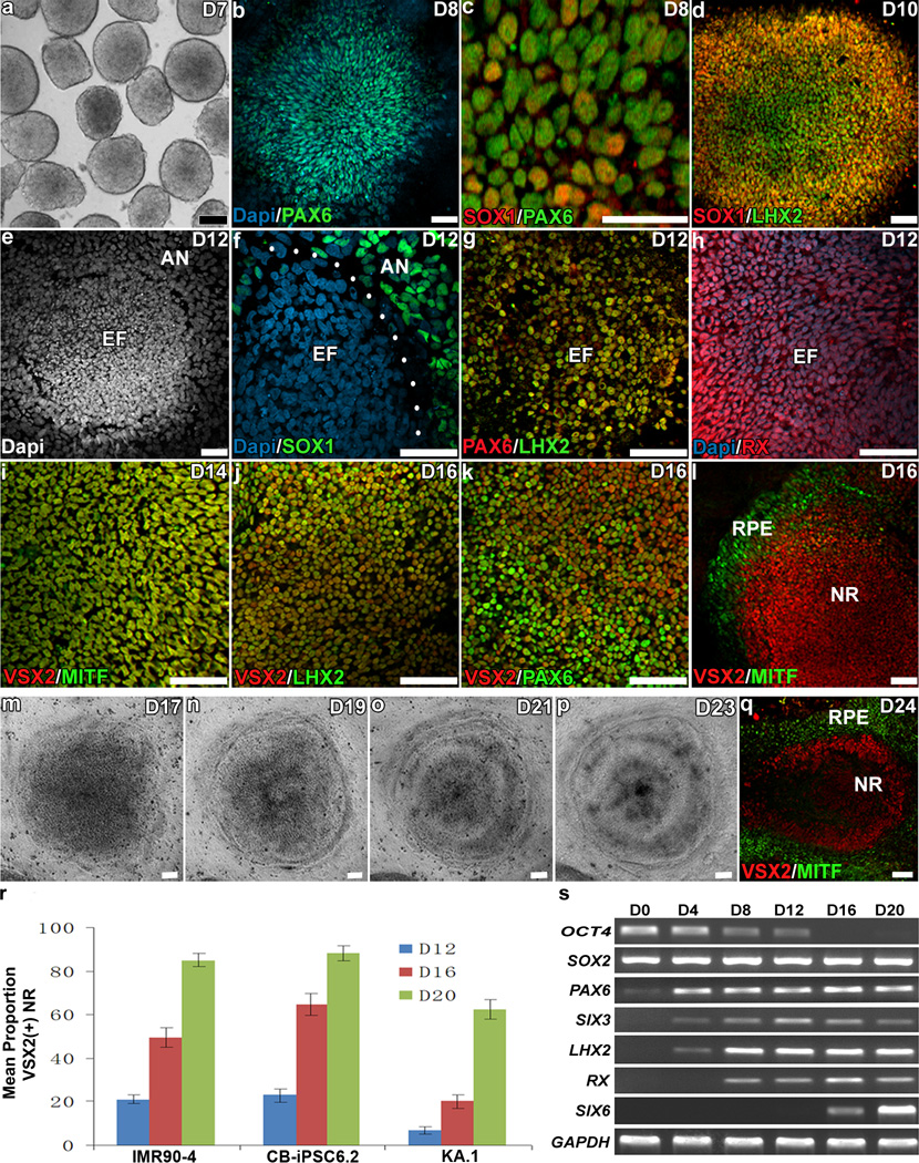
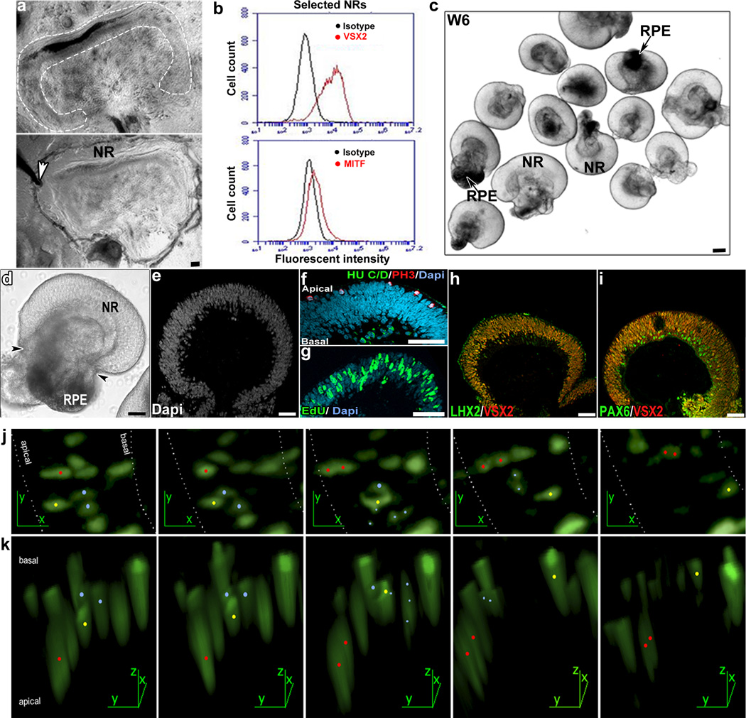
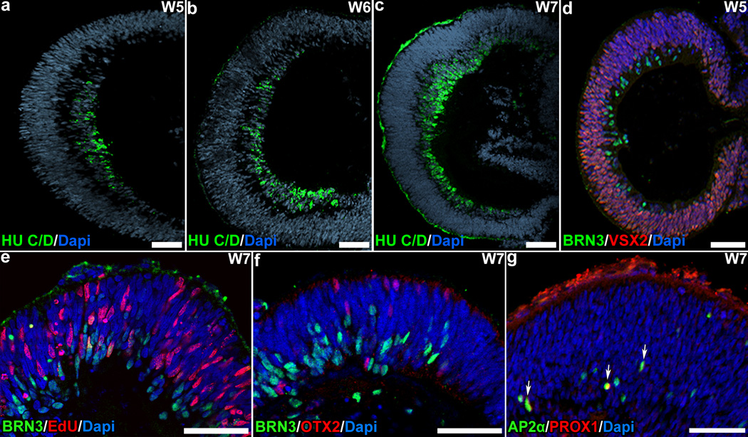
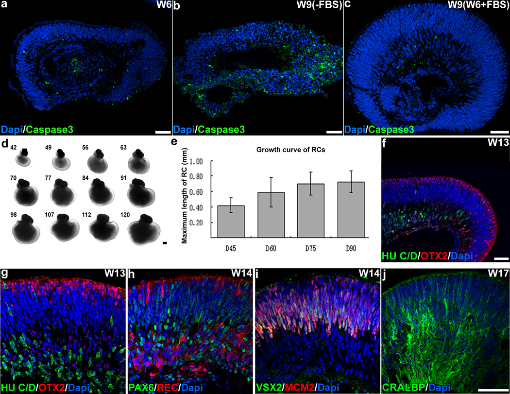
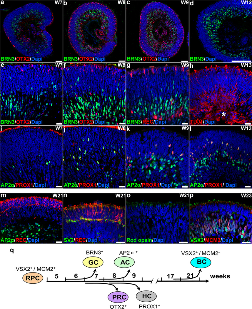
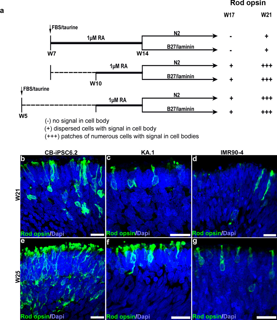
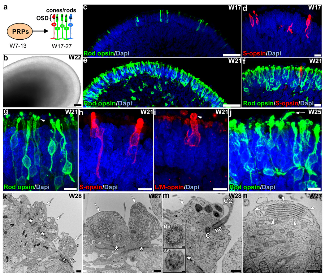
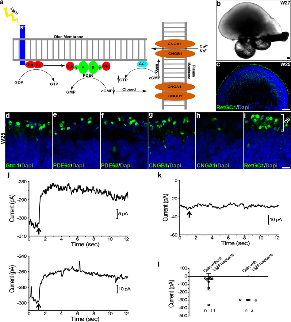
Many forms of blindness result from the dysfunction or loss of retinal photoreceptors. Induced pluripotent stem cells (iPSCs) hold great potential for the modelling of these diseases or as potential therapeutic agents. However, to fulfill this promise, a remaining challenge is to induce human iPSC to recreate in vitro key structural and functional features of the native retina, in particular the presence of photoreceptors with outer-segment discs and light sensitivity. Here we report that hiPSC can, in a highly autonomous manner, recapitulate spatiotemporally each of the main steps of retinal development observed in vivo and form three-dimensional retinal cups that contain all major retinal cell types arranged in their proper layers. Moreover, the photoreceptors in our hiPSC-derived retinal tissue achieve advanced maturation, showing the beginning of outer-segment disc formation and photosensitivity. This success brings us one step closer to the anticipated use of hiPSC for disease modelling and open possibilities for future therapies.


Conflict of interest statement

Conflict of Interest Statement

Under a licensing agreement between Life Technologies Corporation and the Johns Hopkins University (JHU), E.T.Z. is entitled to a share of royalty received by the University for licensing of stem cells. The terms of this arrangement are managed by JHU in accordance with its Conflict of Interest policies. This does not alter authors’ adherence to journal policies on sharing data and materials. Requests for CB-iPSC6.2 and KA.1 cell lines should be addressed to E.T.Z. (ezambid1@jhmi.edu). The rest of the authors declare no competing financial interests.

Figures

Figure 1
Figure 1. hiPSC-derived retinal progenitors self-organized into eye field-like domains (EF) and subsequently differentiated into neural retina (NR) and retinal pigment epithelium (RPE)
a–d, hiPSC-derived, free-floating aggregates (a) seeded in matrigel-coated dishes acquired an anterior neuroepithelial (AN) fate characterized by SOX1/PAX6 expression (b–c); subsequently, retinal progenitors (LHX2-positive) first appeared in the center of the aggregates (d). e–h, By D12, well-defined EF domains (e, f) expressing PAX6, LHX2 and RX (g, h) could be observed surrounded by AN cells (f). i–l, As differentiation progressed, cells within the EF domains co-expressed VSX2 and MITF (i), and afterward differentiated into a central VSX2/LHX2/PAX6-positive NR domain (j–l) and a peripheral RPE domain expressing MITF but not VSX2 (l). m–q, The NR domain progressively acquired an optic-cup-like shape. r, Efficiency of NR-domain formation among three hiPSC lines (mean ± SD, 3 experiments/time point/cell line). s, RT-PCR analysis showing progressive acquisition of retinal fate. Scale bars: 100µm (a and m–p); 50µm (b–l, and q).
Figure 2
Figure 2. Formation of 3-D retinal cups (RCs)
a, One NR domain (top panel) is being detached with a tungsten needle (arrowhead in bottom panel). b, FACS analysis of collected NR domains showed enrichment in NR progenitors (VSX2-positive) compared to retinal pigment epithelium (RPE) progenitors (MITF-positive). c, Detached NR domains cultured in suspension formed 3-D RCs, composed of a NR epithelium and RPE (arrow). d, Higher magnification of a typical 3-D RC with a NR epithelium continuous with the adjacent RPE bundled at the tip (arrowheads). e–f, The pseudostratified neural epithelium within the RC showed the typical polarity, with mitosis (PH3-positive) occurring at the apical side, and postmitotic neuronal precursors (HU C/D-positive) accumulating at the basal side. g–i, NR cells proliferated actively (EdU-positive, g) and co-expressed transcription factors characteristic of neural retina progenitor cells (h–i). j–k, Retinal progenitors within the NR epithelium underwent interkinetic nuclear migration. j, Time-lapse imaging of retinal progenitors expressing nuclear GFP. k, 3-D volume rendering of the cells shown in (j). red dot: cell undergoing mitosis; yellow dot: cell nucleus migrating from the apical to the basal side of the neuroepithelium; blue dots: cells undergoing apoptosis. Scale bars: 100 µm (a and c); 50µm (d–i).
Figure 3
Figure 3. Retinal progenitors within hiPSC-derived retinal cups underwent spontaneous differentiation
Retinal progenitors differentiated following the typical central-to-peripheral pattern (a–c) and acquired early-born cell fates, beginning with generation of ganglion cells (BRN3-positive/EdU-negative, d–f), followed by photoreceptors (OTX2-positive, f), amacrine cells (AP2α-positive, g) and horizontal cells (AP2α/PROX1-positive, arrows in g). Scale bars: 50µm.
Figure 4
Figure 4. Long-term suspension culture of hiPSC-derived retinal cups (RCs)
a–c, After W7, RCs progressively lost their histological organization due to increasing cell death (caspase 3-positive, a–b), which was avoidable by supplementation with FBS, taurine and retinoic acid from the beginning of W7 onward (c). d–e, Under these conditions, RCs maintained their shape, cellular organization, and steady growth during long-term culture (bars: mean ± SD; 15–20 RCs per time point). f–i, By W13-14, RCs showed distinguishable layers containing the precursors of most of the major neuronal cell types, including ganglion, amacrine and horizontal cells (HU C/D- and PAX6-positive), and photoreceptors (OTX2- and recoverin-positive); as in the developing human retina, cells expressing OTX2 and recoverin were observed in the developing outer nuclear layer as well as in the inner side of the retinal epithelium,. A conspicuous neuroblastic layer containing mitotic retinal progenitors (VSX2/MCM2-positive) was still present by W14 (i). j, Müller cells expressing CRALBP were first seen by W17. Scale bars: 200µm (d); 50µm (a–c and f–j).
Figure 5
Figure 5. hiPSC-derived neural retina (NR) progenitors within the 3-D retinal cups recapitulated the spatiotemporal pattern of NR differentiation in vivo
a–l, Cells within the RCs differentiated and migrated to their corresponding layers, with ganglion cells (GC; BRN3-positive, a–g; TUJ1-positive, h where (*) indicates a developing nerve-fiber-like layer) appearing first, followed by photoreceptor cells (PRC; OTX2/recoverin-positive, a–c, e–g), amacrine cells (AC; AP2α-positive, i–l), and horizontal cells (HC; PROX1-positive, i–l). m–p, By W21-W23, RCs presented a well-established outer nuclear layer (REC-positive, m–n) delineated by a developing outer plexiform layer (SV2-positive, n) and containing rod-opsin-positive photoreceptors (o), and a developing bipolar cell layer containing bipolar cells (BC; VSX2-positive/MCM2-negative, p) intermingled with remaining retinal progenitor cells (RPC; VSX2-positive/MCM2-positive, p). q, Timeline of retinogenesis in hiPSC-derived 3-D RCs. Scale bars: 100µm (a–d); 20µm (e–p).
Figure 6
Figure 6. Effect of retinoic acid on the differentiation of hiPSC-derived photoreceptors
a, Two windows of exposure to 1 µM retinoic acid (RA) were tested in combination with slight modifications to the culture media in CB-iPSC6.2-derived retinal cups (RCs). RA treatment in W7-W14 led to low levels of rod-opsin expression in the distal part of photoreceptor cells as observed in the original culture conditions, except for some RCs that showed few cells with rod-opsin expression in the cell bodies at W21. In contrast, RCs treated during W10-W14 showed dispersed cells with strong rod-opsin expression in their cell bodies already at W17, and forming large patches at W21. We did not observe significant differences associated with modifications of the culture media after RA treatment or presence of FBS/Taurine from W5. Similar results were observed in 3 independent experiments, 5 RCs per treatment per time window. b–g, When KA.1- and IMR90-4-derived RCs were subjected to RA treatment in W10-W14, induction of rod-opsin expression was also observed, although less efficiently than in CB-iPSC6.2-derived RCs (3 independent experiments, 8–22 RCs per cell line). Scale bars: 20µm.
Figure 7
Figure 7. hiPSC-derived rod and cone photoreceptors achieved an advanced level of differentiation including outer-segment disc formation
a, During normal development photoreceptor precursors (PRPs) differentiate into rods, L/M-, and S-cones (in green, red and blue color respectively). b, A W22 CB-iPSC6.2-derived retinal cup that has been exposed to 1 µM retinoic acid in W10-W14. c–j, Under this condition, photoreceptors expressing high levels of rod opsin in the entire cell body were first observed by W17 (c), significantly increasing in number and forming large patches by W21 (e–f). S-opsin expression could be observed in some rod-opsin-negative photoreceptors (d, f). High-magnification images of W21 retinal cups showing rods (g), S- (h) and L/M- (i) cones with a morphology and a topological organization similar to those of the in vivo retina, including structures reminiscent of short, nascent outer segments (arrowheads). By W25, elongated structures resembling more developed outer segments were rarely observed (j, arrow). k–n, Transmission-EM analysis revealed the presence of an outer limiting membrane (*), inner segments (arrows), basal bodies (BB), connecting cilia (CC) and stacks of outer-segment-discs (demarcated by arrowheads). BB and CC presented the photoreceptor-specific microtubule arrangement consisting of 9×3+0 and 9×2+0 respectively (inserts in m). C, centriole. Scale bars: 50µm (b, c, and e); 10µm (d, f, and g–j); 0.5µm (k–n); 0.05 µm (m inserts).
Figure 8
Figure 8. hiPSC-derived photoreceptors expressed proteins of the phototransduction pathway and occasionally showed light response in vitro
a, Diagram of rod phototransduction pathway in vivo. b, 3-D retinal cups (RCs) still maintained their structural organization in W27. c–i, Photoreceptors within the RCs showed expression of phototransduction proteins with the appropriate cellular distributions (OS: putative developing outer segments). j–l, Perforated-patch electrophysiological recordings from CB-iPSC6.2-derived photoreceptors, showing the flash-triggered responses (single trial) from two light-sensitive cells (j), the lack of flash response from one light-insensitive cell (k), and collected data from 13 cells (l). Cells were voltage-clamped at −50 mV. Inward current is negative. Flash (indicated by arrow) was 40 ms in duration and 2.46 × 10⁵ µW cm−2 in intensity (white light from a mercury arc lamp). In panel l, open circles indicate individual cells, solid circles indicate mean values, and error bars indicate SD. Scale bars: 50µm (b and c); 5µm (d–i).

References

    1. Hartong DT, Berson EL, Dryja TP. Retinitis pigmentosa. Lancet. 2006;368:1795–1809. - PubMed
    1. Goodwin P. Hereditary retinal disease. Current opinion in ophthalmology. 2008;19:255–262. - PubMed
    1. Hamel CP. Cone rod dystrophies. Orphanet J Rare Dis. 2007;2:7. - PMC - PubMed
    1. Viczian AS. Advances in retinal stem cell biology. J Ophthalmic Vis Res. 2013;8:147–159. - PMC - PubMed
    1. Stern JH, Temple S. Stem cells for retinal replacement therapy. Neurotherapeutics. 2011;8:736–743. - PMC - PubMed

Publication types