. 2014 Jun 10;9(6):e98073.
doi: 10.1371/journal.pone.0098073. eCollection 2014.

Anticancer properties of peptide fragments of hair proteins

Affiliations

Anticancer properties of peptide fragments of hair proteins

Sergiusz Markowicz et al. PLoS One. .

Abstract

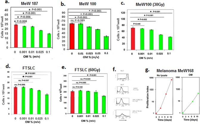
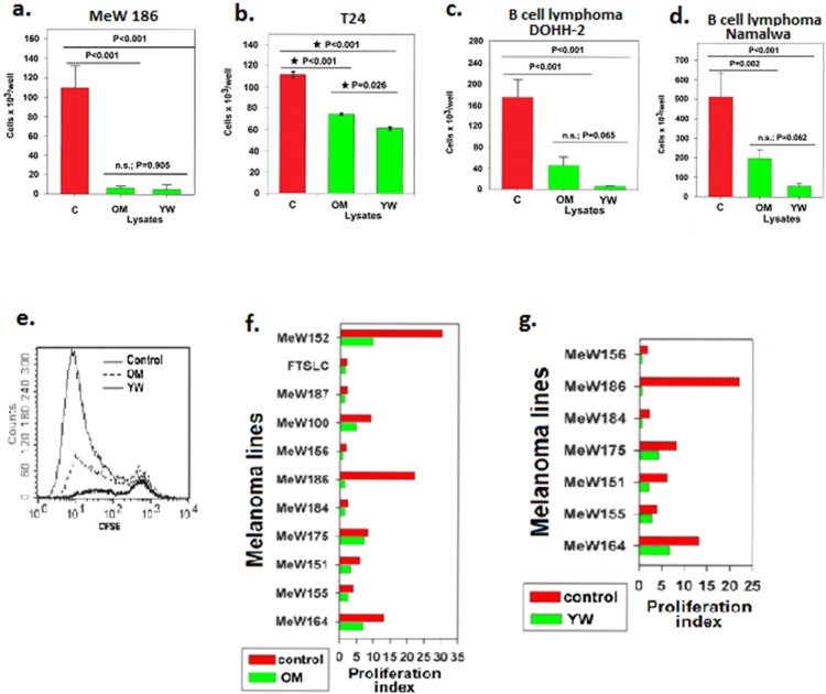
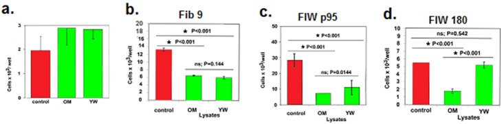
The primary function of hair and fur covering mammalian skin is to provide mechanical and thermal protection for the body. The proteins that constitute hair are extremely resistant to degradation by environmental factors. However, even durable materials can be slowly broken down by mechanical stresses, biodegradation mediated by endogenous enzymes in the skin or host microbes. We hypothesised that the biodegradation products of hair may possess bioprotective properties, which supplement their physical protective properties. Although evolutionary processes have led to a reduction in the amount of hair on the human body, it is possible that the bioprotective properties of hair biodegradation products have persisted. The human skin is exposed to various environmental carcinogenic factors. Therefore, we hypothesised that the potential bioprotective mechanisms of hair degradation products affect melanoma growth. We used pepsin to partially digest hair enzymatically, and this process produced a water-soluble lysate containing a mixture of peptides, including fragments of keratin and keratin-associated proteins. We found out that the mixtures of soluble peptides obtained from human hair inhibited the proliferation of human melanoma cells in vitro. Moreover, the hair-derived peptide mixtures also inhibited the proliferation of B lymphoma cells and urinary bladder cancer cells. Normal human cells varied in their susceptibility to the effects of the lysate; the hair-derived peptide mixtures modulated the proliferation of normal human fibroblasts but did not inhibit the proliferation of human mesenchymal cells derived from umbilical cord stromal cells. These results suggest that hair-derived peptides may represent a new class of anti-proliferative factors derived from basically structural proteins. Identification of active regulatory compounds and recognition of the mechanism of their action might pave the way to elaboration of new anticancer drugs.


Conflict of interest statement

Competing Interests: The authors have declared that no competing interests exist.

Figures

Figure 1
Figure 1. The human hair lysate obtained from the OM male donor inhibited the proliferation of early and late-passage human melanoma cells.
Gamma irradiation-resistant melanoma sublines generated from these cells remained susceptible to the inhibitory effects of the hair lysate. The viable cell count was evaluated in 3-day cultures of MeW187 melanoma cells plated at 13×103 cells/well with or without the lysate following 3 previous passages (a); 4-day cultures of the MeW100 melanoma cells plated at 7×10³ cells/well following 38 previous passages (b); 4-day cultures of the Mew100 subline pre-selected by irradiation with a 137Cs source (30 Gy) and plated at 10×103 cells/well (c); 3-day cultures of FSTLC melanoma cells plated at 15×103 cells/well after 53 passages (d); and 3-day cultures of the FSTLC subline preselected by irradiation (60 Gy) and plated at 15×103 cells/well (e). Fresh medium with or without the lysate was added to the cultures once daily. The groups of cultures treated with the lysate were compared to the control cultures using Dunnett’s test. Arrows mark the initial cell number in the cultures, and the bars represent the mean values ± s.d. of the viable cell count in triplicate cultures. Flow cytometry analysis of CFSE-labelled cells demonstrated that there were fewer cell divisions in the melanoma cell cultures supplemented with the lysate than in the control group (f). The histograms of the CFSE dilutions in MeW155 melanoma cells cultured for 5 days with or without the lysate are representative of triplicate cultures. The lysate was added only once at the start of the 5-day culture following labelling with CFSE. The proliferation indices (P.I.) were calculated as the proportion of the mean final cell yield to the initial cell number in the cultures. The effects of the lysate were evaluated in the prolonged passaged cultures (g). Proliferation indices were determined for MeW168 melanoma cells cultured with or without OM lysate on days 3 and 7 when the cells were passaged, and on day 11. Fresh medium containing the lysate at a concentration of 0.1% (m/v) was added on days 0, 3, 7, and 9.
Figure 2
Figure 2. The OM and YW human hair lysates obtained from two different donors inhibited the proliferation of human melanoma cells and other types of human cancer cells in vitro.
The viable cell count was evaluated in 7-day cultures of MeW186 melanoma cells plated after 3 passages at 5×10³ cells/well with or without the hair lysates (a); 4-day cultures of urinary bladder cancer T24 cells plated at 5×10³ cells/well (b); 4-day cultures of the B cell lymphoma line Namalwa plated at 120×103 cells/well (c); and 4-day cultures of the B cell lymphoma line DOHH-2 plated at 120×103 cells/well (d). The lysates were added at a concentration of 0.1% (m/v). Fresh medium with or without the lysate was added to the MeW186 melanoma cell cultures on days 0, 3, 4, and 5. Medium with or without the lysate was changed once daily for cultures of the urinary bladder cancer T24 cells and for cultures of lymphoma cells. The bars represent the mean values ± s.d. of the viable cell count in triplicate cultures. Groups of cultures were compared using the LSD ANOVA test. The profiles of the CFSE dilution in DOHH-2 cells indicated that the lymphoma cells divided in the presence of the lysate, although the yield of viable cells was substantially reduced in comparison to cells cultured in the absence of the lysate (d); the representative histograms for cultures shown in Fig. 2d. A comparison of the proliferation indices showing that both the OM lysate (f) and the YW lysate (g) inhibited the proliferation of various melanoma lines generated from different patients. The proliferation index was calculated as the proportion of the mean viable cell count following culture to the viable cell number at the beginning of the culture.
Figure 3
Figure 3. The effect of human hair lysates on the proliferation of normal human cells.
The OM and YW human hair lysates, when used at a concentration of 0.1% (m/v), did not significantly affect the proliferation of normal human mesenchymal cells derived from the umbilical cord (a). The antiproliferative effect of the lysates on fibroblast lines was dependent on the lysate tested and on the fibroblast line tested (b, c, d). Groups of cultures plated in triplicate were compared using the LSD ANOVA test.

References

    1. Rogers MA, Langbein L, Winter H, Ehmann C, Praetzel S, et al. (2001) Characterization of a Cluster of Human High/Ultrahigh Sulfur Keratin-associated Protein Genes Embedded in the Type I Keratin Gene Domain on Chromosome 17q12-21. J Biol Chem 276: 19440–19451. - PubMed
    1. Rogers M A, Winter H, Langbein L, Wolf C, Schweizer J (2000) Characterization of a 300 kbp region of human DNA containing the type II hair keratin gene domain. J Invest Dermatol 114: 464–472. - PubMed
    1. Rogers M A, Winter H, Wolf C, Heck M, Schweizer J (1998) Characterization of a 190-Kilobase Pair Domain of Human Type I Hair Keratin Genes. J Biol Chem 273: 26683–26691. - PubMed
    1. Fuchs E, Weber K (1994) Intermediate filaments: structure, dynamics, function, and disease. Annu Rev Biochem 63: 345–382. - PubMed
    1. Steinert PM, Steven AC, Roop DR (1985) The molecular biology of intermediate filaments. Cell 42: 411–420. - PubMed

Publication types

MeSH terms