Review
. 2014 Sep-Oct;49(5):361-99.
doi: 10.3109/10409238.2014.925420. Epub 2014 Jun 10.

The cell envelope glycoconjugates of Mycobacterium tuberculosis

Affiliations
Review

The cell envelope glycoconjugates of Mycobacterium tuberculosis

Shiva Kumar Angala et al. Crit Rev Biochem Mol Biol. 2014 Sep-Oct.

Abstract

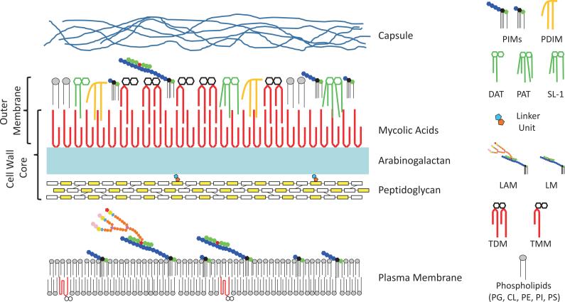
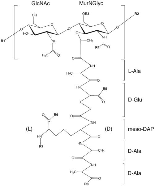
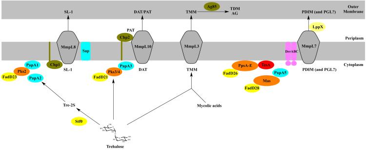
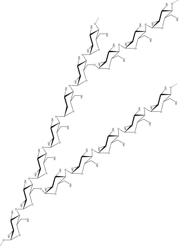
Tuberculosis (TB) remains the second most common cause of death due to a single infectious agent. The cell envelope of Mycobacterium tuberculosis (Mtb), the causative agent of the disease in humans, is a source of unique glycoconjugates and the most distinctive feature of the biology of this organism. It is the basis of much of Mtb pathogenesis and one of the major causes of its intrinsic resistance to chemotherapeutic agents. At the same time, the unique structures of Mtb cell envelope glycoconjugates, their antigenicity and essentiality for mycobacterial growth provide opportunities for drug, vaccine, diagnostic and biomarker development, as clearly illustrated by recent advances in all of these translational aspects. This review focuses on our current understanding of the structure and biogenesis of Mtb glycoconjugates with particular emphasis on one of the most intriguing and least understood aspect of the physiology of mycobacteria: the translocation of these complex macromolecules across the different layers of the cell envelope. It further reviews the rather impressive progress made in the last 10 years in the discovery and development of novel inhibitors targeting their biogenesis.

Keywords: (lipo)polysaccharides; Acyltrehaloses; arabinogalactan; flippase; glycosyltransferase; lipoarabinomannan; peptidoglycan; phosphatidylinositol mannosides.


Figures

Figure 1
Figure 1. Schematic representation of the Mtb cell envelope
Many of the classes of lipids and glycolipids discussed in the review are represented schematically and are shown in probable locations in the cell envelope. The overall schematic and individual structures are not drawn to scale. Proteins and peptides are not shown for the sake of clarity. The color code used in the representation of LM and LAM is the same as in Fig. 6. PE, phosphatidylethanolamine, PI, phosphatidyl-myo-inositol; CL, cardiolipin; PS, phosphatidylserine; PG, phosphatidylglycerol.
Figure 2
Figure 2. Structures of a representative monomer of mycobacterial PG prior to peptide trimming
R1, N-glycolylmuramic acid residue of another monomer; R2, N-acetylglucosamine residue of another monomer; R3, H or the linker unit of AG; R4, H, COCH3 (N-acetyl) or COCH2OH (N-glycolyl); R5, R6, R8, OH, NH2 or OCH3; R7, H, or cross-linked to penultimate D-Ala or to the D-center of another meso-DAP residue.
Figure 3
Figure 3. Structure of arabinogalactan
See text for details.
Figure 4
Figure 4. Schematic diagram of arabinogalactan biosynthesis
The synthesis of AG begins with the cytoplasmic formation of the linker unit on a decaprenyl monophosphate carrier lipid followed by the addition of Galf residues still on the cytosolic face of the plasma membrane and that of Araf residues and other decorating motifs (e.g., GalN motif) on the periplasmic side of the membrane. See text for details.
Figure 5
Figure 5. Structures of the two major tetracylated forms of PIM2 and PIM6
The forms of PIM2 and PIM6 represented here both harbor three palmitic and one tuberculostearic acyl chains.
Figure 6
Figure 6. Structures of LM and LAM
See text for details. MPI, mannosylated phosphatidyl-myo-inositol anchor.
Figure 7
Figure 7. Schematic diagram of PIM, LM and LAM biosynthesis
The biosynthesis of PIM, LM and LAM is initiated on the cytoplasmic side of the plasma membrane by GDP-Manp-utilizing ManTs that catalyze attachment of mannosyl residues to the myo-inositol ring of PI. Di- or tri-mannosylated forms of PIMs are then flipped to the periplamic face of the membrane where they undergo further elongation catalyzed by integral membrane polyprenyl-monophospho-mannose-dependent ManTs and β-D-arabinofuranosyl-1-monophosphoryl-decaprenol (DPA)-dependent AraTs to generate polar forms of PIMs, LM, and ManLAM. See text for details.
Figure 8
Figure 8. Structures of the acyltrehaloses of Mtb
In TMM and TDM, trehalose is here shown esterified with alpha-mycolic acid chains. In SL-I (2,3,6,6’-tetraacyl α–α’-trehalose-2’-sulfate), trehalose is sulfated at the 2’ position and esterified with palmitic acid and the multimethyl-branched phthioceranic and hydroxyphthioceranic acids. In DAT (2,3-di-O-acyltrehalose), trehalose is esterified with palmitic acid and the multimethyl-branched mycosanoic acid. In PAT (penta-acyltrehalose), trehalose is esterified with stearic acid and the multimethyl-branched mycolipenic acids. The oligosaccharide of the LOS of Mtb Canettii strains consists of 2-O-methyl-α-L-Fucp-(1,3)-β–D-Glcp-(1,3)-2-O-methyl-α-L-Rhap-(1,3)-2-O-methyl-L-Rhap-(1,3)-β-D-Glcp-(1,3)-4-O-methyl-α-L-Rhap-(1,3)-6-O-methyl-α-D-Glc-(1,1)-α-D-Glc. R are 2,4-dimethylhexadecanoic acid and 2,4,6,8-tetramethyloctadecanoic acid residues.
Figure 9
Figure 9
A schematic representation of the SL, DAT/PAT and LOS biosynthetic gene clusters of Mtb H37Rv (SL; DAT/PAT) and Mtb canettii (LOS).
Figure 10
Figure 10. Biogenesis of SL, DAT/PAT and PDIM/PGL in Mtb
The enzymes and transporters that have been involved in the elongation, assembly and export of SL, DAT/PAT and PDIM/PGL and their localization in the bacterium are represented. FadD enzymes are fatty acyl-AMP ligases; PapA and Chp enzymes are acyltransferases; Pks, Mas and PpsA-E are the polyketide synthases responsible for the elongation of the polymethyl-branched fatty acids found in DAT/PAT, SL and PDIM/PGL; TesA is a thioesterase; Stf0 is a sulfotransferase; Antigens 85 (Ag85) are mycolyltransferases; DrrABC is an ABC-transporter; LppX is a periplasmic lipoprotein required for the translocation of PDIM to the outer membrane; MmpL proteins are integral membrane RND superfamily transporters required for the translocation of acyltrehaloses and PDIM to the periplasmic space and outer membrane. The precise extent of (glyco)lipid translocation mediated by MmpL proteins, LppX and DrrABC has has not yet been defined. See text for further details.
Figure 11
Figure 11. Structures of the phthiocerol dimycocerosates (PDIM), phenolic glycolipids (PGL) and p-hydroxybenzoic acid derivatives (p-HBADs) of Mtb
The lipid core of PGL from Mtb is composed of phenolphthiocerol esterified by mycocerosic acids (p, p’=3-5; n, n’=16-18; m2=15-17 ; m1= 20-22; R= CH2-CH3 or CH3). The trisaccharide substituent of PGL and p-HBAD-II (i.e., the fully elaborated form of p-HBAD produced by Mtb) consists of 2,3,4-tri-O-methyl-α-L-Fucp-(1,3)-α–L-Rhap-(1,3)-2-O-methyl-α-L-Rhap.
Figure 12
Figure 12
Structure of the predominant mannosyl-β-1-phosphomycoketide from Mtb H37Rv.
Figure 13
Figure 13
Structure of the capsular α-D-glucan of Mtb.
Figure 14
Figure 14. Biosynthesis of α-D-glucans in Mtb
See text for details.

References

    1. AFONSO-BARROSO A, CLARK SO, WILLIAMS A, ROSA GT, NOBREGA C, SILVA-GOMES S, VALE-COSTA S, UMMELS R, STOKER N, MOVAHEDZADEH F, VAN DER LEY P, SLOOTS A, COT M, APPELMELK BJ, PUZO G, NIGOU J, GEURTSEN J, APPELBERG R. Lipoarabinomannan mannose caps do not affect mycobacterial virulence or the induction of protective immunity in experimental animal models of infection and have minimal impact on in vitro inflammatory responses. Cell Microbiol. 2012;15:660–674. - PubMed
    1. ALAIMO C, CATREIN I, MORF L, MAROLDA CL, CALLEWAERT N, VALVANO MA, FELDMAN MF, AEBI M. Two distinct but interchangeable mechanisms for flipping of lipid-linked oligosaccharides. EMBO J. 2006;25:967–976. - PMC - PubMed
    1. ALBESA-JOVE D, GIGANTI D, JACKSON M, ALZARI PM, GUERIN ME. Structure-function relationships of membrane-associated GT-B glycosyltransferases. Glycobiology. 2014;24:108–24. - PMC - PubMed
    1. ALDERWICK LJ, DOVER LG, SEIDEL M, GANDE R, SAHM H, EGGELING L, BESRA GS. Arabinan-deficient mutants of Corynebacterium glutamicum and the consequent flux in decaprenylmonophosphoryl-D-arabinose metabolism. Glycobiology. 2006b;16:1073–81. - PubMed
    1. ALDERWICK LJ, LLOYD GS, GHADBANE H, MAY JW, BHATT A, EGGELING L, FUTTERER K, BESRA GS. The C-terminal domain of the Arabinosyltransferase Mycobacterium tuberculosis EmbC is a lectin-like carbohydrate binding module. PLoS Pathog. 2011b;7:e1001299. - PMC - PubMed

Publication types

MeSH terms