. 2014 Sep;1842(9):1638-47.
doi: 10.1016/j.bbadis.2014.06.002. Epub 2014 Jun 8.

Myocardial ischemic reperfusion induces de novo Nrf2 protein translation

Affiliations

Myocardial ischemic reperfusion induces de novo Nrf2 protein translation

Beibei Xu et al. Biochim Biophys Acta. 2014 Sep.

Abstract

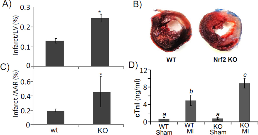
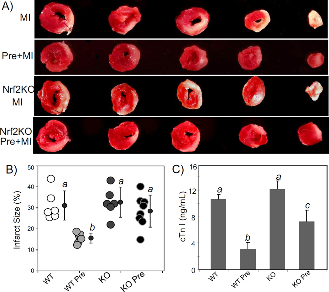
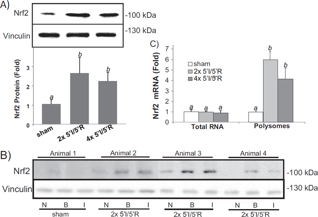
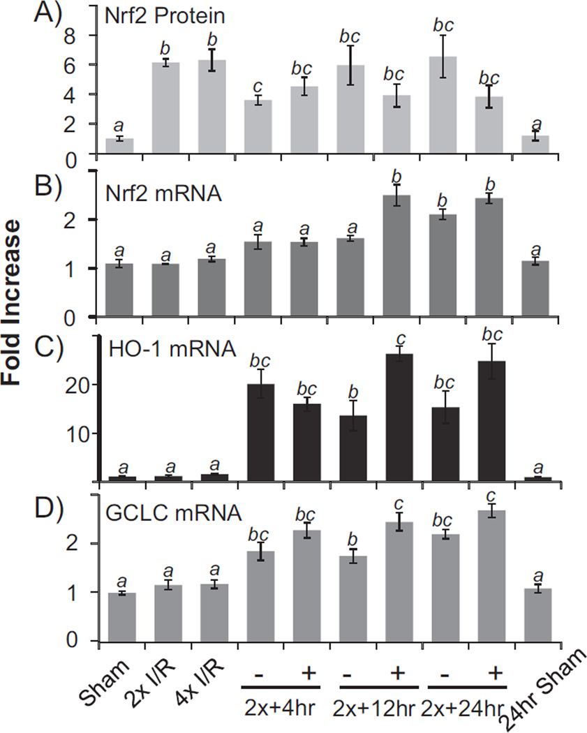
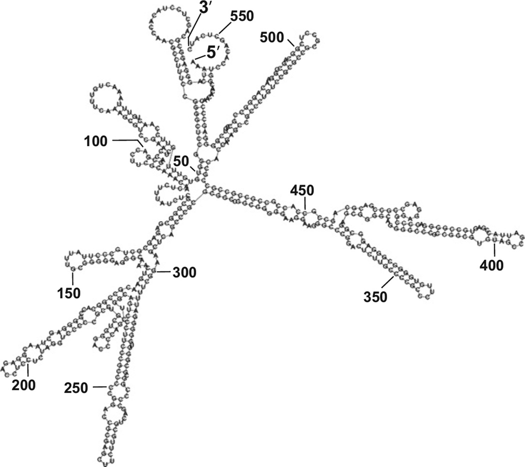
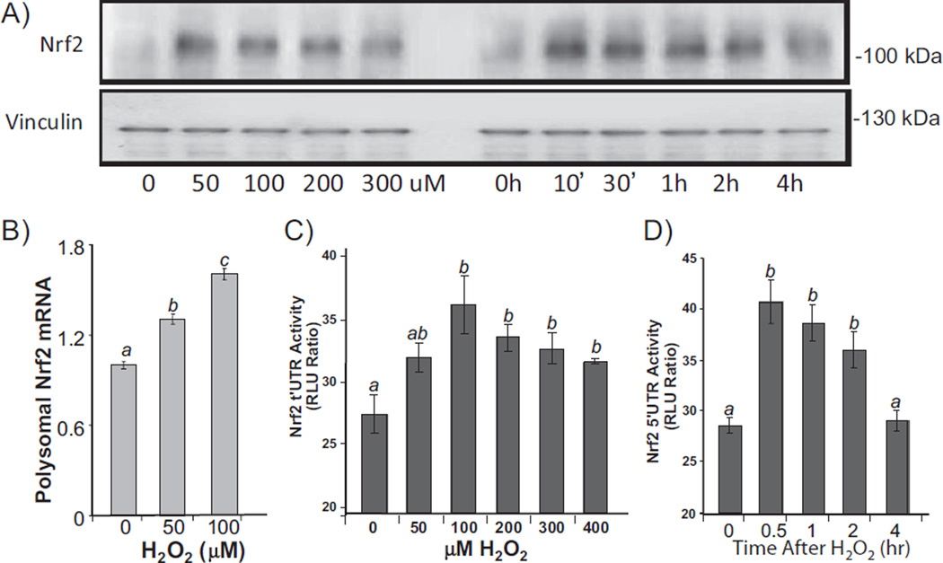
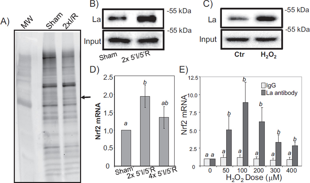
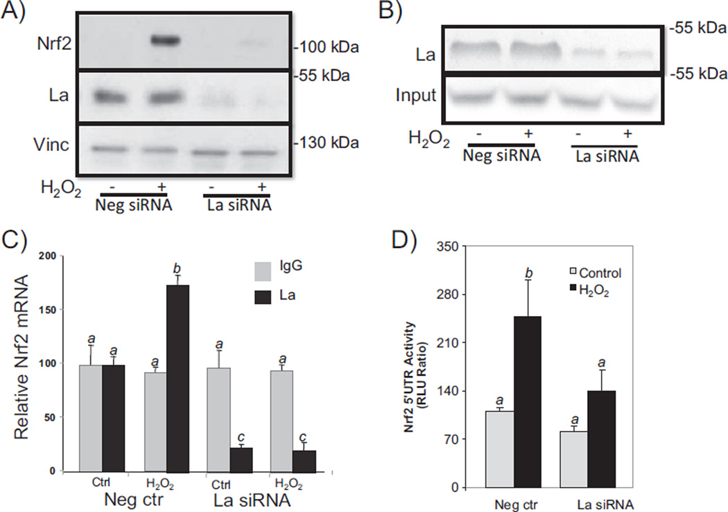
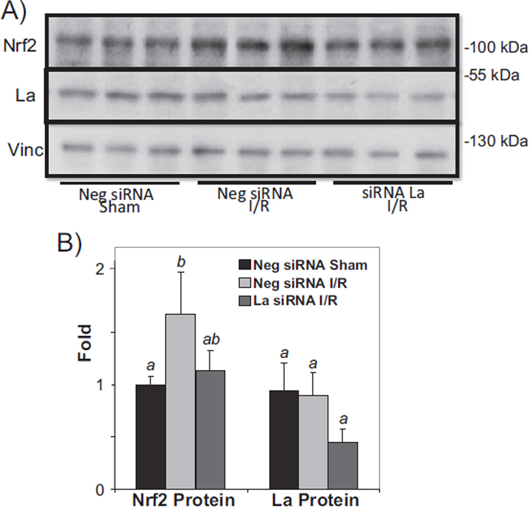
Nrf2 is a bZIP transcription factor regulating the expression of antioxidant and detoxification genes. We have found that Nrf2 knockout mice have an increased infarction size in response to regional ischemic reperfusion and have a reduced degree of cardiac protection by means of ischemic preconditioning. With cycles of brief ischemia and reperfusion (5'I/5'R) that induce cardiac protection in wild type mice, an elevated Nrf2 protein was observed without prior increases of Nrf2 mRNA. When an mRNA species is being translated into a protein, it is occupied by multiple ribosomes. The level of ribosome-associated Nrf2 mRNA increased following cycles of 5'I/5'R, supporting de novo Nrf2 protein translation. A dicistronic reporter assay indicated a role of the 5' untranslated region (5' UTR) of Nrf2 mRNA in oxidative stress induced Nrf2 protein translation in isolated cardiomyocytes. Western blot analyses after isolation of proteins binding to biotinylated Nrf2 5' UTR from the myocardium or cultured cardiomyocytes demonstrated that cycles of 5'I/5'R or oxidants caused an increased association of La protein with Nrf2 5' UTR. Ribonucleoprotein complex immunoprecipitation assays confirmed such association indeed occurring in vivo. Knocking down La using siRNA was able to prevent Nrf2 protein elevation by oxidants in cultured cardiomyocytes and by cycles of 5'I/5'R in the myocardium. Our data point out a novel mechanism of cardiac protection by de novo Nrf2 protein translation involving interaction of La protein with 5' UTR of Nrf2 mRNA in cardiomyocytes.

Keywords: Cardiac protection; Ischemic stress; Protein translation; RNA binding.


Figures

Fig. 1
Fig. 1
Nrf2 KO mice are more sensitive to ischemic reperfusion injury. Nrf2 KO mice and wild type littermates were subjected to 30 min ischemia by LAD coronary artery occlusion followed by 24 h reperfusion before quantification of infarct size or infarct over area-at-risk (AAR) following trypan blue perfusion and TTC staining. The data represent average ± standard deviations from 4 animals in each group. When two groups of means are compared, a statistical difference (p < 0.05) was determined by Student's t test and is labeled with *. When multiple groups of means were compared, ANOVA was used to determine statistical differences (p < 0.05). The means labeled “a” are significantly different from the means labeled “b” or “c”, while the difference between means labeled with “b” and “c” is statistically significant.
Fig. 2
Fig. 2
Nrf2 KO mice lose preconditioning induced cardiac protection. Wild type (WT) and Nrf2 KO mice at 8–10 weeks old were used for LAD coronary artery occlusion surgery. For preconditioning, mouse hearts were subjected to 5 min occlusion and 5 min release of the LAD coronary artery for 2 cycles (2× 5′I/5′R). For control group not having 2× 5′I/5′R or preconditioning group with 2× 5′I/5′R, the coronary artery was permanently occluded. At 24 h after permanent occlusion, the hearts and blood were collected for TTC staining to measure areas of infarction (A, B) and for cTnI concentration assay (C) respectively. A series of transverse sections of representative hearts downstream of the ligature are shown (A). Total myocardial areas or areas of infarct were quantified by using NIH Image J software. Each open circle represents the measurement from one animal and average means with standard deviations from 6 to 8 animals are shown with filled-in circles (B). ANOVA was used to determine the significant difference (p < 0.05) between the means. The means labeled “a” are significantly different from that labeled “b” or “c”, whereas the label “b” indicates significant difference from the mean labeled “c”.
Fig. 3
Fig. 3
Brief cycles of I/R cause elevation of Nrf2 protein. Mouse hearts were subjected to 2× or 4× 5′I/5′R. Tissue extracts from the whole hearts were used for Western blots (80 µg protein/lane) with vinculin as a loading control (A). The bar graph represents the relative density of the Nrf2 band over vinculin in the same sample as means ± standard deviations from 13 animals (A). Ischemic (I) versus non-ischemic (N) areas and 1–2 mm border zone (B) were rapidly dissected following Evan's blue dye perfusion for Western blot (B). A sham operated control was used for collecting areas corresponding to I, N or B (B). Total RNA or polysomal RNA was extracted from the whole heart tissue lysates for real time RT-PCR (C). The data represent means ± standard deviations from 3 (total RNA) or 4 animals (polysomal RNA, C). ANOVA was used to determine the significant difference (p < 0.05) between the means. The means labeled “a” are significantly different from that labeled “b”.
Fig. 4
Fig. 4
Transcription of Nrf2 downstream genes following elevation of Nrf2 protein. Mouse hearts were subjected to 2× or 4× 5′I/5′R. Immediately following 2× 5′I/5′R, the LAD coronary artery was permanently occluded for 4, 12, or 24 h for the + groups, whereas the – groups serve as sham operated controls, which had 2× 5′I/5′R but not permanent coronary artery occlusion. Tissue extracts from the whole hearts were used for Western blot (A) or harvesting total RNA for real time RT-PCR (B–D). The data represent means ± standard deviations from 3 animals. ANOVA was used to compare means and statistical differences were indicated by different letters. The means labeled “a” are significantly different from the means labeled “b”, “c” or “bc”, whereas “b” indicates significant difference of the mean from that labeled “c”. The mean labeled “bc” is not significantly different from that of “b” or “c”.
Fig. 5
Fig. 5
Nrf2 5′ UTR secondary structure predicted by Zucker's MFOLD algorithm.
Fig. 6
Fig. 6
Nrf2 5′ UTR mediated protein translation. Primary cultured rat cardiomyocytes were treated with H2O2 at indicated doses for 10 min before harvesting at 1 h (A, B, C) or with 100 µM H2O2 for 10 min before harvesting at indicated time (A, D). Levels of Nrf2 protein were measured by Western blot (30 µg protein/lane) with vinculin as a loading control (A). Ribosome-associated RNA was isolated for real time RT-PCR (B). Cells in 6-well culture dishes were transfected with 1 µg DNA of pRL–luc–Nrf2 5′ UTR-FL-luc dicistronic vector. At 48 h after transfection and 24 h culture in 0.5% FBS/DMEM, cells were treated with various doses of H2O2 for 10 min and harvested at 1 h later (C), or with 100 µM H2O2 for 10 min and harvested at indicated time (D) for dual luciferase assays. ANOVA was used to compare means and statistically significant differences (p < 0.05) are indicated by different letters. The mean labeled “a” is significantly different from the means labeled “b” or “c”, while “b” indicates statistical difference from that labeled “c”. The mean labeled “ab” is not significantly different from “a” or “b”.
Fig. 7
Fig. 7
La autoantigen interacts with Nrf2 5′ UTR. Biotinylated Nrf2 5′ UTR generated via in vitro transcription was used as a bait to isolate bound proteins from myocardial tissues (A, B) or cardiomyocytes (C). Bound proteins were eluted for SDS gel electrophoresis (A) or Western blot to detect La autoantigen (B, C). The lysates from the whole hearts with 2× or 4× 5′I/5′R, or from primary cultured cardiomyocytes harvested at 1 h after various doses of H2O2 treatment for 10 min were used for immunoprecipitation with an antibody specific to La autoantigen (D, E). The immunoprecipitates were used for real time RT-PCR to detect Nrf2 mRNA (D, E). The data are means ± standard deviations from triplicates of one experiment representative of three. ANOVA was used to compare means and statistically significant differences (p < 0.05) are indicated by different letters. The mean labeled “a” is significantly different from that labeled “b”, while “ab” indicates no significant difference from that labeled “a” or “b”.
Fig. 8
Fig. 8
La autoantigen regulates H2O2 induced Nrf2 protein translation in cardiomyocytes. Cardiomyocytes were transfected with 300 nM siRNA sequence against La autoantigen or scrambled sequence (negative siRNA). At 48 h after transfection, cells were treated with 100 µM H2O2 for 10 min before harvesting 1 h later for Western blot (A), in vitro biotinylated Nrf2 5′ UTR binding then Western blot (B), or immunoprecipitation of La complex then real time RT-PCR to measure Nrf2 mRNA (C). La siRNA was cotransfected with pRL–luc–Nrf2 5′ UTR-FL-luc dicistronic vector for measurement of Nrf2 5′ UTR driven Firefly luciferase over Renilla luciferase 1 h after 10 min treatment of 100 µM H2O2 (D). ANOVA was used to compare means and the statistically significant differences (p < 0.05) are indicated by different letters. The means labeled “a” are significantly different from the means labeled “b” or “c”, while“b” indicates significant difference from the means labeled “c”.
Fig. 9
Fig. 9
La autoantigen regulates Nrf2 protein induction in vivo. siRNA or negative control siRNA (15 µg) was injected via the jugular vein 24 h before surgery to induce 2× 5′I/5′R (C). Tissue lysates from the whole heart were used for Western blot (80 µg protein/lane), with each lane representing one sample from one animal (A). The intensities of the bands were quantified by NIH image J software and presented as means ± SD from 3 animals (B). ANOVA was used to compare means and the statistically significant differences (p < 0.05) are indicated by different letters. The means labeled “a” are significantly different from the means labeled “b”, while “ab” indicates no significant difference from that labeled “a” or “b”.

References

    1. Jackson RJ, Hellen CU, Pestova TV. The mechanism of eukaryotic translation initiation and principles of its regulation. Nat. Rev. Mol. Cell Biol. 2010;11:113–127. - PMC - PubMed
    1. Holcik M, Sonenberg N. Translational control in stress and apoptosis. Nat. Rev. Mol. Cell Biol. 2005;6:318–327. - PubMed
    1. Yamasaki S, Anderson P. Reprogramming mRNA translation during stress. Curr. Opin. Cell Biol. 2008;20:222–226. - PMC - PubMed
    1. Baird SD, Turcotte M, Korneluk RG, Holcik M. Searching for IRES. RNA. 2006;12:1755–1785. - PMC - PubMed
    1. Spriggs KA, Bushell M, Mitchell SA, Willis AE. Internal ribosome entry segment-mediated translation during apoptosis: the role of IRES-trans-acting factors. Cell Death Differ. 2005;12:585–591. - PubMed

MeSH terms

LinkOut - more resources