

Correlation between serum tryptase, mast cells positive to tryptase and microvascular density in colo-rectal cancer patients: possible biological-clinical significance
- PMID: 24915568
- PMCID: PMC4051753
- DOI: 10.1371/journal.pone.0099512
Correlation between serum tryptase, mast cells positive to tryptase and microvascular density in colo-rectal cancer patients: possible biological-clinical significance
Abstract
Background: Tryptase is a serin protease stored and released from mast cells (MCs) that plays a role in tumour angiogenesis. In this study we aimed to evaluate serum tryptase levels in colo-rectal cancer (CRC) patients before (STLBS) and after (STLAS) radical surgical resection. We also evaluated mast cell density positive to tryptase (MCDPT) and microvascular density (MVD) in primary tumour tissue.
Methods: A series of 61 patients with stage B and C CRC (according to the Astler and Coller staging system) were selected. Serum blood samples were collected from patients one day before and one day after surgery. Tryptase levels were measured using the UniCAP Tryptase Fluoroenzymeimmunoassay (Pharmacia, Uppsala, Sweden). Tumour sections were immunostained with a primary anti-tryptase antibody (clone AA1; Dako, Glostrup, Denmark) and an anti CD-34 antibody (QB-END 10; Bio-Optica Milan, Italy) by means of immunohistochemistry and then evaluated by image analysis methods.
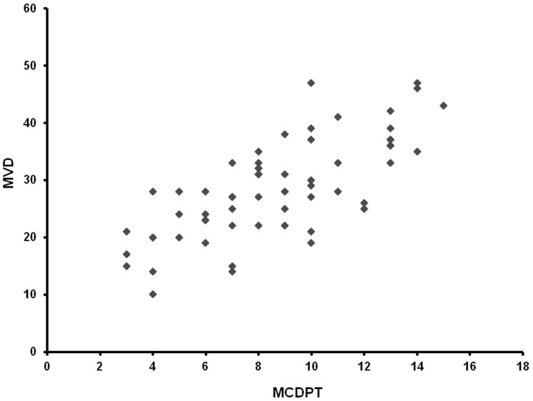
Results: The mean ± s.d. STLBS and STLAS was 5.63±2.61 µg/L, and 3.39±1.47 µg/L respectively and a significant difference between mean levels was found: p = 0.000 by t-test. The mean ± s.d. of MCDPT and MVD was 8.13±3.28 and 29.16±7.39 respectively. A strong correlation between STLBS and MVD (r = 0.83, p = 0.000); STLBS and MCDPT (r = 0.60, p = 0.003); and MCDPT and MVD (r = 0.73; p = 0.001) was found.
Conclusion: Results demonstrated higher STLBS in CRC patients, indicating an involvement of MC tryptase in CRC angiogenesis. Data also indicated lower STLAS, suggesting the release of tryptase from tumour-infiltrating MCs. Serum tryptase levels may therefore play a role as a novel bio-marker predictive of response to radical surgery. In this context tryptase inhibitors such as Gabexate and Nafamostat Mesilate might be evaluated in adjuvant clinical trials as a new anti-angiogenic approach.
Conflict of interest statement
Figures


References
-
- Ranieri G, Labriola A, Achille G, Florio G, Zito AF, et al. (2002) Microvessel density, mast cell density and thymidine phosphorylase expression in oral squamous carcinoma. Int J Oncol 21: 1317–1323. - PubMed
-
- Ranieri G, Ammendola M, Patruno R, Celano G, Zito FA, et al. (2009) Tryptase-positive mast cells correlate with angiogenesis in early breast cancer patients. Int J Oncol 35: 115–120. - PubMed
-
- Weidner N, Semple JP, Welch WR, Folkman J (1991) Tumour angiogenesis and metastasis correlation in invasive breast carcinoma. N Engl J Med 324: 1–8. - PubMed
-
- Kankkunen JP, Harvima IT, Naukkarinen A (1997) Quantitative analysis of tryptase and chymase containing mast cells in benign and malignant breast lesions. Int J Cancer 72: 385–388. - PubMed
-
- Soucek L, Lawlor ER, Soto D, Shchors K, Swigart LB, et al. (2007) Mast cells are required for angiogenesis and macroscopic expansion of Myc-induced pancreatic islet tumours. Nat Med 13: 1211–1218. - PubMed
MeSH terms
Substances
LinkOut - more resources
Full Text Sources
Other Literature Sources
Medical
