. 2014 Jun 10;5(3):76.
doi: 10.1186/scrt465.

Mesenchymal stem cell-derived exosomes promote hepatic regeneration in drug-induced liver injury models

Mesenchymal stem cell-derived exosomes promote hepatic regeneration in drug-induced liver injury models

Cheau Yih Tan et al. Stem Cell Res Ther. .

Abstract

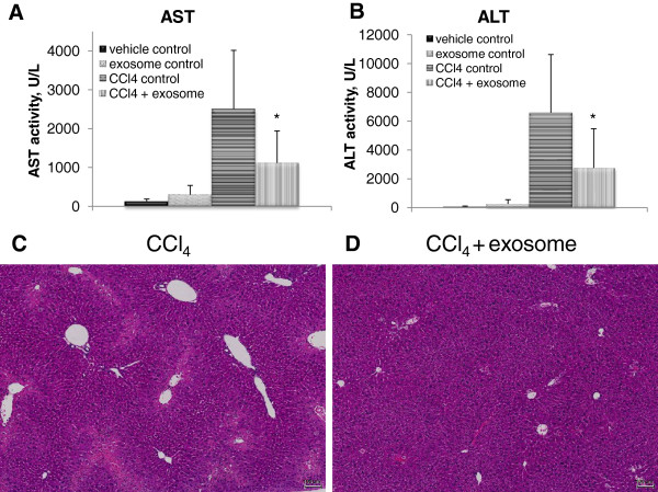
Introduction: Mesenchymal stem cell-conditioned medium (MSC-CM) has been shown to have protective effects against various cellular-injury models. This mechanism of protection, however, has yet to be elucidated. Recently, exosomes were identified as the active component in MSC-CM. The aim of this study is to investigate the effect of MSC-derived exosomes in an established carbon tetrachloride (CCl4)-induced liver injury mouse model. This potential effect is then validated by using in vitro xenobiotic-induced liver-injury assays: (1) acetaminophen (APAP)- and (2) hydrogen peroxide (H2O2)-induced liver injury.

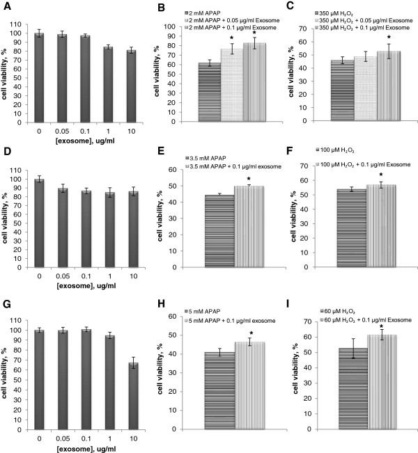
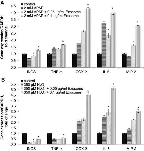
Methods: The exosomes were introduced concurrent with CCl4 into a mouse model through different routes of administration. Biochemical analysis was performed based on the blood and liver tissues. Subsequently the exosomes were treated in APAP and H2O2-toxicants with in vitro models. Cell viability was measured, and biomarkers indicative of regenerative and oxidative biochemical responses were determined to probe the mechanism of any hepatoprotective activity observed.

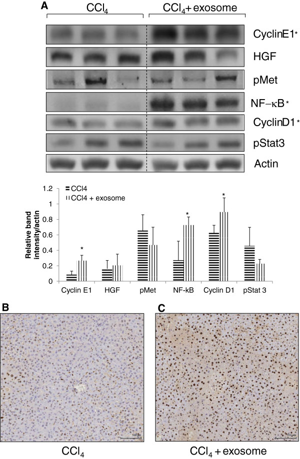
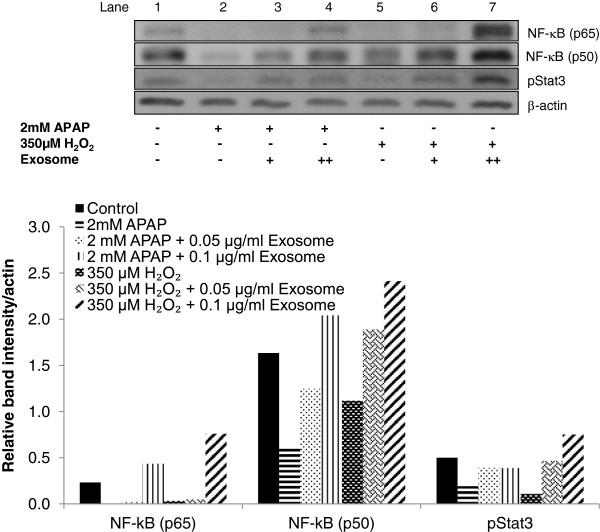
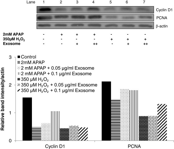
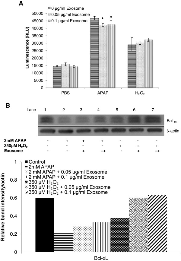
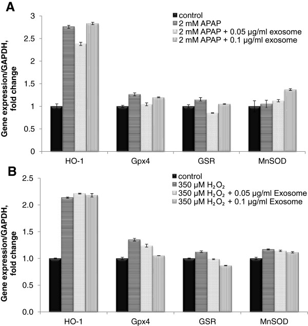
Results: In contrast to mice treated with phosphate-buffered saline, CCl4 injury in mice was attenuated by concurrent-treatment exosomes, and characterized by an increase in hepatocyte proliferation, as demonstrated with proliferating cell nuclear antigen (PCNA) elevation. Significantly higher cell viability was demonstrated in the exosomes-treated group compared with the non-exosome-treated group in both injury models. The higher survival rate was associated with upregulation of the priming-phase genes during liver regeneration, which subsequently led to higher expression of proliferation proteins (PCNA and cyclin D1) in the exosomes-treated group. Exosomes also inhibited the APAP- and H2O2-induced hepatocytes apoptosis through upregulation of Bcl-xL protein expression. However, exosomes do not mitigate hepatocyte injury via modulation of oxidative stress.

Conclusions: In summary, these results suggest that MSC-derived exosomes can elicit hepatoprotective effects against toxicants-induced injury, mainly through activation of proliferative and regenerative responses.


Figures

Figure 1
Figure 1
Effect of exosomes on biochemical parameters and hepatocyte injury after CCl4 treatment in vivo. Serum (A) aspartate aminotransferase (AST) and (B) alanine aminotransferase (ALT) levels were measured after 24-hour dosing of CCl4 with or without exosomes administration (n = 6 per group; *P < 0.05 versus of CCl4 control for both AST and ALT). Standard hematoxylin and eosin (H&E) staining for liver tissue harvested after 24 hours of (C) CCl4 administration and (D) CCl4 with exosomes treatment. Obvious hepatic necrosis appeared in the section from a CCl4-treated animal. Original magnification × 80; scale bar, 100 μm.
Figure 2
Figure 2
Effect of exosomes on hepatocyte proliferation after CCl4-induced injury in mice. (A) Expression of cyclin E, HGF, pMet, NF-κB p65, cyclin D1, and phosphorylated STAT3 was determined by immunoblotting after 24 hours of CCl4 administration with or without exosomes (n = 6 per group (three per group were shown); *P < 0.05 versus CCl4 control). The relative densities of all the proteins bands were normalized to actin in the same samples. Livers were processed for immunohistochemistry on PCNA to quantify hepatocyte proliferation after 24 hours of (B) CCl4 administration and (C) CCl4 with exosomes treatment. Original magnification × 200; scale bar, 100 μm.
Figure 3
Figure 3
Effect of exosomes in cell viability after APAP- and H2O2-induced injury. Experiments were performed in TAMH (A-C), THLE-2 (D-F), and Huh-7 (G-I) hepatocytes. (A, D, G) Cytotoxicity of exosomes at different concentrations in respective cell lines. 0.05 μg/ml and 0.1 μg/ml of exosomes were added to respective concentration of APAP (B, E, H) and H2O2(C, F, I), and MTT were performed 24 or 72 hours later. Cell viability was normalized against vehicle control group and expressed in percentage (n = 6 per group; *P < 0.05 versus APAP or H2O2 non-exosomes-treatment group.
Figure 4
Figure 4
Effect of exosomes in hepatoregenerative-expression genes on APAP- and H2O2-induced injury in hepatocytes. Expression of regenerative-response genes after 24 hours of exosomes in (A) APAP and (B) H2O2 treatment was determined by quantitative real-time PCR and normalized against GAPDH expression of the same sample and presented as fold-increase over the controls. Bars represent means ± SEM. *P < 0.05 versus APAP or H2O2 nonexosomes treatment group.
Figure 5
Figure 5
Effect of exosomes in G1phase of the cell cycle after APAP- or H2O2-induced injury in hepatocytes. Expression of NF-κB (p65 and p50) and phosphorylated STAT3 was determined by immunoblotting after 24-hour treatment in APAP- or H2O2-injury models. The relative densities of NF-κB and phosphorylated STAT3 bands were normalized to actin in the same samples.
Figure 6
Figure 6
Effect of exosomes in cell proliferation after APAP- or H2O2-induced injury in hepatocytes. Expression of cyclin D1 and PCNA was determined by immunoblotting after 24-hour treatment in the APAP- or H2O2-injury model. The relative densities of cyclin D1 and PCNA bands were normalized to actin in the same samples.
Figure 7
Figure 7
Effect of exosomes on antiapoptosis in APAP- or H2O2-induced injury in hepatocytes. (A) Caspase 3/7 was measured after 24-hour treatment of exosomes in APAP or H2O2 injury. (B) Expression of Bcl-xL was determined by immunoblotting after 24-hour treatment in the APAP- or H2O2-injury model. *P < 0.05 versus APAP or H2O2 non-exosomes treatment group. The relative densities of Bcl-xL bands were normalized to actin in the same samples.
Figure 8
Figure 8
Effect of exosomes on antioxidation in APAP- or H2O2-induced injury in hepatocytes. Liver stress/defense-related genes expression in TAMH cells treated with or without exosomes concurrently in (A) 2 mM APAP or (B) 350 μM H2O2 for 24 hours. All the expressions were determined by quantitative real-time PCR, normalized against GAPDH expression of the same sample and presented as fold-increase over the controls. Bars represent mean ± SEM.

References

    1. Thorgeirsson SS. Hepatic stem cells in liver regeneration. FASEB J. 1996;10:1249–1256. - PubMed
    1. Navarro VJ, Senior JR. Drug-related hepatotoxicity. N Engl J Med. 2006;354:731–739. - PubMed
    1. Daniels D, Grytdal S, Wasley A. Centers for Disease Control and Prevention: surveillance for acute viral hepatitis, United States, 2007. MMWR Surveill Summ. 2009;58:1–27. - PubMed
    1. Song L, Tuan RS. Transdifferentiation potential of human mesenchymal stem cells derived from bone marrow. FASEB J. 2004;18:980–982. - PubMed
    1. Le Blanc K, Pittenger M. Mesenchymal stem cells: progress toward promise. Cytotherapy. 2005;7:36–45. - PubMed

Publication types

MeSH terms