

Comment
doi: 10.7554/eLife.03285.
All Purkinje cells are not created equal
Affiliations
- PMID: 24916160
- PMCID: PMC4049172
- DOI: 10.7554/eLife.03285
Item in Clipboard
Comment
All Purkinje cells are not created equal
Elife.
.
Abstract
Although the wiring of the cerebellar cortex appears to be uniform, the neurons in this region of the brain behave more differently from each other than previously thought.
Keywords: Purkinje cells; TRPC3; cerebellar modules; cerebellum; neural circuits; zebrin II.
Copyright © 2014, Albergaria and Carey.
Conflict of interest statement
Figures
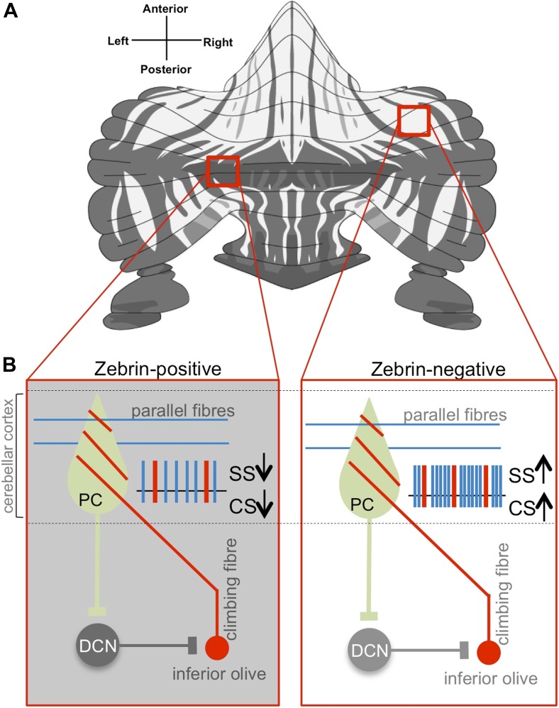
(A) The alternation of zebrin-positive (dark) and zebrin-negative (light) zones gives the cerebellum a striped pattern. (B) Purkinje cells (PC) are the only neurons that carry signals out of the cerebellar cortex; they receive input signals from thousands of parallel fibres (blue) and one single climbing fibre (red). Climbing fibres arise from a region of the brainstem called the inferior olive. The inputs from parallel fibres (integrated with other inputs, not shown) lead to high-frequency simple spikes (SS, blue) in the Purkinje cell. The climbing fibre produces infrequent complex spikes (CS, red). Purkinje cells inhibit neurons in the deep cerebellar nuclei (DCN), which in turn inhibit neurons in the inferior olive. Zhou, Lin et al. found that both simple and complex spike firing frequency of Purkinje cells throughout the cerebellar cortex was decreased in zebrin-positive zones (shown on the left) when compared to zebrin-negative zones (shown on the right).
Comment on
-
Cerebellar modules operate at different frequencies.Elife. 2014 May 7;3:e02536. doi: 10.7554/eLife.02536. Elife. 2014. PMID: 24843004 Free PMC article.
References
-
- Bloedel JR. 1992. Functional heterogeneity with structural homogeneity: how does the cerebellum operate? Behavioral and Brain Sciences 15:666–678. doi: 10.1017/S0140525X00068862 - DOI
Publication types
MeSH terms
LinkOut - more resources
Full Text Sources
Other Literature Sources
