. 2014 Sep;128(3):423-37.
doi: 10.1007/s00401-014-1299-6. Epub 2014 Jun 12.

TDP-43 pathology and neuronal loss in amyotrophic lateral sclerosis spinal cord

Affiliations

TDP-43 pathology and neuronal loss in amyotrophic lateral sclerosis spinal cord

Johannes Brettschneider et al. Acta Neuropathol. 2014 Sep.

Abstract

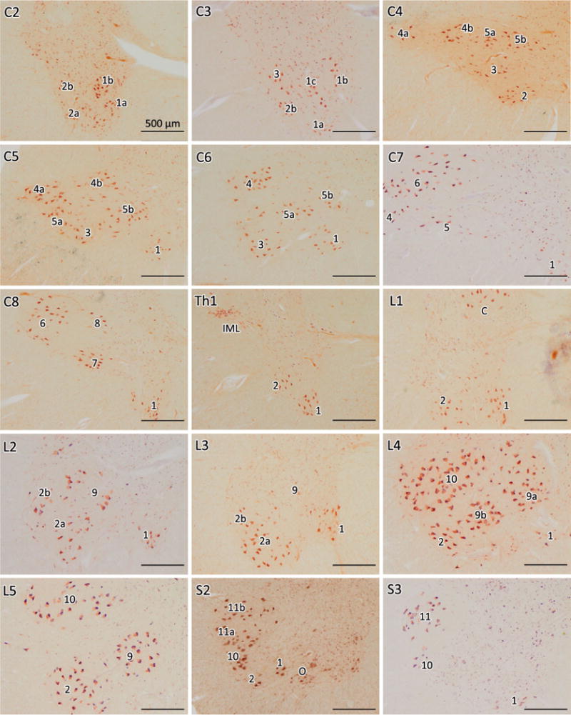
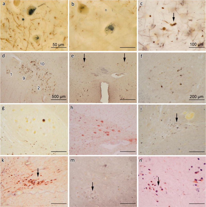
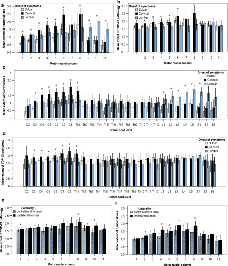
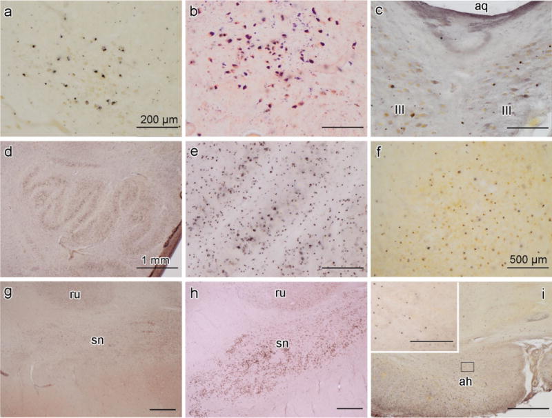
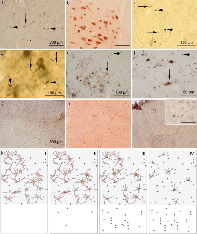
We examined the phosphorylated 43-kDa TAR DNA-binding protein (pTDP-43) inclusions as well as neuronal loss in full-length spinal cords and five selected regions of the central nervous system from 36 patients with amyotrophic lateral sclerosis (ALS) and 10 age-matched normal controls. The most severe neuronal loss and pTDP-43 lesions were seen in lamina IX motor nuclei columns 4, 6, and 8 of lower cervical segments and in columns 9-11 of lumbosacral segments. Severity of pTDP-43 pathology and neuronal loss correlated closely with gray and white matter oligodendroglial involvement and was linked to onset of disease, with severe involvement of columns 4, 6, and 8 of upper extremity onset cases and severe involvement of columns of 9, 10, and 11 in cases with lower extremity onset. Severe TDP-43 lesions and neuronal loss were observed in stage 4 cases and sometimes included Onuf's nucleus. Notably, three cases displayed pTDP-43 aggregates in the midbrain oculomotor nucleus, which we had not seen previously even in cases with advanced (i.e., stage 4) pathology. pTDP-43 aggregates were observed in neurons of Clarke's column in 30.6 % of cases but rarely in the intermediolateral nucleus (IML). Gray matter oligodendroglial pTDP-43 inclusions were present in areas devoid of neuronal pTDP-43 aggregates and neuronal loss. Taken together, our findings indicate that (1) the dorsolateral motor nuclei columns of the cervical and lumbosacral anterior horn may be the earliest foci of pTDP-43 pathology in the spinal cord, (2) gray matter oligodendroglial involvement is an early event in the ALS disease process that possibly heralds subsequent involvement of neurons by pTDP-43 pathology, and (3) in some very advanced cases, there is oculomotor nucleus involvement, which may constitute an additional neuropathological stage (designated here as stage 5) of pTDP-43 pathology in ALS.


Figures

Fig. 1
Fig. 1
Motor nuclei columns of the spinal cord anterior horn in controls. Pigment-Nissl staining (aldehyde fuchsin and Darrow red) of motor nuclei columns of lamina IX in different segments of spinal cord from C2 to S3 in control cases using the organizational scheme proposed by [50]. The right anterior horn is shown in all sections. IML intermediolateral nucleus, C Clarke’s column (posterior thoracic nucleus), O Onuf’s nucleus. Scale bar in C2 is valid for all segments
Fig. 2
Fig. 2
pTDP-43 pathology in motor and non-motor areas of the spinal cord of ALS. a, b Detailed view of skein-like lesions and large rounded aggregates in α-motoneurons using double-labeling IHC with the pTDP-43 antibody (dark blue) and an antibody to the heavy neurofilament subunit (brown) c Skein-like neuronal aggregates (arrow). d Example of pTDP-43 pathology in motor nuclei columns of ALS showing the spinal cord anterior horn at segment L4; numbers indicate the respective motor nuclei columns at this level. e–i, k Images show involvement by pTDP-43 pathology of non-motor areas in the spinal cord of ALS. e Bilateral involvement of Clarke’s column (arrows) at segment L1. f and g show pTDP-43 positive aggregates in neurons of Clarke’s column from segment L2 at higher resolution. h While pTDP-43 aggregates were observed in Clarke’s column of some ALS cases, there was no neuronal loss there: pigment-Nissl staining shows intact neurons of Clarke’s columns at segment L2 in the same ALS case as in g. i, k Involvement of the IML (arrow) at segment Th6 (i) where aggregates are mild in pTDP-43 IHC without detectable neuronal loss in pigment-Nissl staining (k). m, n Mild pTDP-43 IHC (arrow) in Onuf’s nucleus at segment S2 without neuronal loss (arrow) as shown by pigment-Nissl staining (n). Scale bar in (a) is valid for (b). Scale bar in (f) also applies to (g–m). Scale bar in d is valid for e
Fig. 3
Fig. 3
Graphic showing burden of pTDP-43 pathology and severity of neuronal loss in 11 motor nuclei columns at different segments of the spinal cord anterior horn in ALS. a Barplots show severity of neuronal loss and b burden of pTDP-43 pathology in 11 motor nuclei columns of lamina IX anterior horn for bulbar (white bar), cervical (black bar), and lumbar (blue bar) types of ALS onset. c Barplots show severity of neuronal loss and d burden of pTDP-43 pathology from segments C2 to S3 for bulbar (white), cervical upper extremity (black), and lumbar lower extremity (blue) types of ALS onset. e Barplots show the burden of pTDP-43 pathology and f severity of neuronal loss in bulbar (white), cervical (black), and lumbar (blue) bulbar (white), cervical (black), and lumbar (blue) motor nuclei columns of lamina IX of the anterior horn depending on the site of clinical onset of paresis. e, f Blue bars show extent of pathology on the side contralateral to that of the first clinical manifestation of paresis; black bars indicate the extent of pathology on the side ipsilateral to first clinical manifestation. Bars indicate mean and 95 % confidence interval throughout. Significant difference in Wilcoxon Mann–Whitney Test (p < 0.05) is indicated by an asterisk
Fig. 4
Fig. 4
Late-stage pTDP-43 pathology in ALS. Protein aggregation of pTDP-43 and extensive neuronal loss of late-stage ALS (possible ALS stage 5). a Involvement of the oculomotor nucleus in the midbrain. b Pigment-Nissl staining of the same area as shown in a. c Bilateral involvement of the oculomotor nucleus (III), aq—cerebral aqueduct. d Section through the medulla oblongata at the level of the hypoglossal nucleus and inferior olivary complex showing, at higher resolution in e, severe involvement of the inferior olivary complex. f pTDP-43 pathology in the dentate gyrus of the cerebellum. g, h Midbrain sections containing the red nucleus (rn) and substantia nigra (sn). g Substantia nigra from an ALS stage 5 case with severe loss of melanized neurons. h Pigment-Nissl-stained section with intact substantia nigra from an ALS stage 1 case for purposes of comparison. i pTDP-43 IHC shows markedly atrophic anterior horn (ah) with a complete loss of α-motoneurons and widespread gray as well as white matter oligodendroglial lesions from a late-stage ALS case. Framed insert shows gray matter oligodendroglial with pTDP-43-positive aggregates at higher resolution. Scale bar in a applies to b, c, e and framed area in i. Scale bar in d applies to g and h, and scale bar in f is also valid for i
Fig. 5
Fig. 5
Oligodendroglial pTDP-43 pathology in ALS spinal cord. a, b Early involvement of gray matter oligodendroglia (small dot-like/rounded aggregates) at the level of S2 indicated by arrowheads, whereas α-motoneurons (arrow) and white matter oligodendroglia are largely unaffected, and no neuronal loss is detectable in b (pigment-Nissl staining). c Double-labeling IHC of segment C6 anterior horn (asterisk marks a normal α-motoneuron) using an OSP antibody (brown) and pTDP-43 antibody (dark blue). Arrowheads indicate oligodendrocytes containing aggregated pTDP-43, arrows point to oligodendrocytes without pTDP-43 aggregates. d Double-labeling IHC of segment L5 anterior horn using an oligo2 antibody (brown) and pTDP-43 antibody (dark blue). Arrowheads show oligodendrocytes with pTDP-43 aggregates, arrows point to intact oligodendrocyte devoid of pTDP-43 pathology. Micrographs in (e) and (f) show aggregates both in α-motoneurons (arrows) and gray matter oligodendrocytes (arrowheads) demonstrated by pTDP-43 IHC in segments L4 (e) and C7 (f). g C7 anterior horn with massive gray and white matter oligodendroglial pTDP-43 involvement. h Pigment-Nissl staining of same region as in (g) with severe loss of α-motoneurons. i White matter oligodendroglial pTDP-43 pathology in dorsal (sensory) white matter areas at the level of segment Th8. Framed inset shows dorsal white matter oligodendroglia at higher resolution. Scale bar in a also applies to b, scale bar in f is valid for framed area in i, and scale bar in g applies to h, i. k Schematic drawing illustrating the hypothetical sequence of oligodendroglial and neuronal pTDP-43 pathology in gray (upper half of each panel) or white (lower half of each panel) matter of the spinal cord: I—oligodendroglial pTDP-43 inclusions (indicated by brown dots in each panel) are present in the anterior horn gray matter of cases lacking neuronal pTDP-43 aggregates and without evident loss of α-motoneurons or detectable white matter oligodendroglial involvement. II, III—With increasing severity of neuronal pTDP-43 lesions (II) and progressive loss of α-motoneurons (III), oligodendroglial pTDP-43 aggregates are increasingly observed in the spinal cord white matter. IV—In cases with severe neuronal pTDP-43 pathology and extensive α-motoneuron loss, extensive oligodendroglial pTDP-43 pathology is observed across both gray and white matter

References

    1. Braak H, Alafuzoff I, Arzberger T, et al. Staging of Alzheimer disease-associated neurofibrillary pathology using paraffin sections and immunocytochemistry. Acta Neuropathol. 2006;112:389–404. - PMC - PubMed
    1. Braak H, Braak E. Neuropathological stageing of Alzheimer-related changes. Acta Neuropathol. 1991;82:239–259. - PubMed
    1. Braak H, Brettschneider J, Ludolph AC, et al. Amyotrophic lateral sclerosis-a model of corticofugal axonal spread. Nat Rev Neurol. 2013;9:708–714. - PMC - PubMed
    1. Braak H, Del Tredici K. The pathological process underlying Alzheimer’s disease in individuals under thirty. Acta Neuropathol. 2011;121:171–181. - PubMed
    1. Braak H, Del Tredici K. Where, when, and in what form does sporadic Alzheimer’s disease begin? Curr Opin Neurol. 2012;25:708–714. - PubMed

Publication types

MeSH terms