. 2014 Jun;71(6):380-94.
doi: 10.1002/cm.21181. Epub 2014 Jun 26.

Arf1 and Arf6 promote ventral actin structures formed by acute activation of protein kinase C and Src

Affiliations

Arf1 and Arf6 promote ventral actin structures formed by acute activation of protein kinase C and Src

Juliane P Caviston et al. Cytoskeleton (Hoboken). 2014 Jun.

Abstract

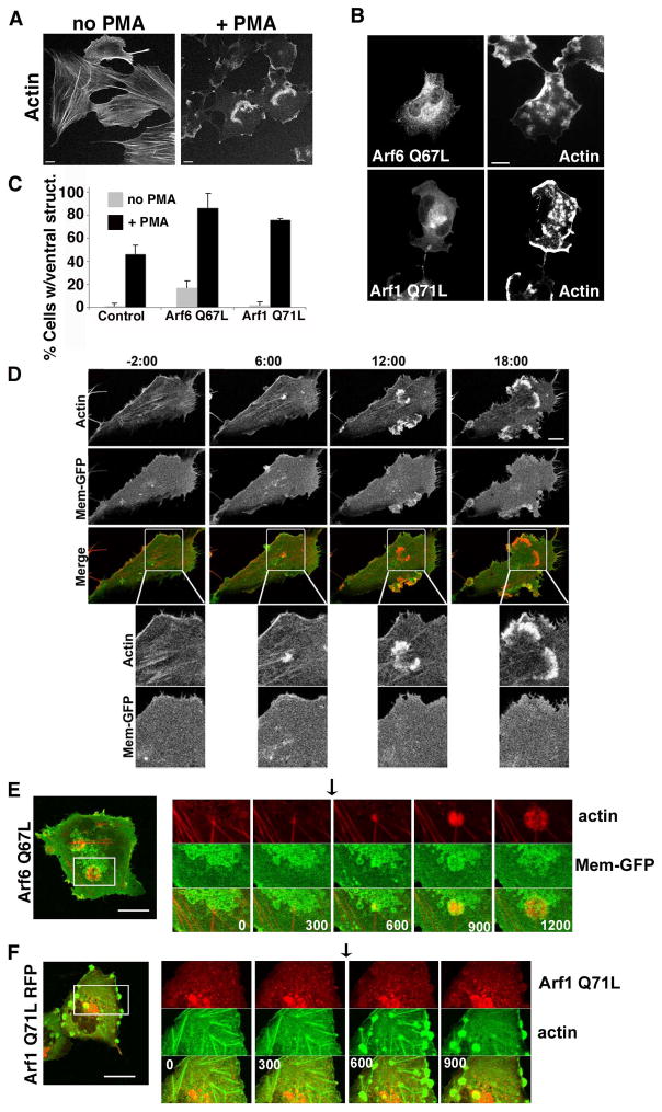
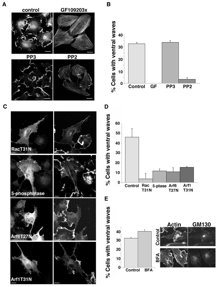
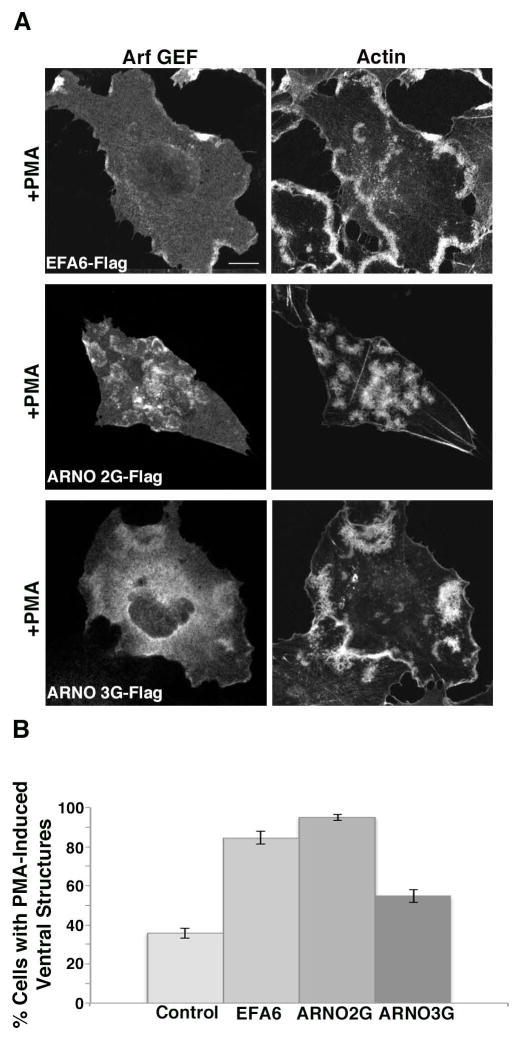
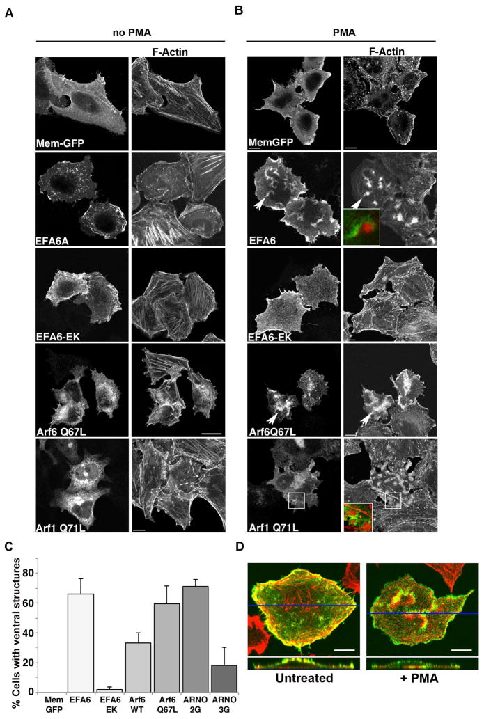
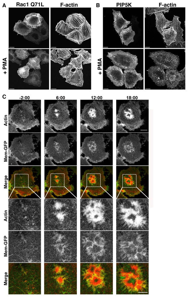
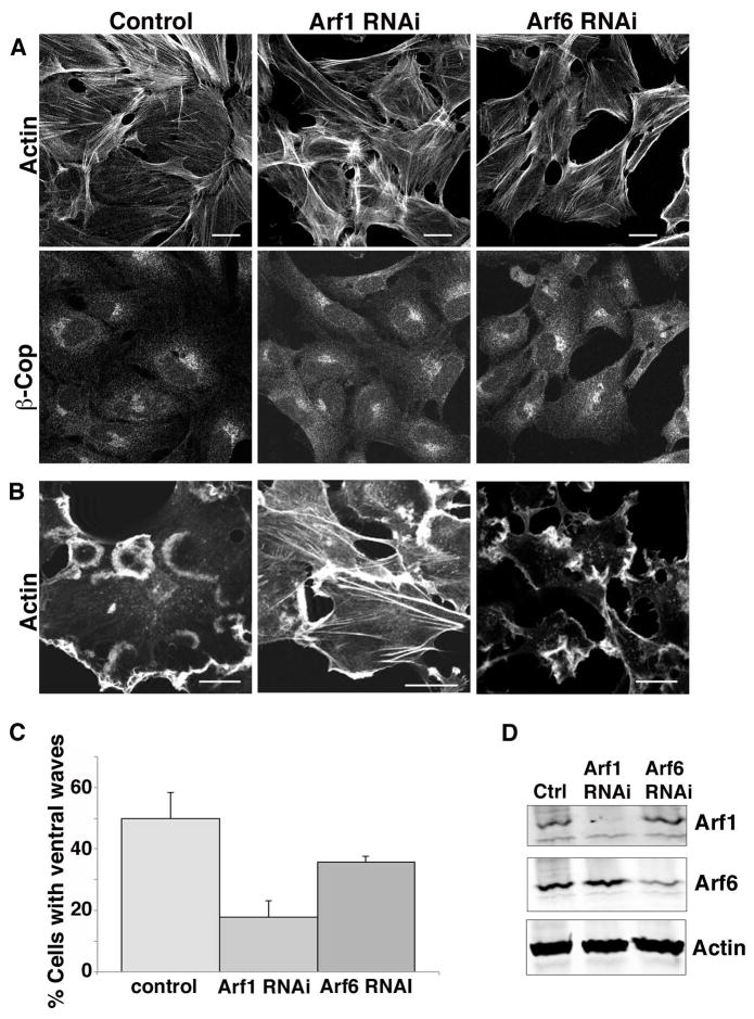
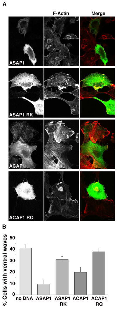
Arf proteins regulate membrane traffic and organelle structure. Although Arf6 is known to initiate actin-based changes in cell surface architecture, Arf1 may also function at the plasma membrane. Here we show that acute activation of protein kinase C (PKC) induced by the phorbol ester PMA led to the formation of motile actin structures on the ventral surface of Beas-2b cells, a lung bronchial epithelial cell line. Ventral actin structures also formed in PMA-treated HeLa cells that had elevated levels of Arf activation. For both cell types, formation of the ventral actin structures was enhanced by expression of active forms of either Arf1 or Arf6 and by the expression of guanine nucleotide exchange factors that activate these Arfs. By contrast, formation of these structures was blocked by inhibitors of PKC and Src and required phosphatidylinositol 4, 5-bisphosphate, Rac, Arf6, and Arf1. Furthermore, expression of ASAP1, an Arf1 GTPase activating protein (GAP) was more effective at inhibiting the ventral actin structures than was ACAP1, an Arf6 GAP. This study adds to the expanding role for Arf1 in the periphery and identifies a requirement for Arf1, a "Golgi Arf," in the reorganization of the cortical actin cytoskeleton on ventral surfaces, against the substratum.

Keywords: Arf1; PKC; Src; cortical actin; ventral surface.


Figures

Figure 1
Figure 1. PMA treated Beas-2b cells form ventral actin waves
(A) Untransfected Beas-2b cells were treated with vehicle (no PMA) or 200 nM PMA for 30 min prior to fixation and staining with rhodamine phalloidin. (B) Beas-2b cells were transfected with plasmids encoding untagged Arf6 Q67L (top) or Arf1 Q71L-GFP (bottom) and treated with 200 nM PMA for 30 min prior to fixation and labeling with antibodies to Arf6 for Arf6 detection and actin. Bars, 10 μm. (C) The fraction of transfected cells with 1 or more ventral waves was quantified and is expressed as the average percentage obtained from three independent experiments. Error bars represent standard deviation from the means. GFP was used as a control for transfection. One-way ANOVA test of the PMA-treated Control vs. Arf6Q67L- and Arf1Q71L-transfected cells were significant (p<0.05). (D) Beas-2b cells transfected with Mem-GFP and RFP-LifeAct were imaged as described in Materials and Methods. PMA was added after 2 min, designated time 0, and frames were captured every 30 seconds thereafter. Selected stills from the movie at −2, 6,12 and 18 min are shown (see Movie 1 in Supplementary materials). No membrane folds are associated with actin structures. Bar, 10μM. Boxed regions are shown at higher magnification below each time point. Bar, 5μM. (E) Beas-2b cells were transfected with plasmids encoding untagged Arf6 Q67L (in background), Mem-GFP to mark vacuolar membranes in transfected cells, and LifeAct-RFP (to visualize actin). A cell was imaged for 20 min; at 5 min, 200 nM PMA was added (arrow) to induce ventral wave formation (Movie 2 in Supplementary materials). (F) Beas-2b cells were transfected with plasmids encoding Arf1-Q71L-RFP and GFP-Actin. A cell was imaged for 15 minutes; at 5 min 200 nM PMA was added (arrow) to induce ventral wave formation (Movie 3 in Supplementary materials). Shown for each movie is an image of the entire cell taken at the end of the movie with a square around the region shown in the movie. A series of frames from the movie with time indicated in seconds from the beginning of the movie is also shown. Black arrows indicate when PMA was added. Bars,10 μm.
Figure 2
Figure 2. Ventral actin wave formation in Beas-2b cells requires PKC, Src, Rac activity, PIP2, and Arf
(A) Beas-2b cells were treated for 30 min with 200 nM PMA in the absence or presence of 10 μM GF 109303x (inhibitor of PKC), 10 μM PP3 (inactive analog of PP2) or 10 μM PP2 (inhibitor of Src) as indicated. Following PMA treatment, cells were fixed and stained with rhodamine phalloidin as described. Bars,10 μm. (B) Fraction of control or drug-treated cells with 1 or more ventral waves visible in phalloidin stain was quantified and is expressed as the average percentage from three independent experiments. Error bars represent standard deviation from the mean. Tukey multiple comparison test showed that GF and PP2 differed from the Control (p<0.001). (C) Beas-2b cells expressing untagged Rac T31N, the p72 PI(4,5)P2 5-phosphatase (myc tagged), Arf6 T27N, or Arf1 T31N-HA were treated with 200 nM PMA 30 min prior to fixation and immunofluorescence staining as described in the Material and Methods section with an appropriate primary antibody to detect the transfected protein and Alexa 488 conjugated secondary antibody. Cells were co-stained with rhodamine phalloidin (right panels). (D) Fraction of transfected cells with 1 or more ventral waves visible in phalloidin stain was quantified and is expressed as the average percentage from three independent experiments. Error bars represent standard deviation from the mean. One way ANOVA test showed all treatments differed from Control (p<0.001). (E) Untransfected Beas-2b cells were left untreated (control) or pretreated for 2 h with 5 μg/ml brefeldin A (BFA). Both sets of cells were treated with 200 nM PMA for 30 min prior to fixation and staining with rhodamine phalloidin and antibody to GM130. The percentage of cells with 1 or more ventral waves was quantified and is expressed as the average percentage of three independent experiments. Error bars represent one standard deviation from the mean. Student's T-test showed Control vs. BFA treated was not statistically significant. Bars,10 μm.
Figure 3
Figure 3. Arf GEFs enhance ventral actin structure formation in Beas-2b cells
(A) Beas 2B cells were transfected with plasmids encoding Flag-EFA6 (Arf6 GEF), or the Arf1 GEFS Flag-ARNO 2G (PIP3 binding) or Flag-ARNO 3G (PIP2 binding). Cells were treated with PMA, fixed and immunostained with antibody against Flag and rhodamine phalloidin. Bar, 10 μm. (B) Percentage of cells exhibiting one or more ventral actin structures was quantified. With the exception of Flag-EFA6, in total, 200 cells were counted from three independent experiments. For Flag-EFA6, in total, 123 cells were counted from three independent experiments. Error bars represent standard error for proportional data, P<0.0001 as determined by Fisher’s Exact test (two-sided) indicating that GEF expression resulted in increase in cells with ventral actin structures.
Figure 4
Figure 4. Active Arf6 or Arf1 promotes ventral actin structure formation in HeLa cells upon PMA treatment
HeLa cells were transfected with plasmids encoding Mem-GFP, FLAG-EFA6, FLAG-EFA6-EK, untagged Arf6 Q67L or Arf1Q71L-GFP. 18 hours following transfection, cells were untreated (A) or treated for 30 min with 200 nM PMA (B) prior to fixation and immunofluorescence staining as described in Materials and Methods. F-actin was labeled with rhodamine phalloidin, and antibodies to FLAG, Arf6 and Arf1 were detected with an Alexa 488-conjugated secondary antibody. In B arrow points to ventral ruffles, which are enlarged in inset. Bars,10 μm. (C) Fraction of cells expressing Mem-GFP, FLAG-EFA6, FLAG-EFA6-EK, Arf6 Q67L, Arf1Q71L, FLAG-ARNO 2G or FLAG-ARNO 3G with 1 or more PMA-induced ventral actin structure visible by phalloidin staining was quantified and is expressed as the average percentage of transfected cells from three independent experiments with 100 cells scored in each experiment. Error bars represent one standard deviation from the mean. (D) Z-sections of HeLa cells, transfected with Flag-EFA6 that were untreated or treated with PMA for 15 min, fixed and stained with an antibody against Flag (green) and Rhodamine phalloidin (red). A cross-section of the cell shows that Flag-EFA6, which associates with the plasma membrane, shows increased concentration on the ventral (bottom) surface upon stimulation with PMA, where the ventral actin structure creates a membrane fold, or ruffle. A blue line denotes the location of the cross section.
Figure 5
Figure 5. Rac1 and PIP5-kinase are not sufficient for ventral actin structures and actin and membrane dynamics in HeLa cells treated with PMA
HeLa cells expressing Rac1Q71L (A) or PIP5-kinase (PIP5K) (myc tagged) (B) were treated with 200 nm PMA for 30 min prior to fixation and immunofluorescence staining as described. Bars 10 μM. (C). HeLa cells coexpressing FLAG-EFA6 (in background), Mem-GFP (to label the membrane) and Life-Act-RFP (to label F-actin) were imaged live before and after addition of 100 nM PMA. PMA was added after 2 min, designated time 0, and frames were captured every 30 seconds thereafter. Selected stills from the movie at −2, 6,12 and 18 min are shown (see Movie 4 in Supplementary materials). Nascent actin structures visible at −2 min expand into larger actin structures after addition of PMA and by 6 min and can be seen pushing a membrane fold outward. Bar, 10μM. Boxed regions are shown at higher magnification below each time point. Bar, 5 μM.
Figure 6
Figure 6. Arf1 and Arf6 are required for ventral wave formation in Beas-2b cells
(A) Beas-2b cells were transfected with siRNA to knockdown Arf1 or Arf6, or mock treated (control), as described in the Materials and Methods. 72 h after transfection, cells were fixed and stained by immunofluorescence as described in the Materials and Methods with an antibody against β-COP, and co-stained with rhodamine phalloidin. (B) Control and Arf1-or Arf6-depleted cells were treated with 200 nM PMA for 30 min prior to fixation and staining with rhodamine phalloidin. Bars,10 μm. (C) The fraction of 200 cells with 1 or more visible ventral waves was quantified and is expressed as the average percentage from three independent experiments. Error bars represent one standard deviation of the mean. One-way ANOVA revealed that both siRNA-depletions differed from control (p<0.01). (D) 5 X 10⁵ cells were collected, lysed, and run on a SDS page gel, and immunoblotted with antibodies against Arf1, Arf6 and actin. Arf1 was depleted by 90% and Arf6 by 75%.
Figure 7
Figure 7. Expression of Arf GAPs inhibits ventral actin wave formation in Beas-2b cells
(A) Flag-ASAP1, Flag-ASAP1 RK, Flag-ACAP1 or Flag-ACAP1 RQ was transfected into Beas-2b cells. Cells were treated with PMA and stained with anti-Flag antibody (left column) and rhodamine phalloidin to detect F-actin (middle column). ASAP1 and ACAP1 transfected cells largely did not exhibit ventral actin waves, while cells transfected with enzymatically inactive control constructs ASAP1 RK and ACAP1RQ retained ventral actin waves. Bar, 10μm. (B) Quantification of percentage of control or transfected cells exhibiting ventral actin waves after PMA treatment. A minimum of 100 cells in total was counted from at least 3 independent experiments. Bars represent standard error for proportional data. Percentage of cells transfected with ASAP1 or ACAP1 was significantly reduced compared to control cells (P<0.0001) as assessed by calculating P-values using Fisher’s Exact test (two-tailed.) Percentage of cells expressing ASAP1 RK or ACAP1RQ exhibiting ventral waves was not statistically different than control cells.

Similar articles

Cited by

References

    1. Aikawa Y, Martin TFJ. ARF6 regulates a plasma membrane pool of phosphatidylinositol(4,5)bisphosphate required for regulated exocytosis. J Cell Biol. 2003;162(4):647–659. - PMC - PubMed
    1. Albiges-Rizo C, Destaing O, Fourcade B, Planus E, Block MR. Actin machinery and mechanosensitivity in invadopodia, podosomes and focal adhesions. J Cell Sci. 2009;122(Pt 17):3037–49. - PMC - PubMed
    1. Balasubramanian N, Scott DW, Castle JD, Casanova JE, Schwartz MA. Arf6 and microtubules in adhesion-dependent trafficking of lipid rafts. Nat Cell Biol. 2007;9(12):1381–91. - PMC - PubMed
    1. Beemiller P, Hoppe AD, Swanson JA. A phosphatidylinositol-3-kinase-dependent signal transition regulates ARF1 and ARF6 during Fcgamma receptor-mediated phagocytosis. PLoS Biol. 2006;4(6):e162. - PMC - PubMed
    1. Bharti S, Inoue H, Bharti K, Hirsch DS, Nie Z, Yoon HY, Artym V, Yamada KM, Mueller SC, Barr VA, et al. Src-dependent phosphorylation of ASAP1 regulates podosomes. Mol Cell Biol. 2007;27(23):8271–83. - PMC - PubMed

Publication types