. 2014 Oct;5(10):761-9.
doi: 10.1007/s13238-014-0074-8. Epub 2014 Jun 11.

Gluconate 5-dehydrogenase (Ga5DH) participates in Streptococcus suis cell division

Affiliations

Gluconate 5-dehydrogenase (Ga5DH) participates in Streptococcus suis cell division

Zhongyu Shi et al. Protein Cell. 2014 Oct.

Abstract

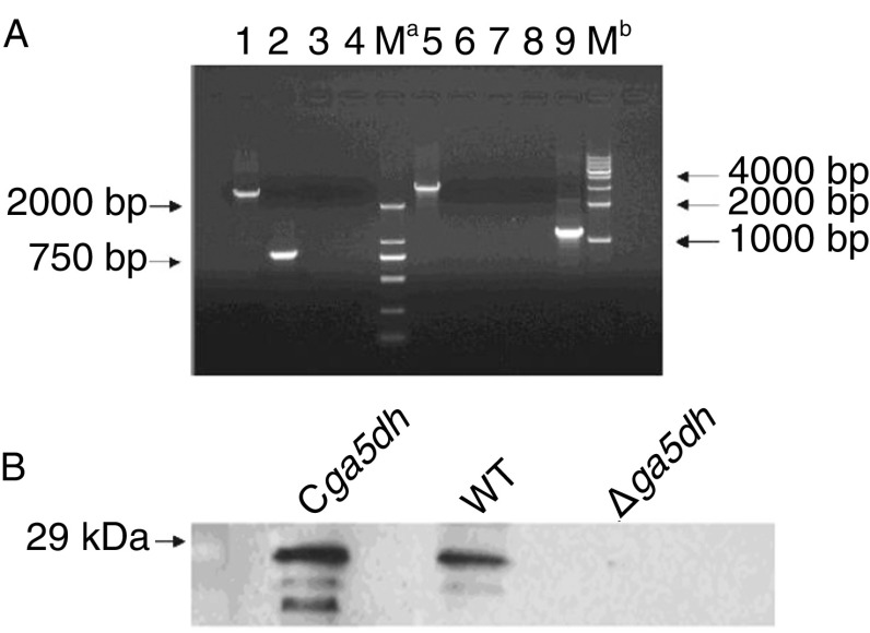
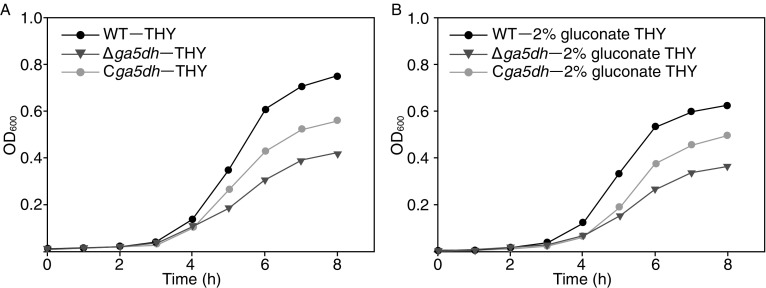
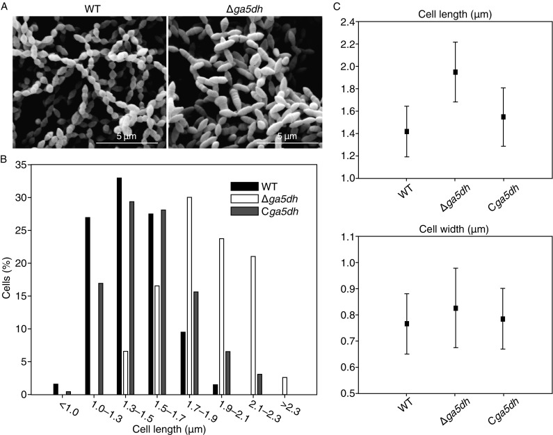
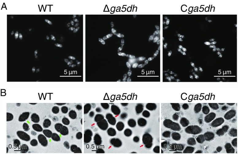
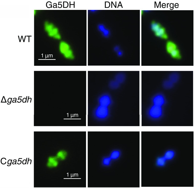
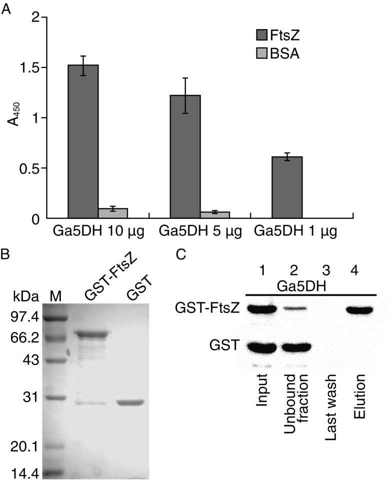
Bacterial cell division is strictly regulated in the formation of equal daughter cells. This process is governed by a series of spatial and temporal regulators, and several new factors of interest to the field have recently been identified. Here, we report the requirement of gluconate 5-dehydrogenase (Ga5DH) in cell division of the zoonotic pathogen Streptococcus suis. Ga5DH catalyzes the reversible reduction of 5-ketogluconate to D-gluconate and was localized to the site of cell division. The deletion of Ga5DH in S. suis resulted in a plump morphology with aberrant septa joining the progeny. A significant increase was also observed in cell length. These defects were determined to be the consequence of Ga5DH deprivation in S. suis causing FtsZ delocalization. In addition, the interaction of FtsZ with Ga5DH in vitro was confirmed by protein interaction assays. These results indicate that Ga5DH may function to prevent the formation of ectopic Z rings during S. suis cell division.


Figures

Figure 1
Figure 1
Construction and confirmation of the Δ ga5dh mutant S. suis and its complemented strain by multiplex PCR and Western blot analyses. (A) Genomic DNA extracts from the WT or the deletion mutant were used as templates: WT strain (lanes 1–4) and Δga5dh mutant (lanes 5–9). Two different DNA ladder markers, which are marked as Ma and Mb respectively, were used and labeled. The primer pairs and the theoretical size (bp) of the indicated PCR product are as follows: lanes 1: LU/RD, 2892 bp; 2: ga5dh-F/ga5dh-R, 835 bp; 3: ga5dh-F/spc-R, (-); 4: spc-F/ga5dh-R, (-); 5: LU/RD, 3209 bp; 6: ga5dh-F/ga5dh-R, (-); 7: ga5dh-F/spc-R, (-); 8: spc-F/ga5dh-R, (-); and 9: spc-F/spc-R, 1130 bp. (B) Western blot assay characterizing the protein expression of Ga5DH. The indicated S. suis cells were probed with Ga5DH polyclonal antibodies, and the expression profiles are shown
Figure 2
Figure 2
Ga5DH is required for S. suis growth. Comparative analyses of growth curves from three S. suis strains (WT, the Δga5dh mutant, and the complemented strain Cga5dh). The strains were grown in THY liquid medium (A) or in THY liquid medium supplemented with 2% (w/v) gluconate (B). Each point represents the mean ± SD OD600 value from triplicate experiments
Figure 3
Figure 3
Micrographs of WT, Δ ga5dh mutant cells, and C ga5dh cells. (A) Scanning electron micrographs of the WT and the Δga5dh mutant strains. (B) Histogram statistics of the WT (n = 200, black), Δga5dh (n = 200, white), and Cga5dh (n = 200, gray) cell lengths after growth in THY medium. To determine the cell length, we measured the longitudinal length of single membrane-stained cells. Δga5dh cells were significantly longer than WT cells. (C) Cell size parameters of the WT, Δga5dh, and Cga5dh cells. The cell length of Δga5dh cells was significantly different from that of the WT strain (P < 0.01; two-tailed t-test)
Figure 4
Figure 4
Micrographs of the WT, Δ ga5dh , and C ga5dh strains. (A) FM4-64 staining; scale bar, 5 μm. (B) Transmission electron micrographs; scale bar, 0.5 μm. Green arrows highlight the correct septum placement in WT cells. Red arrows mark the aberrant septum placement in Δga5dh cells. Cell morphology of the WT strain revealed the characteristic ellipsoid shape and normal division pattern of S. suis, with correct septum placement and symmetric daughter cells. The mutant cells displayed an increased length and multiple aberrant septa with incorrect placement
Figure 5
Figure 5
Subcellular localization of Ga5DH. DNA was visualized with DAPI (blue). Ga5DH was visualized using an anti-Ga5DH polyclonal antibody and an anti-mouse IgG secondary antibody coupled to Alexa Fluor 488 (green). The merged pictures show the overlay of Ga5DH and DAPI staining. Scale bar, 1 μm
Figure 6
Figure 6
Ga5DH interacts with FtsZ in vitro. (A) Solid-phase binding assay for FtsZ and Ga5DH. Wells of a microtiter plate were coated with FtsZ or BSA and incubated with various concentrations of Ga5DH protein as indicated. The bound Ga5DH protein was immunodetected with Ga5DH polyclonal antibodies by ELISA. (B) Inputs of GST-FtsZ and GST in the GST pull-down assay detected by SDS-PAGE. (C) GST pull-down assay characterizing the interaction between FtsZ and Ga5DH. Lane 1: the input His-tagged Ga5DH in the pull-down assay. Lane 2: the unbound fraction of Ga5DH. Lane 3: the last wash of the unbound fraction of Ga5DH. Lane 4: the eluted protein fraction using 20 mmol/L glutathione. All samples were detected by Western blotting using an anti-His monoclonal antibody
Figure 7
Figure 7
Delocalization of FtsZ rings in S. suis cells lacking Ga5DH. The morphology of FtsZ was detected by immunolabeling with anti-FtsZ polyclonal antibodies and an anti-mouse IgG secondary antibody coupled to Alexa Fluor 488 (green). DNA was visualized with DAPI (blue). The merged pictures show the overlay of FtsZ and DAPI staining. Scale bar = 1 μm

Similar articles

Cited by

References

    1. Addinall SG, Lutkenhaus J. FtsZ-spirals and -arcs determine the shape of the invaginating septa in some mutants of Escherichia coli. Mol Microbiol. 1996;22:231–237. doi: 10.1046/j.1365-2958.1996.00100.x. - DOI - PubMed
    1. Albarracin Orio AG, Pinas GE, Cortes PR, Cian MB, Echenique J. Compensatory evolution of pbp mutations restores the fitness cost imposed by β-lactam resistance in Streptococcus pneumoniae. PLoS Pathog. 2011;7:e1002000. doi: 10.1371/journal.ppat.1002000. - DOI - PMC - PubMed
    1. Bi EF, Lutkenhaus J. FtsZ ring structure associated with division in Escherichia coli. Nature. 1991;354:161–164. doi: 10.1038/354161a0. - DOI - PubMed
    1. Dziedzic R, Kiran M, Plocinski P, Ziolkiewicz M, Brzostek A, Moomey M, Vadrevu IS, Dziadek J, Madiraju M, Rajagopalan M. Mycobacterium tuberculosis ClpX interacts with FtsZ and interferes with FtsZ assembly. PloS One. 2010;5:e11058. doi: 10.1371/journal.pone.0011058. - DOI - PMC - PubMed
    1. Elbaz M, Ben-Yehuda S. The metabolic enzyme ManA reveals a link between cell wall integrity and chromosome morphology. PLoS Genet. 2010;6:e1001119. doi: 10.1371/journal.pgen.1001119. - DOI - PMC - PubMed

Publication types