. 2014 Aug;3(4):470-83.
doi: 10.1002/mbo3.181. Epub 2014 Jun 10.

Unusual interaction of RNA polymerase with the bacteriophage Mu middle promoter Pm in the absence of its activator protein Mor

Affiliations

Unusual interaction of RNA polymerase with the bacteriophage Mu middle promoter Pm in the absence of its activator protein Mor

Yongkai Mo et al. Microbiologyopen. 2014 Aug.

Abstract

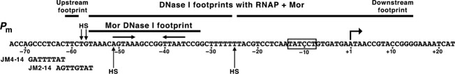
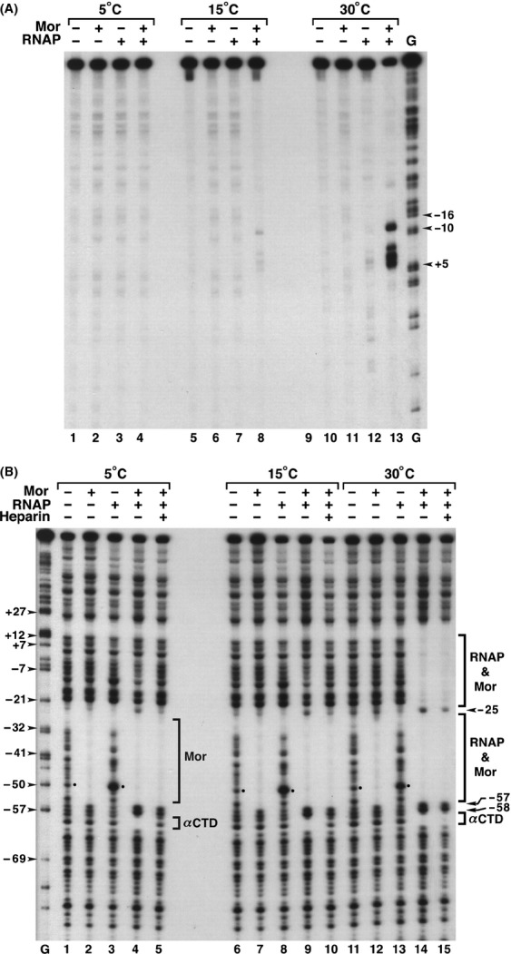
The bacteriophage Mu Mor activator protein is absolutely required for transcription from the Mu middle promoter P(m). However, when RNA polymerase (RNAP) was incubated with P(m) DNA in the absence of Mor, a band at promoter position -51 was hypersensitive to DNase I cleavage, demonstrating an interaction of RNAP with the promoter DNA. The hypersensitivity was similar at four different lengths of P(m) DNA assayed from -62 to +10, -62 to +46, -96 to +10, and -96 to +46. The hypersensitivity occurred equally well at 5 °C, 15 °C, and 30 °C, indicating that it did not require open complex formation, which only occurred at 30 °C. The -51 hypersensitivity at 5 °C and 15 °C was eliminated by the addition of heparin, consistent with the possibility that it arose by formation of unstable closed complexes of RNAP bound to P(m) DNA. Generation of the hypersensitive band required the complete RNAP with its αCTDs, but neither the αCTD nor intact α were sufficient for the interaction and resulting hypersensitivity. There was no correlation between the level of hypersensitivity observed in vitro and the level of Pm activity in vivo, as assayed by the Mor-dependent production of β-galactosidase from a P(m)-lacZ fusion. In an "order of addition" experiment, preincubation of P(m) DNA with Mor followed by addition of RNAP led to the fastest open complex formation, whereas preincubation of P(m) DNA with RNAP gave the slowest. These results support the conclusion that Mor recruits RNAP to P(m) rather than reposition a prebound RNAP, as occurs for C-dependent repositioning of RNAP at the Mu late promoter Pmom .

Keywords: Bacteriophage Mu; Mor activator protein; Mu middle promoter Pm; RNAP-promoter interactions; prokaryotic transcription.


Figures

Figure 1
Figure 1
Model for Pm promoter initiation complex. The horizontal line represents Pm DNA with relevant positions marked below it. Mor is shown as a dimer bound to the dyad-symmetric element indicated by inverted arrows centered at −43.5. The −10 hexamer is shown as a black box, and the transcription start (+1) is designated by a bent arrow. The Mor-αCTD and Mor-σCTD interactions are designated by black ovals. The RNAP subunits are labeled; the position of the second αCTD is not known. This figure is based on observations of the Mor-RNAP interaction paper from the Howe lab (Artsimovitch et al. 1996).
Figure 2
Figure 2
The Pm sequence with DNase I footprints. The sequence of Pm from −73 to +23 is shown with dots indicating 10-base intervals that are assigned “−” numbers upstream and “+” numbers downstream of +1, the initiation site. The bars indicate the bases protected from DNase I digestion by the proteins shown. Inverted arrows correspond to the position of the dyad-symmetric Mor binding site; vertical arrows indicate the locations of hypersensitive sites (HS) cleaved by DNase I; the −10 hexamer is in a box, and the bent arrow designates the start of the RNA transcript at +1. The altered sequences present in two mutants, JM2-14 and JM4-14, are aligned directly below the corresponding positions in Pm.
Figure 3
Figure 3
DNase I footprinting of probes containing different lengths of wild-type Pm DNA. Panel (A) contains linear, top strand, 5' end-labeled DNA fragments (0.42 nmol/L) containing different lengths of Pm DNA (shaded bar) and flanking plasmid vector DNA (open bar) were incubated with Mor (800 nmol/L) and/or RNAP (56 nmol/L) at 30°C for 20 min, then treated with DNase I as described in Methods. When both Mor and RNAP were used, Mor was added to DNA first and the reaction was incubated for 5 min prior to addition of RNAP. The length of Pm DNA in the probe is given above the bracket for each panel. The relative β-galactosidase (β-gal) activity produced in vivo by a Pm-lacZ fusion plasmid carrying those sequences is given just below the bracket and is the average derived from three independent assays. The presence or absence of each protein is indicated by a “+” or “−”, respectively, above each lane. The positions of relevant bands are indicated by arrowheads to the left of lane 1. The bar to the left of lane 2 identifies the Mor footprint, whereas those to the left of lane 4 indicate footprints arising in the presence of both Mor and RNAP. Panel (B) contains DNA treated as in panel (A) with an additional lane containing a G-ladder and was derived from a different experiment. Lane 19 contains a G-ladder with arrowheads marking the promoter positions of specific G-ladder bands. These bands migrate 1.5 nucleotides faster than corresponding DNase I fragments (Sollner-Webb and Reeder 1979).
Figure 4
Figure 4
DNase I and KMnO4 footprinting at different temperatures. Binding reactions were generated with probe containing Pm sequence −98 to +46 and flanking vector DNA, as described for Figure3 except that samples were incubated at 5°C, 15°C, and 30°C. For KMnO4 footprinting in panel (A) the probe was labeled at the 5′ end of the bottom strand. After 5 min incubation with Mor and 6 min incubation with RNAP, the samples were subjected to KMnO4 modification and cleavage. Arrowheads mark promoter positions of specific G-ladder bands. For DNase I footprinting in panel (B) lanes 5, 10, and 15 also received heparin to 100 ngL, and the mixture was incubated for 1 min prior to DNase I digestion. Bars indicate the extent of the footprints generated by Mor alone, or by Mor and RNAP together. Arrowheads indicate the promoter positions of G-ladder bands. Dots mark the bands at position −51. Arrows and arrowheads marked HS identify positions −25, −57, −58 that are hypersensitive to DNase I digestion.
Figure 5
Figure 5
DNase I footprinting with RNA polymerases and different forms of the αCTD at 5°C. The DNA probe was linear, top strand and 5′ end-labeled with Pm sequences from −98 to +46 and flanking plasmid vector DNA. (A) The probe was preincubated with or without Mor (800 nmol/L) for 5 min, then RNAP or RNAP-ΔαCTD was added and the reactions incubated for 20 min prior to DNase I digestion. The presence and absence of the proteins are designated by “+” and “−” signs above each lane. The upstream footprint is identified with a bracket, and dots mark the bands for position −51. Arrowheads mark the positions of G ladder bands, which migrate 1.5 nucleotides faster than bands generated by DNase I cleavage (Sollner-Webb and Reeder ; Artsimovitch et al. 1996). (B) The same DNA probe was incubated with His-α (9 μmol/L), His-αCTD (35 μmol/L), and αCTD (35 μmol/L) at 5°C for 20 min prior to DNase I digestion. Labeling follows that in panel (A). (C) The same DNA probe was incubated with (+) and without (−) Mor (800 nmol/L) and/or His-αCTD at 35 μmol/L (+), 175 μmol/L (++), and 350 μmol/L (+++) or RNAP (56 nmol/L) prior to treatment with DNase I. The positions of band −51 are identified on the left, and the positions of the −59 to −61 upstream footprint are identified on the right.
Figure 6
Figure 6
DNase I footprinting with wild-type and mutant Pm probes. All probe DNAs were 5′-end labeled and contained Pm sequences from −98 to +10 and flanking plasmid vector DNA. Probes JM4-14 and JM2-14 contain the mutations shown in Figure2. Binding reactions were performed and the figure labeled as described for Figure3.
Figure 7
Figure 7
Order of addition DNase I and KMnO4 footprinting. The probe contained wild-type Pm sequence from −98 to +46 (and flanking plasmid vector DNA). For DNase I digestions, the DNA probe was labeled at the 5′ end of the top strand. For KMnO4 footprinting, the DNA probe was labeled at the 5′ end of the bottom strand. The three sets of reactions differed in which two components were incubated together (Mixed first) prior to addition of the third component (Added last). The amounts of each protein and probe used were the same as for Figure3. After addition of the third component, samples were subjected to DNase I digestion (A) or KMnO4 treatment and cleavage (B) at the times indicated above each lane. In the left set of reactions (lanes 1–7), Mor was preincubated with the Pm probe for 5 min before RNAP addition; in the middle set (lanes 8–14), RNAP was preincubated with the probe for 6 min before Mor addition; in the right set (lanes 15–20), Mor and RNAP were preincubated together for 6 min before addition of Pm probe. The positions of the hypersensitive band at −51 and the upstream footprint are indicated. Arrowheads mark the promoter positions of specific G-ladder bands.

Similar articles

Cited by

References

    1. Artsimovitch I. Howe MM. Transcription activation by the bacteriophage Mu Mor protein: analysis of promoter mutations in Pm identifies a new region required for promoter function. Nucleic Acids Res. 1996;24:450–457. - PMC - PubMed
    1. Artsimovitch I, Murakami K, Ishihama A. Howe MM. Transcription activation by the bacteriophage Mu Mor protein requires the C-terminal regions of both alpha and sigma70 subunits of Escherichia coli RNA polymerase. J. Biol. Chem. 1996;271:32343–32348. - PubMed
    1. Balke V, Nagaraja V, Gindlesperger T. Hattman S. Functionally distinct RNA polymerase binding sites in the phage Mu mom promoter region. Nucleic Acids Res. 1992;20:2777–2784. - PMC - PubMed
    1. Barne KA, Bown JA, Busby SJW. Minchin SD. Region 2.5 of the Escherichia coli RNA polymerase sigma70 subunit is responsible for the recognition of the ‘extended-10’ motif at promoters. EMBO J. 1997;16:4034–4040. - PMC - PubMed
    1. Belyaeva TA, Bown JA, Fujita N, Ishihama A. Busby SJ. Location of the C-terminal domain of the RNA polymerase alpha subunit in different open complexes at the Escherichia coli galactose operon regulatory region. Nucleic Acids Res. 1996;24:2242–2251. - PMC - PubMed

Publication types

Substances

LinkOut - more resources