. 2014 Jul;76(1):108-19.
doi: 10.1002/ana.24195. Epub 2014 Jun 27.

Acute mechanisms underlying antibody effects in anti-N-methyl-D-aspartate receptor encephalitis

Affiliations
Free PMC article

Acute mechanisms underlying antibody effects in anti-N-methyl-D-aspartate receptor encephalitis

Emilia H Moscato et al. Ann Neurol. 2014 Jul.
Free PMC article

Abstract

Objective: A severe but treatable form of immune-mediated encephalitis is associated with antibodies in serum and cerebrospinal fluid (CSF) against the GluN1 subunit of the N-methyl-D-aspartate receptor (NMDAR). Prolonged exposure of hippocampal neurons to antibodies from patients with anti-NMDAR encephalitis caused a reversible decrease in the synaptic localization and function of NMDARs. However, acute effects of the antibodies, fate of the internalized receptors, type of neurons affected, and whether neurons develop compensatory homeostatic mechanisms were unknown and are the focus of this study.

Methods: Dissociated hippocampal neuron cultures and rodent brain sections were used for immunocytochemical, physiological, and molecular studies.

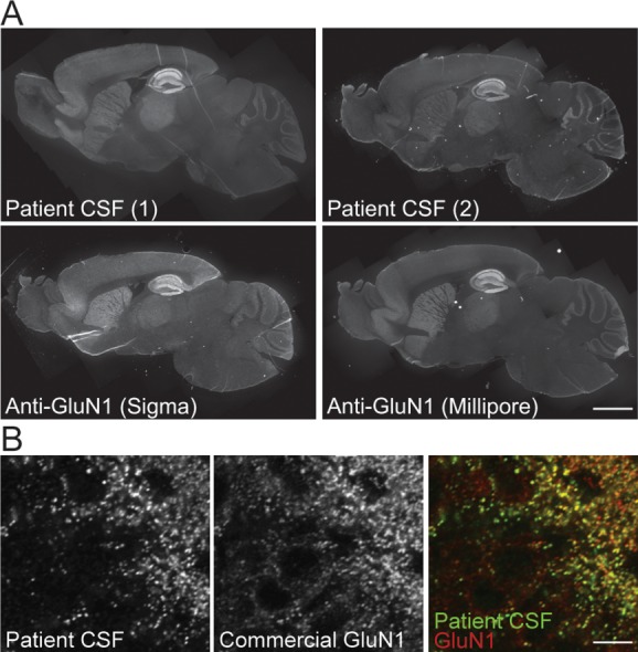
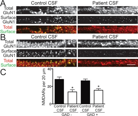
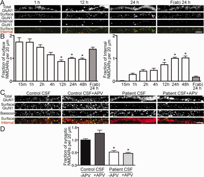
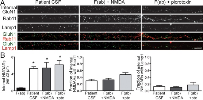
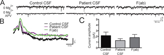
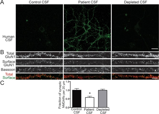
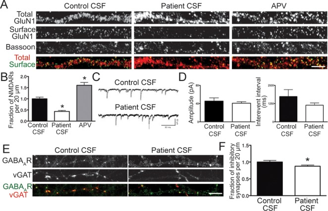
Results: Patient antibodies bind to NMDARs throughout the rodent brain, and decrease NMDAR cluster density in both excitatory and inhibitory hippocampal neurons. They rapidly increase the internalization rate of surface NMDAR clusters, independent of receptor activity. This internalization likely accounts for the observed decrease in NMDAR-mediated currents, as no evidence of direct blockade was detected. Once internalized, antibody-bound NMDARs traffic through both recycling endosomes and lysosomes, similar to pharmacologically induced NMDAR endocytosis. The antibodies are responsible for receptor internalization, as their depletion from CSF abrogates these effects in hippocampal neurons. We find that although anti-NMDAR antibodies do not induce compensatory changes in glutamate receptor gene expression, they cause a decrease in inhibitory synapse density onto excitatory hippocampal neurons.

Interpretation: Our data support an antibody-mediated mechanism of disease pathogenesis driven by immunoglobulin-induced receptor internalization. Antibody-mediated downregulation of surface NMDARs engages homeostatic synaptic plasticity mechanisms, which may inadvertently contribute to disease progression.


Figures

FIGURE 1
FIGURE 1
Patient and commercial GluN1 antibodies were similarly distributed after immunostaining. (A) Sagittal mouse brain sections immunostained for GluN1 with 2 patient cerebrospinal fluid (CSF) samples (top) or 2 commercial anti-GluN1 antibodies against C-terminal epitopes (bottom). The pattern of N-methyl-D-aspartate receptor (NMDAR) localization is similar, with the greatest intensity of immunoreactivity in the hippocampus and less in cortex, striatum, and cerebellum. Scale bar = 1mm. (B) Cellular localization in hippocampal dentate gyrus neurons stained with patient CSF (left, green in overlay) or commercial anti-GluN1 (middle, red in overlay), demonstrating colabeling of NMDAR clusters throughout the neuropil. Scale bar = 20μm.
FIGURE 2
FIGURE 2
Patient antibodies decreased surface N-methyl-D-aspartate receptors (NMDARs) on excitatory and inhibitory hippocampal neurons. (A, B) Hippocampal neurons immunostained for surface GluN1, total GluN1, and glutamic acid decarboxylase 6 (GAD6) after treatment for 24 hours with control or patient cerebrospinal fluid (CSF) and imaged with confocal microscopy. (A) GAD, excitatory neurons; (B) GAD+, inhibitory neurons. Surface NMDARs were defined as the colocalization of nonpermeabilized patient antibody staining, which recognized an extracellular epitope, and permeabilized commercial GluN1 staining, which recognized an intracellular epitope. Scale bar = 5μm. (C) Quantification of surface NMDAR density on excitatory, GAD neurons, and inhibitory, GAD+ neurons (n = 12–28 cells per condition, 3 independent experiments). Treatment with patient CSF caused a similar, significant reduction in surface NMDAR clusters on both excitatory and inhibitory neurons compared to control CSF treatment (excitatory, 27.97 ± 2.67 vs 13.71 ± 2.51; inhibitory, 26.38 ± 1.96 vs 15.6 ± 1.83). *p < 0.05, 1-way analysis of variance.
FIGURE 3
FIGURE 3
Patient antibodies increased the rate of N-methyl-D-aspartate receptor (NMDAR) internalization in a time-dependent and activity-independent manner. (A) Hippocampal neurons were treated for various lengths of time with patient cerebrospinal fluid (CSF) or F(ab) fragments and then immunostained for surface antibody-bound GluN1, internalized antibody-bound GluN1, and total GluN1. F(ab) fragments were used to monitor the constitutive, antibody-independent turnover of NMDARs. Coverslips were imaged, and NMDAR cluster density was analyzed. Scale bar = 5μm. (B) Quantification of surface and internalized NMDARs following treatment (n = 17–55 cells per condition). Surface NMDAR density was significantly decreased after 12 hours of patient CSF treatment compared with patient antibody-derived F(ab) fragments (86.02 ± 7.46% vs 140.2 ± 8.68%), after which surface levels reached a plateau. This was paralleled by an increase over time in the density of internalized NMDARs (by 12 hours, 72.01 ± 9.67% vs 17.83 ± 4.26%). *p < 0.05, 1-way analysis of variance (ANOVA). (C) Hippocampal neurons were treated for 24 hours with control or patient CSF with or without amino-phosphonovaleric acid (APV), then immunostained for surface GluN1, total GluN1, and presynaptic terminal marker bassoon. Scale bar = 5μm. (D) Quantification of synaptic NMDAR density (n = 12–18 cells per condition, 3 independent experiments). Patient CSF caused a significant reduction in NMDAR density in both the presence (46.86 ± 4.17% of control CSF) and absence (52.18 ± 6.19% of control CSF) of APV. *p < 0.05, 1-way ANOVA.
FIGURE 4
FIGURE 4
Patient antibody-bound N-methyl-D-aspartate receptors (NMDARs) trafficked through recycling endosomes and lysosomes. (A) Hippocampal neurons were treated with 1 of the following: F(ab) fragments generated from patient antibodies, patient cerebrospinal fluid (CSF; full immunoglobulin G), F(ab) plus NMDA and glycine, or F(ab) plus picrotoxin. Neurons were then immunostained for internalized antibody-bound GluN1, Rab11 (to mark recycling endosomes), and Lamp1 (to mark lysosomes). Scale bar = 5μm. (B) Quantification of intracellular trafficking of NMDARs following treatment (n = 15–19 cells per condition, 3 independent experiments). Left panel shows quantification of internalized NMDAR clusters following treatment; middle panel shows quantification of internalized NMDAR clusters colocalized with recycling endosome marker Rab11; right panel shows quantification of internalized NMDAR clusters colocalized with lysosome marker Lamp1. After 24 hours, there was a significant increase in internalized NMDAR cluster density following treatment with patient CSF, F(ab) plus NMDA, and F(ab) plus picrotoxin (ptx) compared with F(ab) fragments alone (5.26 ± 0.45, 5.40 ± 1.31, 6.15 ± 1.03 vs 0.68 ± 0.17). *p < 0.05, 1-way analysis of variance (ANOVA). There were no significant differences in the intracellular localization of NMDARs between the 3 treatments used to induce internalization (recycling endosomes: 29.56 ± 4.54%, 32.46 ± 4.37%, 47.83 ± 6.01%; lysosomes: 12.03 ± 2.80%, 10.16 ± 2.25%, 17.21 ± 8.12%); 1-way ANOVA, p > 0.05.
FIGURE 5
FIGURE 5
Patient antibodies do not acutely antagonize the N-methyl-D-aspartate receptor (NMDAR). (A) Representative traces of α-amino-3-hydroxy-5-methylisoxazole-4-propionic acid receptor (AMPAR)-mediated and NMDAR-mediated miniature excitatory postsynaptic currents (mEPSCs) from whole cell patch clamp recordings of hippocampal neurons following treatment with control or patient cerebrospinal fluid (CSF) for 30 minutes or F(ab) fragments for 24 hours. Recordings were made in the presence of tetrodotoxin, picrotoxin, and 0mM Mg2+ to isolate dual glutamatergic currents. In the lower traces, amino-phosphonovaleric acid (APV) was added to block the NMDAR-mediated portion of the mEPSC. (B) Representative averaged NMDAR-mediated mEPSCs recorded from neurons in different treatment conditions. The difference between the 0mM Mg2+ condition and the 0mM Mg2+ plus APV condition is plotted, showing the NMDAR current. (C) Quantification of NMDAR current amplitude following treatment (n = 5–10 cells per condition). Amplitude was not significantly different: 2.14 ± 0.62pA, control CSF; 1.26 ± 0.34pA, patient CSF; 1.79 ± 0.55pA, F(ab) fragments; 1-way analysis of variance, p > 0.05.
FIGURE 6
FIGURE 6
Patient antibodies are directly pathogenic. (A) Immunoglobulin G (IgG) was removed from patient cerebrospinal fluid (CSF) with protein A and protein G agarose beads. Hippocampal neurons were immunostained with control CSF, patient CSF, or IgG-depleted CSF. Similar to control CSF, depleted CSF did not stain neurons. Scale bar = 20μm. (B) Hippocampal neurons were treated for 24 hours with control CSF, patient CSF, or depleted CSF, then immunostained for surface GluN1, total GluN1, and bassoon. Scale bar = 5μm. (C) Quantification of synaptic N-methyl-D-aspartate receptor (NMDAR) density following treatment (n = 19–21 cells per condition, 3 independent experiments). Depletion of IgG from patient CSF abrogated the reduction in synaptic NMDAR cluster density (depleted CSF, 99.15 ± 6.47% of control CSF; patient CSF, 57.70 ± 7.10% of control CSF). *p < 0.05, 1-way analysis of variance.
FIGURE 7
FIGURE 7
Patient antibodies cause a homeostatic decrease of inhibitory synapse density. (A) Hippocampal neurons were treated for 24 hours with control cerebrospinal fluid (CSF), patient CSF, or amino-phosphonovaleric acid (APV), then immunostained for surface GluN1, total GluN1, and bassoon. Scale bar = 5μm. (B) Quantification of synaptic N-methyl-D-aspartate receptor (NMDAR) density following treatment (n = 18–20 cell per condition, 3 independent experiments). Patient CSF caused a significant decrease in cluster density (43.27 ± 4.48% of control CSF), whereas APV caused a significant increase (160.4 ± 12.69% of control CSF). *p < 0.05, 1-way analysis of variance. (C) Representative traces of γ-aminobutyric acid receptor (GABAAR)-mediated miniature inhibitory postsynaptic currents (mIPSCs) from whole cell patch clamp recordings of hippocampal neurons following treatment with control or patient CSF for 24 hours. Recordings were made in the presence of tetrodotoxin, cyano-nitroquinoxaline-dione (CNQX), and APV. (D) Quantification of mIPSC amplitude and interevent interval following treatment (n = 5–6 cells per condition). Amplitude (45.49 ± 6.97pA, control CSF; 40.74 ± 3.43, patient CSF) and interevent interval (1,734 ± 474.1 milliseconds, control CSF; 1,139 ± 154.3 milliseconds, patient CSF) were not significantly different. Mann–Whitney test, p > 0.05. (E) Hippocampal neurons were treated with control or patient CSF for 24 hours, then immunostained for GABAAR and vesicular γ-aminobutyric acid transporter (vGAT). Scale bar = 5μm. (F) Quantification of inhibitory synapse density onto excitatory neurons following treatment (n = 30 cells per condition, 3 independent experiments). Patient CSF caused a significant decrease in inhibitory synapse density (87.52 ± 3.00% of control treatment). *p < 0.05, Mann–Whitney test.

References

    1. Lau CG, Zukin RS. NMDA receptor trafficking in synaptic plasticity and neuropsychiatric disorders. Nat Rev Neurosci. 2007;8:413–426. - PubMed
    1. Kessels HW, Malinow R. Synaptic AMPA receptor plasticity and behavior. Neuron. 2009;61:340–350. - PMC - PubMed
    1. Rosenfeld MR, Dalmau J. Anti-NMDA-receptor encephalitis and other synaptic autoimmune disorders. Curr Treat Options Neurol. 2011;13:324–332. - PMC - PubMed
    1. Lai M, Hughes EG, Peng X, et al. AMPA receptor antibodies in limbic encephalitis alter synaptic receptor location. Ann Neurol. 2009;65:424–434. - PMC - PubMed
    1. Dalmau J, Tüzün E, Wu HY, et al. Paraneoplastic anti-N-methyl-D-aspartate receptor encephalitis associated with ovarian teratoma. Ann Neurol. 2007;61:25–36. - PMC - PubMed

Publication types

MeSH terms