. 2014 Aug;35(26):7598-609.
doi: 10.1016/j.biomaterials.2014.05.032. Epub 2014 Jun 7.

Naturally enveloped AAV vectors for shielding neutralizing antibodies and robust gene delivery in vivo

Affiliations

Naturally enveloped AAV vectors for shielding neutralizing antibodies and robust gene delivery in vivo

Bence György et al. Biomaterials. 2014 Aug.

Abstract

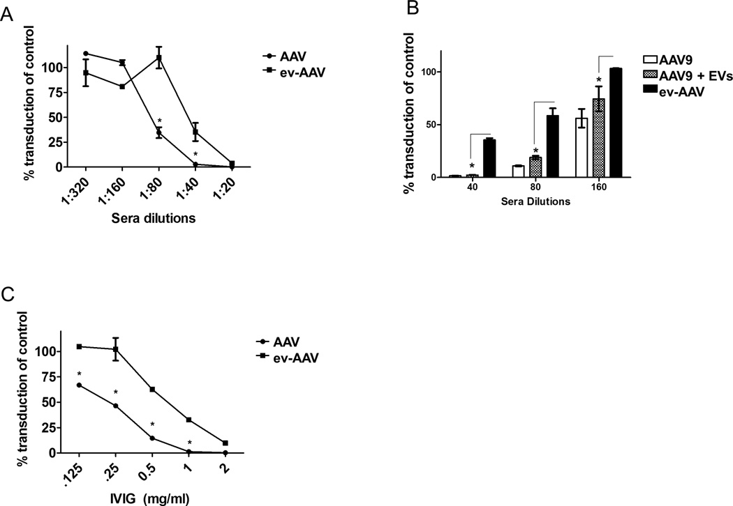
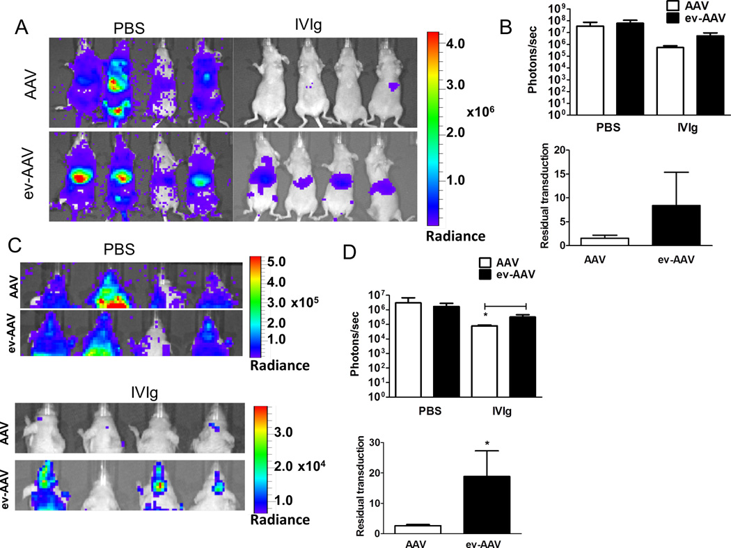
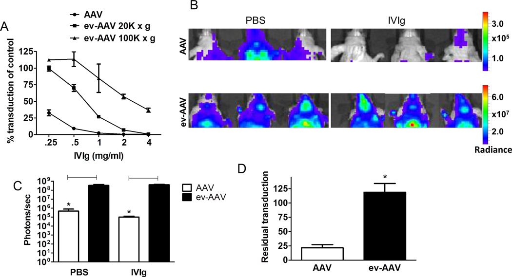
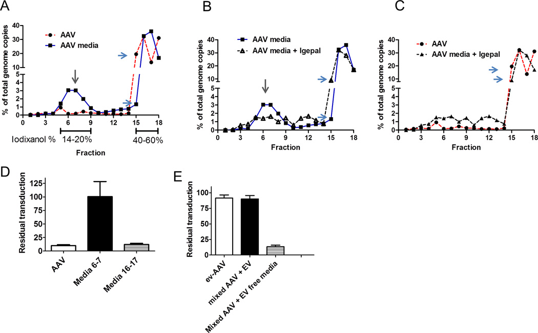
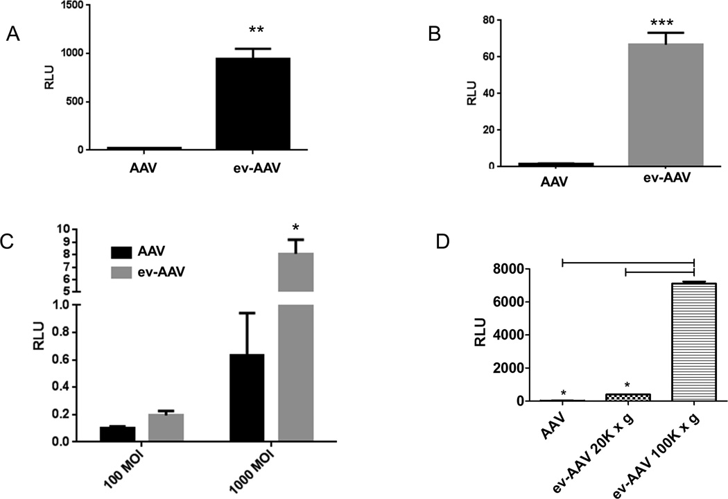
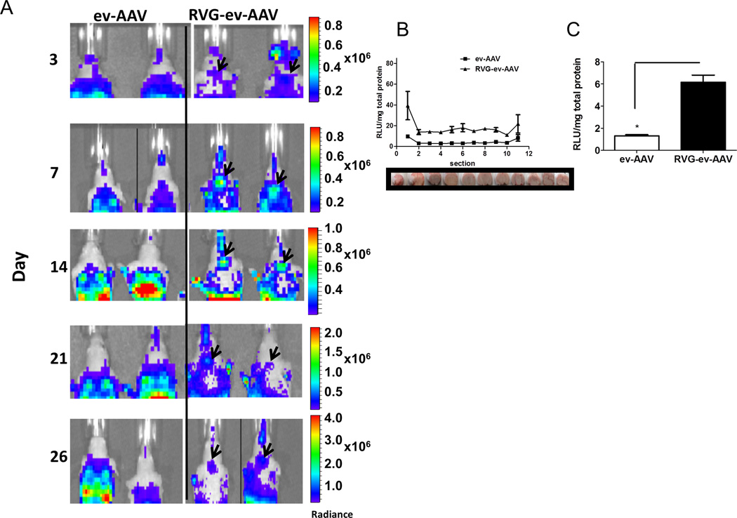
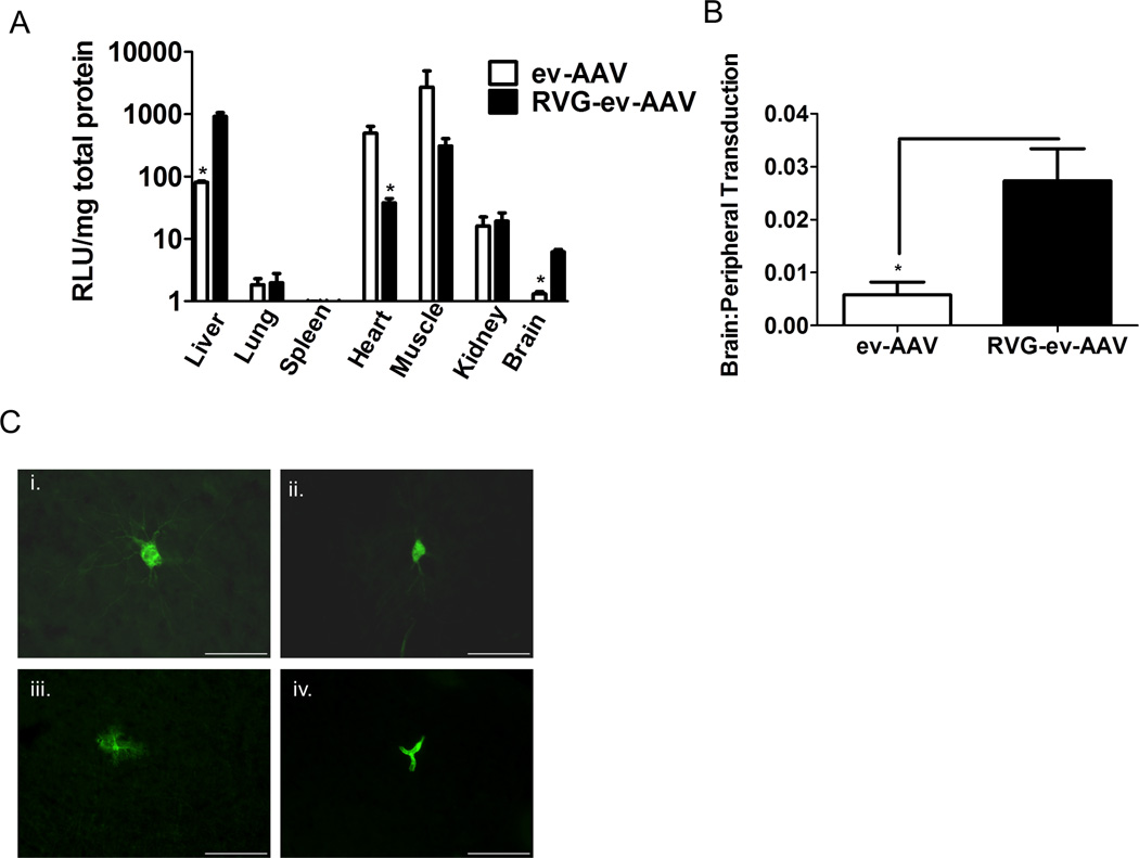
Recently adeno-associated virus (AAV) became the first clinically approved gene therapy product in the western world. To develop AAV for future clinical application in a widespread patient base, particularly in therapies which require intravenous (i.v.) administration of vector, the virus must be able to evade pre-existing antibodies to the wild type virus. Here we demonstrate that in mice, AAV vectors associated with extracellular vesicles (EVs) can evade human anti-AAV neutralizing antibodies. We observed different antibody evasion and gene transfer abilities with populations of EVs isolated by different centrifugal forces. EV-associated AAV vector (ev-AAV) was up to 136-fold more resistant over a range of neutralizing antibody concentrations relative to standard AAV vector in vitro. Importantly in mice, at a concentration of passively transferred human antibodies which decreased i.v. administered standard AAV transduction of brain by 80%, transduction of ev-AAV transduction was not reduced and was 4000-fold higher. Finally, we show that expressing a brain targeting peptide on the EV surface allowed significant enhancement of transduction compared to untargeted ev-AAV. Using ev-AAV represents an effective, clinically relevant approach to evade human neutralizing anti-AAV antibodies after systemic administration of vector.

Keywords: Adeno-associated virus; Exosomes; Extracellular vesicles; Gene delivery; Gene therapy; Microvesicles.


Figures

Figure 1
Figure 1. ev-AAV evade neutralizing antibodies from pooled human serum and purified pooled human intravenous immunoglobulin (IVIg)
(A) Dilutions of pooled human sera (10 donors) were mixed with either standard AAV9-FLuc or ev-AAV9-FLuc. After incubation for 1 h, mixtures were added to HeLa cells and two days later an FLuc assay was performed. FLuc levels were normalized to the levels achieved in the absence of serum. (B) Mixing EVs with standard AAV does not result in significantly enhanced protection from pooled human serum observed with ev-AAV. Standard AAV9-FLuc, AAV9-FLuc mixed with extracellular vesicles, or ev-AAV9-FLuc were mixed with the indicated dilutions of pooled human serum. A neutralization assay was performed on as in (A). (C) Dilutions of IVIg were made and a neutralization assay performed as in (A). *p<0.05.
Figure 2
Figure 2. ev-AAV evades neutralizing anti-AAV antibodies more efficiently than AAV in vivo
Mice were injected with PBS or 0.5 mg IVIg/mouse and 24 h later challenged with 1010 g.c. of either standard AAV9-FLuc or ev-AAV9-FLuc. Seven days later mice were imaged for FLuc expression by bioluminescent imaging. (A) Bioluminescence images of animals in ventral view. (B) Top, photon flux in liver region for each group after PBS pre-treatment or, IVIg pre-treatment; bottom, calculation of residual transduction in liver region of AAV or ev-AAV in the presence of IVIg when compared to transduction after i.p. injection of PBS. n=4, p=0.099. (C) Top, bioluminescence images of all groups of animals in dorsal view. Bottom, IVIG groups shown with lowered scale to clearly indicate higher residual FLuc signal in the head of ev-AAV injected mice. (D) Top, photon flux in head region for each group after PBS pre-treatment or IVIg pre-treatment; bottom, calculation of residual transduction in head region of AAV or ev-AAV in the presence of IVIg when compared to transduction after i.p. injection of PBS. *p=0.0085. Radiance= photons/sec/cm2/sr.
Figure 3
Figure 3. 100k × g ev-AAV9 fraction has enhanced antibody evasion properties
(A) A neutralization assay identical to Fig. 1C with IVIg, using AAV9, 20k × g ev-AAV9, 100k × g ev-AAV9. (B) Mice were injected with PBS or 0.5 mg IVIg/mouse and 24 h later challenged with 1010 g.c. of either standard AAV9-FLuc or 100k × g ev-AAV9-FLuc. Fourteen days later mice were imaged for FLuc expression by bioluminescent imaging. Bioluminescent images of the head region are shown. Note the scale differences between standard AAV and ev-AAV groups. (C) Photon flux of head region for standard AAV9-FLuc or 100k × g ev-AAV9-FLuc with PBS or IVIg pretreatment. (D) Residual transduction in the IVIg groups relative to PBS pretreatment groups. *, p<0.05.
Figure 4
Figure 4. 20k × g depleted media (used for 100k × g ev-AAV isolation) contains an antibody resistant fraction
(A) A AV genome distribution in fractions of a 8–60% iodixanol gradient loaded with standard AAV (red line) or 20k × g depleted media from AAV9 producing-293T cells (blue line). The putative vesicle-associated fractions of AAV is indicated by a gray arrow. Differences in the amount in fraction #15 between the two samples is indicated by blue arrows. (B) Treatment with Igepal detergent (black line) partially disrupts vesicle fraction. Untreated AAV in media is indicated by blue line. (C) Comparison of Igepal treated media sample with standard free AAV. (D) Antibody resistance (IVIG) profile of free standard AAV, or AAV in media fractions 6–7 or 16–17 from (A) (E) Mixing free AAV with EV containing media and centrifuging at 100k × g results in antibody resistant vector. Removing EVs from the same media restores sensitivity to antibody neutralization.
Figure 5
Figure 5. Transduction properties of AAV9 20k × g ev-AAV9 and 100k × g ev-AAV9 fractions
(A) Heparin blocks 20k × g ev-AAV9 transduction. AAV9-FLuc and ev-AAV9-FLuc were incubated with 50 µg/ml heparin and then added to cells. Fluc activity was measured 48 h later and normalized to transduction in the absence of heparin. (B) Triton treatment of 20k × g ev-AAV9 reduces transduction efficiency. AAV9-FLuc and ev-AAV9-FLuc were incubated with 0.3% Triton before diluting and adding to cells. Transduction was normalized to luciferase activity in the absence of Triton. (C) Even higher concentrations of heparin (100 ug/ml) did not inhibit 100k × g ev-AAV9 more than AAV9. (D) Triton treatment had a modest effect on 100k × g ev-AAV9 transduction. (E) Triton treatment breaks open EVs and reduces resistance to antibodies in 20k × g and 100k × g fractions. *** p<0.001,**p<0.006, *p<0.05.
Figure 6
Figure 6. ev-AAV9-FLuc is more efficient in culture at transduction than standard AAV9-FLuc
(A) primary murine neurons (B) SH-SY5Y cells (C) melanocytes. Cells were incubated with AAV9-FLuc or ev-AAV9-FLuc and 48 h later an luciferase assay performed. (D) Comparison of AAV9, and 20k × g and 100k × g fractions of ev-AAV at transduction of HeLa cells. **p=0.006; ***p<0.0001 *; p= 0.012. RLU= Relative light units
Figure 7
Figure 7. RVG-ev-AAV9-FLuc enhances brain transduction compared to untargeted ev-AAV9-Fluc
(A) Bioluminescent images of head region in mice in ev-AAV9-FLuc and RVG-ev-AAV9-FLuc groups. Note the circular pattern of bioluminescence eminating from the brain of RVG-ev-AAV9-FLuc mice (black arrowheads) which indicate this vector is transducing brain better than ev-AAV9-FLuc. (B) Day 32 post-injection mice were sacrificed and brains cryosectioned coronally in 1 mm sections. Each section was homogenized and luciferase activity as well as total protein content determined to give a Fluc expression profile for the entire brain for each group of mice. The brain images below the graph show the approximate location of the section taken for measurement. (C) Average RLU/mg in striatum sections for all three groups. n=4;*, p<0.05. Radiance= photons/sec/cm2/sr. RLU= Relative Light Unit.
Figure 8
Figure 8. RVG-ev-AAV9-FLuc enhances transduction specificity for the brain after i.v. injection
(A) FLuc activity in tissue homogenates. n=4; *=p<0.05. (B) brain targeting specificity calculated from the ratio of brain transduction to peripheral organ transduction. *, p<0.05. (C) RVG-ev-AAV9-GFP transduced cell types in the brain. Vector was injected i.v. and two weeks later fixed sections were immunostained for GFP. GFP positive neurons (i, ii.), astrocytes (iii.), and endothelial cells (iv.) were observed. Scale bar= 55 µm. RLU= Relative Light Unit.

References

    1. Nathwani AC, Tuddenham EG, Rangarajan S, Rosales C, McIntosh J, Linch DC, et al. Adenovirus-associated virus vector-mediated gene transfer in hemophilia B. N Engl J Med. 2011;365(25):2357–2365. - PMC - PubMed
    1. Maguire AM, High KA, Auricchio A, Wright JF, Pierce EA, Testa F, et al. Age-dependent effects of RPE65 gene therapy for Leber’s congenital amaurosis: a phase 1 dose-escalation trial. Lancet. 2009;374(9701):1597–1605. - PMC - PubMed
    1. Haddley K. Alipogene tiparvovec for the treatment of lipoprotein lipase deficiency. Drugs Today (Barc) 2013;49(3):161–170. - PubMed
    1. Muramatsu S, Fujimoto K, Kato S, Mizukami H, Asari S, Ikeguchi K, et al. A phase I study of aromatic L-amino acid decarboxylase gene therapy for Parkinson’s disease. Mol Ther. 2010;18(9):1731–1735. - PMC - PubMed
    1. Scallan CD, Jiang H, Liu T, Patarroyo-White S, Sommer JM, Zhou S, et al. Human immunoglobulin inhibits liver transduction by AAV vectors at low AAV2 neutralizing titers in SCID mice. Blood. 2006;107(5):1810–1817. - PubMed

Publication types

Substances