. 2014 Jun 10;55(7):4338-47.
doi: 10.1167/iovs.14-14061.

Mitigation of oxygen-induced retinopathy in α2β1 integrin-deficient mice

Affiliations

Mitigation of oxygen-induced retinopathy in α2β1 integrin-deficient mice

Aasakiran Madamanchi et al. Invest Ophthalmol Vis Sci. .

Abstract

Purpose: The α2β1 integrin plays an important but complex role in angiogenesis and vasculopathies. Published GWAS studies established a correlation between genetic polymorphisms of the α2β1 integrin gene and incidence of diabetic retinopathy. Recent studies indicated that α2-null mice demonstrate superior vascularization in both the wound and diabetic microenvironments. The goal of this study was to determine whether the vasculoprotective effects of α2-integrin deficiency extended to the retina, using the oxygen-induced retinopathy (OIR) model for retinopathy of prematurity (ROP).

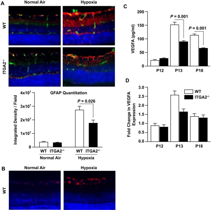
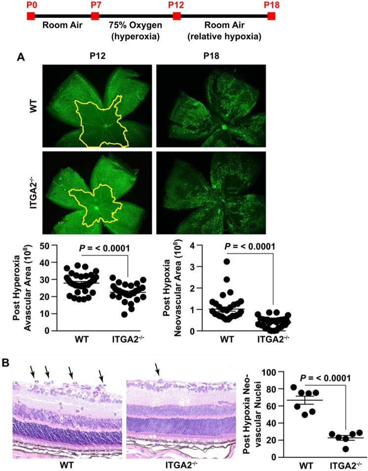
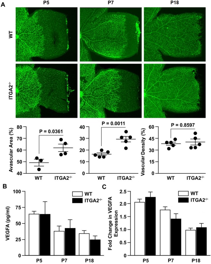
Methods: In the OIR model, wild-type (WT) and α2-null mice were exposed to 75% oxygen for 5 days (postnatal day [P] 7 to P12) and subsequently returned to room air for 6 days (P12-P18). Retinas were collected at postnatal day 7, day 13, and day 18 and examined via hematoxylin and eosin and Lectin staining. Retinas were analyzed for retinal vascular area, neovascularization, VEGF expression, and Müller cell activation. Primary Müller cell cultures from WT and α2-null mice were isolated and analyzed for hypoxia-induced VEGF-A expression.

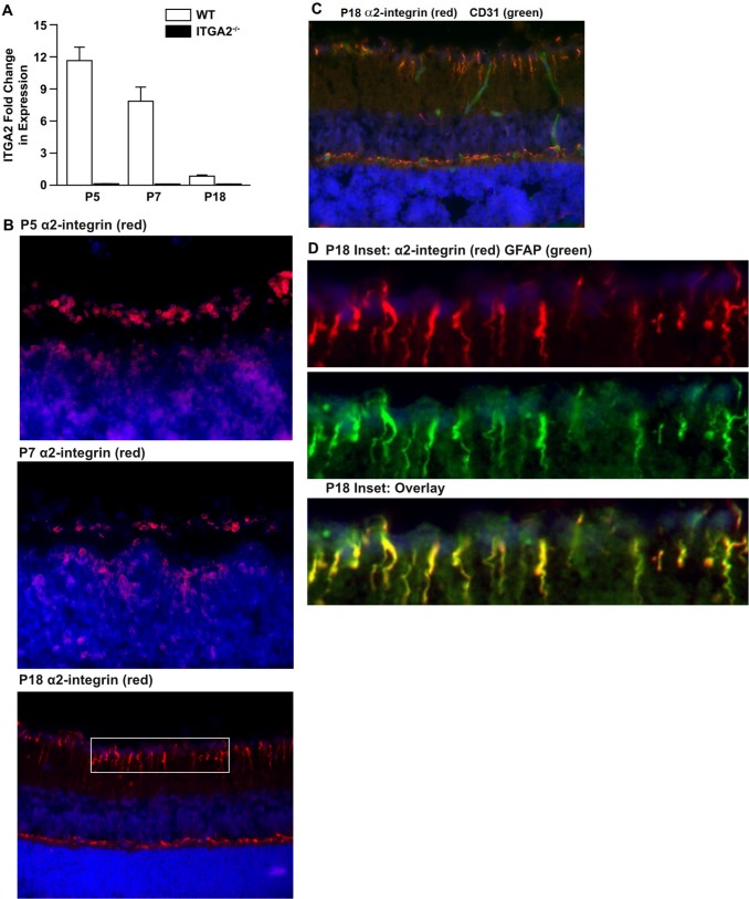
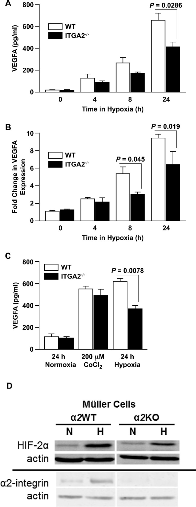
Results: In the retina, the α2β1 integrin was minimally expressed in endothelial cells and strongly expressed in activated Müller cells. Isolated α2-null primary Müller cells demonstrated decreased hypoxia-induced VEGF-A expression. In the OIR model, α2-null mice displayed reduced hyperoxia-induced vaso-attenuation, reduced pathological retinal neovascularization, and decreased VEGF expression as compared to WT counterparts.

Conclusions: Our data suggest that the α2β1 integrin contributes to the pathogenesis of retinopathy. We describe a newly identified role for α2β1 integrin in mediating hypoxia-induced Müller cell VEGF-A production.

Keywords: Müller cells; angiogenesis; integrin; retinopathy.


Figures

Figure 1
Figure 1
α2β1 integrin expression in retinal endothelial and Müller cells. (A) The level of α2 integrin subunit mRNA expression in total retina of WT and ITGA2−/− mice at P5, P7, and P18 was determined by qRT-PCR. Values are normalized to HPRT or RPP30 and displayed relative to WT at P18. Values represent mean ± SEM. (B) Immunofluorescence analysis of α2β1 integrin (red) expression in cross-sections of P5, P7, and P18 WT retinas from unchallenged mice. Integrin expression suggests a pattern indicative of retinal Müller cells. The P5 and P7 images were taken at ×60 magnification. Postnatal day 18 images were taken at ×40 magnification and are representative of five or more retinas taken in three separate trials from separate litters. (C) Immunofluorescence analysis demonstrated limited colocalization of α2β1 integrin expression (red) and CD31 (green) on the vessels in the cross-sections of retinas from P18 WT mice grown in normal air. Nuclei are stained with 4′,6-diamidino-2-phenylindole (DAPI) (blue). Images were taken at ×40 magnification. (D) Analysis of P18 retina cross-section from inset in (B) at ×160 magnification. Staining of α2β1 integrin in red (top) and GFAP in green (middle) are merged to show colocalization in yellow (bottom) of α2β1 integrin and GFAP expression in retinal Müller cells.
Figure 2
Figure 2
α2β1 deletion impairs hypoxia-dependent VEGF production in Müller cells in vitro. (A) Levels of VEGF-A protein in conditioned media from primary WT and ITGA2−/− Müller cells after incubation in hypoxia for 0, 4, 8, or 24 hours, as determined by ELISA. Data are shown as mean ± SEM for a representative experiment of five separate trials taken with separate isolates of primary Müller cells. (B) Fold changes of VEGF-A mRNA expression from primary WT and ITGA2−/− Müller cells incubated in hypoxic conditions for 0, 4, 8, or 24 hours, as determined by qRT-PCR. Fold changes are relative to WT at 0 hours (mean ± SEM). Data are representative of six separate trials with separate isolates of primary Müller cells. (C) Vascular endothelial growth factor-A protein in conditioned media from primary WT and ITGA2−/− Müller cells after 24 hours in normoxic conditions, treatment with 200 μM CoCl2, or hypoxic challenge. Vascular endothelial growth factor-A concentration was determined by ELISA. Data are shown as a mean ± SEM for a representative of three separate trials with separate isolates of primary Müller cells. (D) Immunoblot analysis of primary WT and ITGA2−/− Müller cells for HIF-2α and α2-integrin expression after 24 hours of culture in normal air and hypoxia.
Figure 3
Figure 3
α2β1 integrin-deletion mitigates Müller cell activation and VEGF production in OIR. (A) Cross-sections of retinas from age-matched P18 WT and ITGA2−/− mice raised in normal air or relative hypoxia (6 days after hyperoxia in OIR) were evaluated by immunofluorescence microscopy using anti-GFAP (Müller cells: red) or anti-CD31 (ECs: green) and DAPI (nuclei: blue). Images were taken at ×40 magnification and are representative of three separate trials. The intensity of GFAP staining was quantified and is shown below (mean ± SEM). Images are at ×40 magnification. (B) Cross-sections of retinas from age-matched WT and ITGA2−/− P14 mice in normal air or relative hypoxia (2 days after hyperoxia in OIR) were evaluated by immunofluorescence microscopy using anti-α2β1 integrin (red) and DAPI (nuclei: blue). Images were taken at ×60 magnification. (C) Lysates from age-matched WT and ITGA2−/− mice after OIR injury at P12, P13, and P18 was quantified by ELISA (mean ± SEM). (D) The level of VEGF-A mRNA expression in age-matched WT and ITGA2−/− retinas at P12, P13, and P18 retinas after OIR was determined by qRT-PCR. Fold changes are relative to WT P12 (mean ± SEM). Results in (B, C) are derived from four similar experiments.
Figure 4
Figure 4
α2β1 integrin deletion protects against OIR. The experimental protocol for OIR is shown as a timeline. (A) Representative immunofluorescence images of retinal flat mounts of age-matched WT and ITGA2−/− mice at P12 (after 75% hyperoxia) and P18 (after return to room air and relative hypoxia). Vessels are identified by GS Lectin (green) staining. Quantitation of central avascular area and neovascular area was determined as described and illustrated (mean ± SEM). Images are ×4 magnification. (B) Representative photograph of H&E-stained retinal cross-section of age-matched WT and ITGA2−/− at P18 following OIR injury. Neovascular nuclei are indicated by arrows. The number of nuclei per retinal area was quantitated (mean ± SEM). Images are ×40 magnification.
Figure 5
Figure 5
No significant differences in vascularization or VEGF levels were identified during development. (A) Retinal flat mounts of age-matched WT and ITGA2−/− mice at P5, P7, and P18. Vessels were visualized by GS Lectin (green) via immunofluorescence microscopy. For P5 and P7, vascularization was measured by radial vascular outgrowth, as quantitated by pixel percentage of avascular area at retinal periphery relative to total retina area. At P18, radial vascular outgrowth was complete, and so vascularization was measured by vascular density, as quantitated by pixel percentage of vessel area relative to total retina area. Each data point represents the average result from an experiment with a litter of at least four mice (data represent mean ± SEM). The results are representative of three separate trials. Images are ×4 magnification. (B) The level of VEGF-A protein in retinal lysates of P5, P7, and P18 WT and ITGA2−/− animals under normal conditions, as determined by ELISA (mean ± SD). (C) The fold change in mRNA expression of VEGF-A in developing retinas at P5, P7, and P18 was determined by qRT-PCR. Fold changes are relative to WT day 18 and shown as mean ± SEM. All results (B, C) are representative of three similar experiments.

References

    1. Barnett JM, McCollum GW, Penn JS. Role of cytosolic phospholipase A(2) in retinal neovascularization. Invest Ophthalmol Vis Sci. 2010; 51: 1136–1142 - PMC - PubMed
    1. Steinkuller PG, Du L, Gilbert C, Foster A, Collins ML, Coats DK. Childhood blindness. J AAPOS. 1999; 3: 26–32 - PubMed
    1. Bressler NM, Bressler SB. Preventative ophthalmology. Age-related macular degeneration. Ophthalmology. 1995; 102: 1206–1211 - PubMed
    1. Rahmani B, Tielsch JM, Katz J, et al. The cause-specific prevalence of visual impairment in an urban population. The Baltimore Eye Survey. Ophthalmology. 1996; 103: 1721–1726 - PubMed
    1. Wang GL, Jiang BH, Rue EA, Semenza GL. Hypoxia-inducible factor 1 is a basic-helix-loop-helix-PAS heterodimer regulated by cellular O2 tension. Proc Natl Acad Sci U S A. 1995; 92: 5510–5514 - PMC - PubMed

Publication types

MeSH terms

LinkOut - more resources