. 2014 Jun 10:14:418.
doi: 10.1186/1471-2407-14-418.

B-cell lymphoma 6 protein stimulates oncogenicity of human breast cancer cells

Affiliations

B-cell lymphoma 6 protein stimulates oncogenicity of human breast cancer cells

Qiang Wu et al. BMC Cancer. .

Abstract

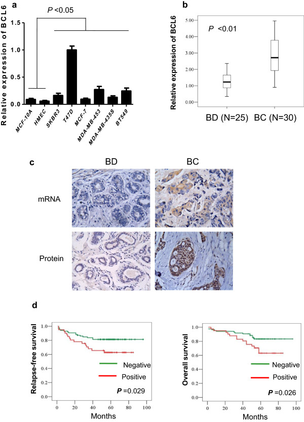
Background: B-cell lymphoma 6 (BCL6) protein, an evolutionarily conserved zinc finger transcription factor, showed to be highly expressed in various human cancers in addition to malignancies in the lymphoid system. This study investigated the role of BCL6 expression in breast cancer and its clinical significance in breast cancer patients.

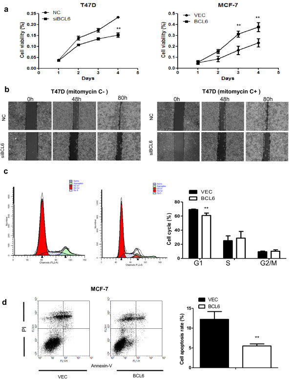
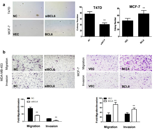
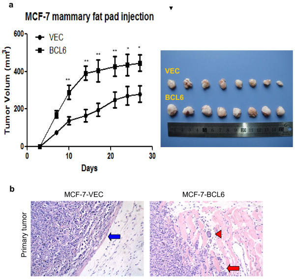
Methods: Expression of BCL6 protein was assessed using in situ hybridization and immunohistochemistry in 127 breast cancer patients and 50 patients with breast benign disease as well as in breast cell lines. Expression of BCL6 was restored or knocked down in two breast cancer cell lines (MCF-7 and T47D) using BCL6 cDNA and siRNA, respectively. The phenotypic change of these breast cancer cell lines was assessed using cell viability MTT, Transwell invasion, colony formation, and flow cytometry assays and in a xenograft mice model. Luciferase reporter gene, immunoblot, and qRT-PCR were used to investigate the molecular events after manipulated BCL6 expression in breast cancer cells.

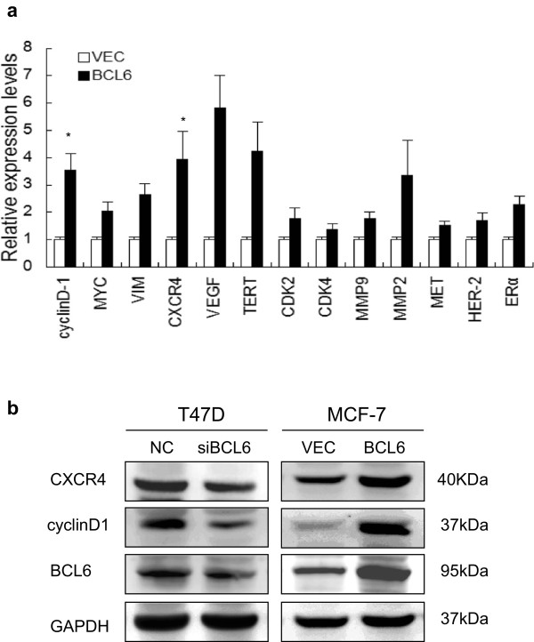
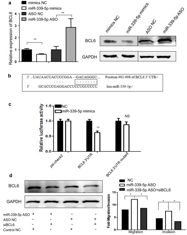
Results: BCL6 protein was highly expressed in breast cancer cell lines and tissue specimens and expression of BCL6 protein was associated with disease progression and poor survival of breast cancer patients. In vitro, the forced expression of BCL6 results in increased proliferation, anchorage-independent growth, migration, invasion and survival of breast cancer cell lines, whereas knockdown of BCL6 expression reduced these oncogenic properties of breast cancer cells. Moreover, forced expression of BCL6 increased tumor growth and invasiveness in a nude mouse xenograft model. At the gene level, BCL6 was a target gene of miR-339-5p. Expression of BCL6 induced expression of CXCR4 and cyclinD1 proteins.

Conclusions: The current study demonstrated the oncogenic property of BCL6 in breast cancer and further study could target BCL6 as a novel potential therapeutic strategy for breast cancer.


Figures

Figure 1
Figure 1
Expression of BCL6 mRNA and protein in human breast cancer cell lines and tissue specimens. (a) qRT-PCR. Level of BCL6 mRNA expression in eight human mammary cell lines was analyzed by qRT-PCR. (b) qRT-PCR. Levels of BCL6 mRNA expression were examined in 30 breast cancer (BC) and 25 breast benign disease tissue specimens (BD) by qRT-PCR. (c) Representative imagines of BCL6 expression analyzed by in situ hybridization and immunohistochemistry (Magnification: ×400). (d) Kaplan-Meier curve of the relapse-free survival (RFS) or overall survival (OS) according to BCL6 expression.
Figure 2
Figure 2
Effects of BCL6 on regulation of breast cancer cell phenotype. (a) Cell viability MTT assay. Cells were transiently transfected with BCL6 siRNA vs. negative control (NC) or BCL6 cDNA vs. control vector (VEC), respectively and then seeded in 96-well plates (3 × 103 per well) and grown for 4 days for MTT assay. (b) Wound healing assay. T47D cells were grown and transiently transfected with BCL6 siRNA or negative control (NC), the wounded monolayers were cultured in the absence (left) or presence (right) of mitomycin C. (c) Flow cytometric analysis of cell cycle distribution in MCF-7 cells after gene transfection. (d) Flow cytometric analysis of apoptosis in MCF-7 cells after gene transfection. The average of apoptosis rate is presented as mean ± SD. All experiments were repeated at least three times. **P < 0.01.
Figure 3
Figure 3
Effects of BCL6 expression on regulation of breast cancer colony formation and migration and invasion capacity. (a) Soft agar assay. After gene transfection, cells were seeded in 0.35% top agarose and 10% FBS in six-well plates in triplicate. The number of colonies was counted after 14 days incubation. (b) Tumor cell migration and invasion assay. MDA-MB-453 cells were grown and transiently transfected with BCL6 siRNA or negative control (NC) for 72 h. MCF-7 cells were grown and transiently transfected with BCL6 cDNA or vector-only (VEC) for 48 h. Cells in the upper chamber were removed and those cells migrated to the lower layer of the inner chamber were stained and counted. **, P < 0.01.
Figure 4
Figure 4
Effects of BCL6 expression on regulation of MCF-7 xenograft growth in nude mice. MCF7-VEC and MCF7-BCL6 cells were transplanted into the mammary fat pad of female BALB/c-nu, respectively. The volume of xenografts was measured twice a week and calculated. (a) Xenograft growth curve of MCF7-VEC and MCF7-BCL6-derived tumors over 27 days. (b) Hematoxylin and eosin staining of tumor xenograft sections. More aggressive behavior was observed in the margin of tumor nodule of MCF-7-BCL6 cells (red arrow) compared to that of MCF-7-VEC cells (blue arrow). Tumor embolus (red arrow head) was visualized in blood vessel (Magnification: ×200). *, P < 0.05; **, P < 0.01.
Figure 5
Figure 5
Effects of BCL6 expression on CXCR4 and cyclinD1 expression. (a) qRT-PCR. MCF-7 cells were transiently transfected with BCL6 cDNA or negative control vector and grown for 2 days. (b) Western blot. MCF-7 and T74D cells were transiently transfected with BCL6 cDNA, BCL6 siRNA, or negative control vector and grown for 2 days and subjected to Western blot. *, P < 0.05.
Figure 6
Figure 6
BCL6 as the direct target gene of miR-339-5p in breast cancer cells. (a) qRT-PCR and Western blot. miR-339-5p mimics or miR-339-5p ASO was transiently transfected into T47D cells and subjected to analysis of BCL6 expression. (b) The binding site of BCL6 3′-UTR and miR-339-5p. (c) Luciferase reporter assay. T47D cells were transfected with psiCHECK-2-BCL6 3′-UTR or psiCHECK-2-BCL6 mutated 3′-UTR plus either miR-339-5p mimics or negative control and subjected to luciferase reporter assay. (d) Tumor cell migration and invasion assay and Western blot. MCF-7 cells were grown and transiently transfected with miR-339-5p ASO, miR-339-5p ASO plus BCL6 siRNA or scrambled sequence oligonucleotides as negative control for 2 days and subjected to migration, invasion and western blot assays. *P < 0.05; **P < 0.01.

References

    1. Siegel R, Naishadham D, Jemal A. Cancer statistics, 2013. CA Cancer J Clin. 2013;63:11–30. doi: 10.3322/caac.21166. - DOI - PubMed
    1. Hanahan D, Weinberg RA. Hallmarks of cancer: the next generation. Cell. 2011;144:646–674. doi: 10.1016/j.cell.2011.02.013. - DOI - PubMed
    1. Chang CC, Ye BH, Chaganti RS, Dalla-Favera R. BCL-6, a POZ/zinc-finger protein, is a sequence-specific transcriptional repressor. Proc Natl Acad Sci U S A. 1996;93:6947–6952. doi: 10.1073/pnas.93.14.6947. - DOI - PMC - PubMed
    1. Jardin F, Ruminy P, Bastard C, Tilly H. The BCL6 proto-oncogene: a leading role during germinal center development and lymphomagenesis. Pathol Biol (Paris) 2007;55:73–83. doi: 10.1016/j.patbio.2006.04.001. - DOI - PubMed
    1. Duan S, Cermak L, Pagan JK, Rossi M, Martinengo C, di Celle PF, Chapuy B, Shipp M, Chiarle R, Pagano M. FBXO11 targets BCL6 for degradation and is inactivated in diffuse large B-cell lymphomas. Nature. 2012;481:90–93. - PMC - PubMed

Publication types