. 2014 Jun;141(12):2446-51.
doi: 10.1242/dev.101188.

Notch signaling functions in lymphatic valve formation

Affiliations

Notch signaling functions in lymphatic valve formation

Aino Murtomaki et al. Development. 2014 Jun.

Abstract

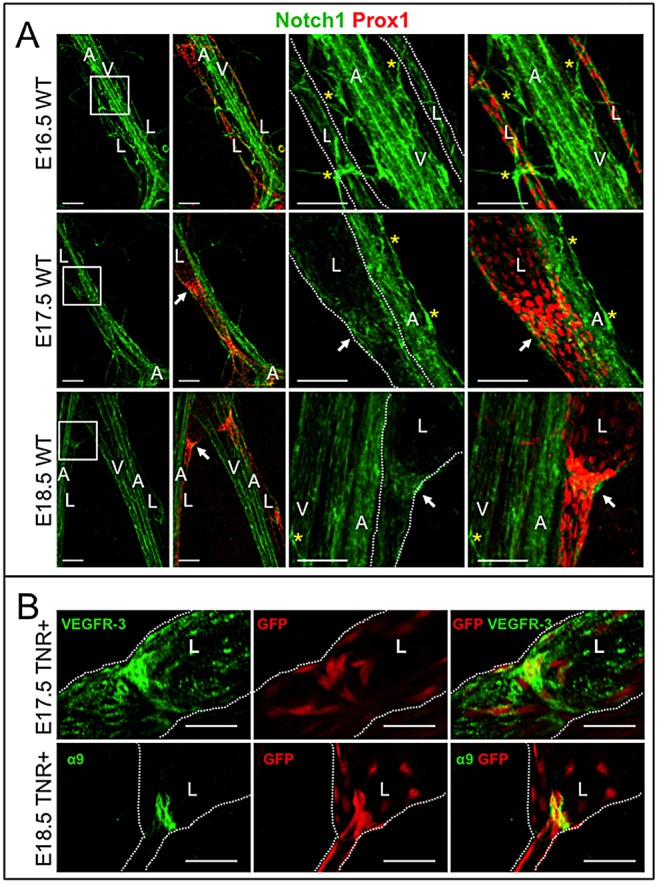
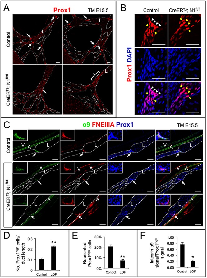
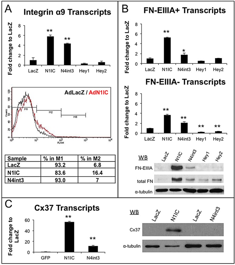
Collecting lymphatic ducts contain intraluminal valves that prevent backflow. In mice, lymphatic valve morphogenesis begins at embryonic day 15.5 (E15.5). In the mesentery, Prox1 expression is high in valve-forming lymphatic endothelial cells, whereas cells of the lymphatic ducts express lower levels of Prox1. Integrin α9, fibronectin EIIIA, Foxc2, calcineurin and the gap junction protein Cx37 are required for lymphatic valve formation. We show that Notch1 is expressed throughout the developing mesenteric lymphatic vessels at E16.5, and that, by E18.5, Notch1 expression becomes highly enriched in the lymphatic valve endothelial cells. Using a Notch reporter mouse, Notch activity was detected in lymphatic valves at E17.5 and E18.5. The role of Notch in lymphatic valve morphogenesis was studied using a conditional lymphatic endothelial cell driver either to delete Notch1 or to express a dominant-negative Mastermind-like (DNMAML) transgene. Deletion of Notch1 led to an expansion of Prox1(high) cells, a defect in Prox1(high) cell reorientation and a decrease in integrin α9 expression at sites of valve formation. Expression of DNMAML, which blocks all Notch signaling, resulted in a more severe phenotype characterized by a decrease in valves, failure of Prox1(high) cells to cluster, and rounding of the nuclei and decreased fibronectin-EIIIA expression in the Prox1(high) cells found at valve sites. In human dermal lymphatic endothelial cells, activation of Notch1 or Notch4 induced integrin α9, fibronectin EIIIA and Cx37 expression. We conclude that Notch signaling is required for proper lymphatic valve formation and regulates integrin α9 and fibronectin EIIIA expression during valve morphogenesis.

Keywords: Integrin α9; Lymphatic valve morphogenesis; Mouse; Notch.


Figures

Fig. 1.
Fig. 1.
Notch1 expression and Notch activity in embryonic lymphatic duct and valve LECs. (A) E16.5, E17.5 and E18.5 wild-type mesenteries were stained for Notch1 and Prox1. Boxed areas are enlarged on the right. White arrows indicate lymphatic valves. Yellow asterisks mark blood capillaries. Scale bars: 100 µm. (B) TNR mesenteries were stained for GFP to detect Notch activity and VEGFR3 (E17.5) or α9 (E18.5). Scale bars: 50 µm. Lymphatic ducts are marked with white dotted lines. A, artery; L, lymphatic collecting duct; V, vein.
Fig. 2.
Fig. 2.
Lymphatic endothelial-specific loss of Notch1 resulted in defective valve morphogenesis and decreased integrin α9 expression. Notch1 was deleted in LECs at E15.5 with tamoxifen and mesenteries isolated at E18.5. (A) N1fl/+ (control) and Prox1CreERT2;N1fl/fl (CreERT2;N1fl/fl) mesenteries stained for Prox1. Arrows indicate Prox1high valves; bracket denotes expansion of Prox1high cells within the duct walls in Prox1CreERT2;N1fl/fl mesenteries. Scale bars: 100 µm. (B) Control and CreERT2;N1fl/fl mesenteries stained for Prox1 and DAPI. White arrowheads indicate Prox1high valve-forming LECs reoriented at least 45° to the duct wall. Yellow arrowheads indicate Prox1high LECs that have failed to reorient. Scale bars: 50 µm. (C) Control and CreERT2;N1fl/fl mesenteries stained for Prox1, α9 and FNEIIIA. White arrows indicate valves magnified in upper left corner. Scale bars: 100 µm. A, artery; L, lymphatic collecting duct; V, vein. (D) Quantification of number of Prox1high cells normalized by duct length. (E) Percentage of reoriented Prox1high cells in Prox1high valve clusters. (F) Quantification of α9-positive signal normalized to Prox1high signal in Prox1high valve LECs. Data are mean±s.e.m. *P<0.05, **P<0.005.
Fig. 3.
Fig. 3.
Lymphatic endothelial-specific loss of Notch signaling resulted in loss and defective valve formation and decreased FNEIIIA expression. DNMAML expression was induced in LECs at (A) E13.5 or (B,C) E15.5 with tamoxifen and mesenteries isolated at E18.5. (A,B) DNMAMLfl/+ (control) and Prox1CreERT2;DNMAMLfl/+ (CreERT2;DNMAMLfl/+) mesenteries stained for Prox1. White arrows indicate Prox1high regions magnified on the right. White arrowheads mark reoriented Prox1high LECs with typical elongated nuclei. Yellow arrowheads indicate Prox1high LECs that have not reoriented and/or display abnormal rounded nuclei. Scale bars: 100 µm. (C) Control and CreERT2;DNMAMLfl/+ mesenteries stained for Prox1, α9 and FNEIIIA. Middle row shows mutant with mild valve phenotype. Bottom row shows severely affected mesenteric lymphatic ducts with no discernible valves. White arrows indicate valves magnified in upper left-hand corner. Scale bars: 100 µm. A, artery; L, lymphatic collecting duct; V, vein. (D) Quantification of Prox1high cells located in clusters. (E) Quantification of Prox1high/α9/FNEIIIA triple-positive valves per duct. (F) Quantification of FNEIIIA-positive signal normalized to Prox1high signal in Prox1high valve LECs. (D-F) Prox1CreERT2;DNMAMLfl/+ (mutant) and DNMAMLfl/+ (control). Data are mean±s.e.m. *P<0.05, **P<0.005.
Fig. 4.
Fig. 4.
Notch activation induced integrin α9, FNEIIIA and Cx37 in HdLECs. HdLECs were adenovirally infected with N1IC, N4int3, Hey1, Hey2 or lacZ-coding viruses. RNA and protein isolated 48 h post-infection. (A) qRT-PCR for α9 and FACs for integrin α9/β1. (B) qRT-PCR for fibronectin transcripts with EIIIA (FN-EIIIA+) and without EIIIA (FN-EIIIA−). Western blots for FN-EIIIA, total fibronectin or α-tubulin. (C) qRT-PCR and western blot for Cx37. Data are mean±s.d. *P≤0.05, **P≤0.005.

References

    1. Andersson E. R., Sandberg R., Lendahl U. (2011). Notch signaling: simplicity in design, versatility in function. Development 138, 3593-3612 10.1242/dev.063610 - DOI - PubMed
    1. Bazigou E., Xie S., Chen C., Weston A., Miura N., Sorokin L., Adams R., Muro A. F., Sheppard D., Makinen T. (2009). Integrin-alpha9 is required for fibronectin matrix assembly during lymphatic valve morphogenesis. Dev. Cell 17, 175-186 10.1016/j.devcel.2009.06.017 - DOI - PMC - PubMed
    1. Borggrefe T., Liefeke R. (2012). Fine-tuning of the intracellular canonical Notch signaling pathway. Cell Cycle 11, 264-276 10.4161/cc.11.2.18995 - DOI - PubMed
    1. Kanady J. D., Dellinger M. T., Munger S. J., Witte M. H., Simon A. M. (2011). Connexin37 and Connexin43 deficiencies in mice disrupt lymphatic valve development and result in lymphatic disorders including lymphedema and chylothorax. Dev. Biol. 354, 253-266 10.1016/j.ydbio.2011.04.004 - DOI - PMC - PubMed
    1. Kang J., Yoo J., Lee S., Tang W., Aguilar B., Ramu S., Choi I., Out H. H., Shin J. W., Dotto G. P., Koh C. J., Detmar M., Hong Y. K. (2010). An exquisite cross-control mechanism among endothelial cell fate regulators directs the plasticity and heterogeneity of lymphatic endothelial cells. Blood 116, 140-150 10.2353/ajpath.2008.080378 - DOI - PMC - PubMed

Publication types

MeSH terms