. 2015 Jan;61(1):108-18.
doi: 10.1002/hep.27242. Epub 2014 Oct 1.

Pnpla3I148M knockin mice accumulate PNPLA3 on lipid droplets and develop hepatic steatosis

Affiliations
Free PMC article

Pnpla3I148M knockin mice accumulate PNPLA3 on lipid droplets and develop hepatic steatosis

Eriks Smagris et al. Hepatology. 2015 Jan.
Free PMC article

Abstract

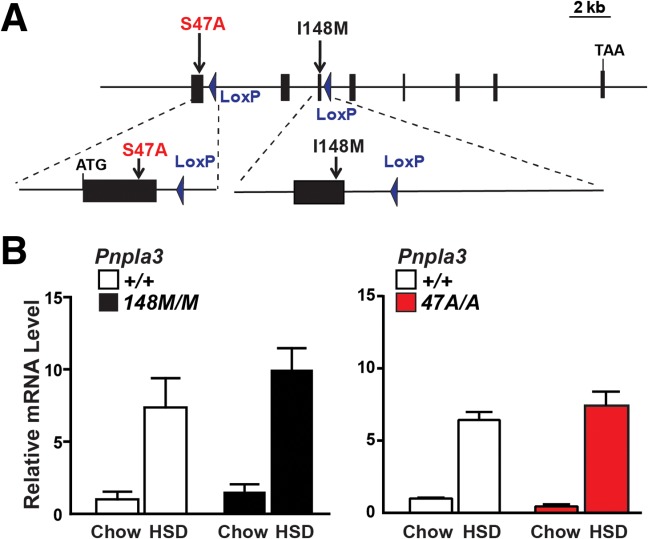
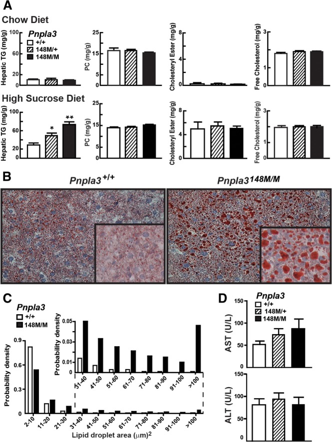
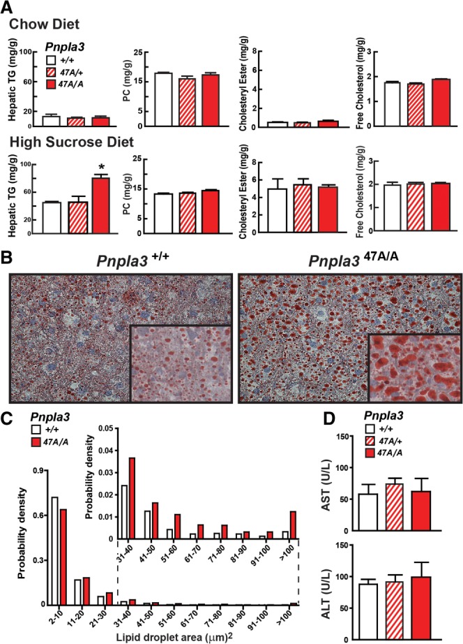
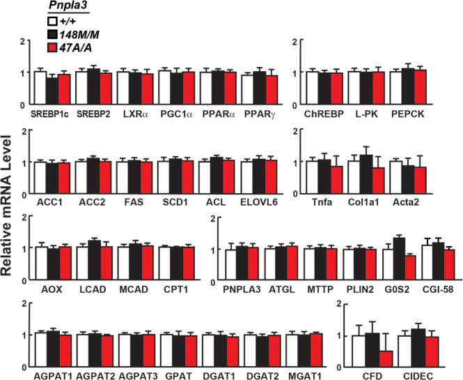
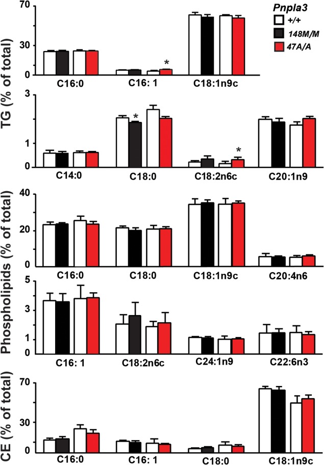
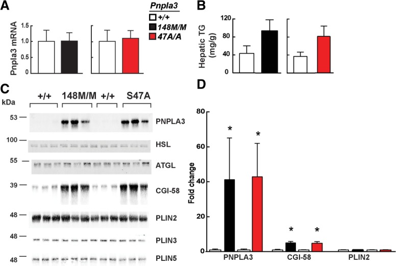
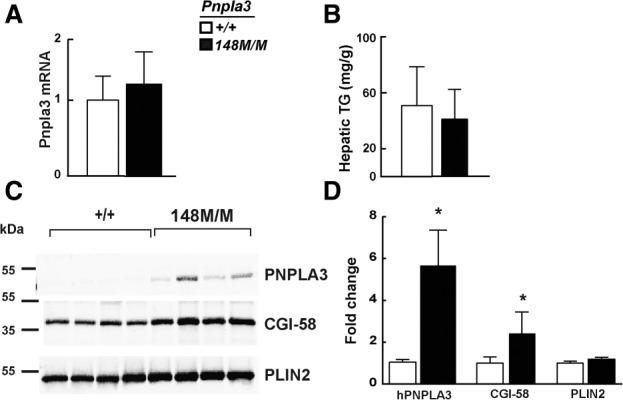
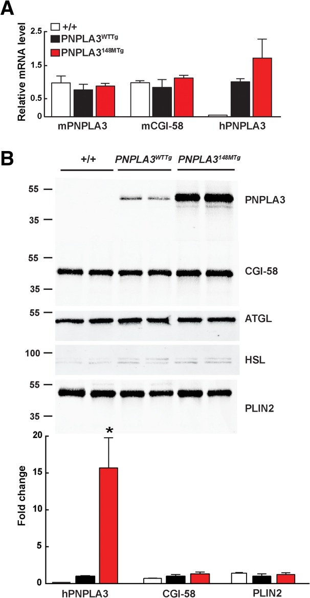
A sequence polymorphism (rs738409, I148M) in patatin-like phospholipid domain containing protein 3 (PNPLA3) is strongly associated with nonalcoholic fatty liver disease (NAFLD), but the mechanistic basis for this association remains enigmatic. Neither ablation nor overexpression of wild-type PNPLA3 affects liver fat content in mice, whereas hepatic overexpression of the human 148M transgene causes steatosis. To determine whether the 148M allele causes fat accumulation in the liver when expressed at physiological levels, we introduced a methionine codon at position 148 of the mouse Pnpla3 gene. Knockin mice had normal levels of hepatic fat on a chow diet, but when challenged with a high-sucrose diet their liver fat levels increased 2 to 3-fold compared to wild-type littermates without any associated changes in glucose homeostasis. The increased liver fat in the knockin mice was accompanied by a 40-fold increase in PNPLA3 on hepatic lipid droplets, with no increase in hepatic PNPLA3 messenger RNA (mRNA). Similar results were obtained when the catalytic dyad of PNPLA3 was inactivated by substituting the catalytic serine with alanine (S47A).

Conclusion: These data provide the first direct evidence that physiological expression of PNPLA3 148M variant causes NAFLD, and that the accumulation of catalytically inactive PNPLA3 on the surfaces of lipid droplets is associated with the accumulation of TG in the liver.


Figures

Fig. 1
Fig. 1
Generation of Pnpla3148M and Pnpla347A KI mice. (A) Diagram of Pnpla3 targeting strategy. Homologous recombination was used to replace either exon 1 (S47A) or exon 3 (I48M) with the corresponding mutant sequences. The selectable marker (Neo) located between two LoxP sites was then excised with Cre recombinase, leaving one LoxP site in the flanking intron. (B) Pnpla3 RNA levels in livers from female mice (n = 4/group) consuming a chow diet or a high-sucrose diet (HSD) for 4 weeks. Livers were collected at the end of the feeding cycle. Levels of Pnpla3 mRNA were determined using real-time PCR and normalized to values of chow-fed wild-type mice.
Fig. 2
Fig. 2
Hepatic TG content in Pnpla3I148M KI mice. Data in panels A-D are from the same experiment (n = 5 mice/group). Tissue and plasma were collected at the end of the feeding cycle. (A) Hepatic lipid levels were measured in 12-week-old female wild-type (+/+), heterozygous (148M/+), and homozygous (148M/M) KI mice fed a chow diet or high-sucrose diet (HSD) for 4 weeks. (B) Liver sections from wild-type (+/+), and 148M/M KI mice on high-sucrose diets were stained with Oil Red O and viewed using a Leica microscope (DM2000) (magnification: 20× and inset 64×). (C) Size distributions of hepatic lipid droplets in wild-type and Pnpla3148M/M mice. Oil Red O-stained slides were analyzed using ImageJ as described in Materials and Methods. (D) Plasma levels of aspartate aminotransferase (AST) and alanine aminotransaminase (ALT) in wild-type, 148M/+, and 148M/M KI mice. Values are means ± SEM. The experiment was performed twice with similar results. *P < 0.05, **P < 0.001.
Fig. 3
Fig. 3
Hepatic TG content in Pnpla3S47A mice. Data in panels A-D are from the same experiment (n = 7 mice/group). Tissue and plasma samples were collected at the end of the feeding cycle. The experiment was repeated with similar results. (A) Hepatic lipid levels were measured in 13-week-old female wild-type (+/+), S47A heterozygous (S47A/+) and homozygous (S47A/A) KI mice fed a chow diet or high-sucrose diet (HSD) for 4 weeks. (B) Liver sections from sucrose-fed wild-type and S47A/A KI mice were stained with Oil Red O and viewed using a Leica microscope (DM2000) (magnification: 20×, inset 64×). (C) Size distributions of hepatic lipid droplets in wild-type and homozygous (S47A/A) KI mice. Oil Red O-stained slides were analyzed using ImageJ as described in the Materials and Methods. (D) Plasma levels of liver enzymes (AST and ALT). Values are means ± SEM. *P = 0.001.
Fig. 4
Fig. 4
Relative mRNA levels of selected genes in livers of Pnpla3148M/M and Pnpla347A/A mice with wild-type littermates (n = 4/group). The mice used in this experiment are described in the legends to Figs. 2 and 3. mRNA levels were quantified by real-time PCR, normalized to levels of 36B4, and expressed relative to levels in wild-type mice. Values are means ± SEM. SREBP1c, sterol regulatory element binding protein 1 isoform C; SREBP2, sterol regulatory element binding protein 2; LXRα, liver X receptor alpha; PGC-1α, PPARγ coactivator 1α; PPARα, peroxisome proliferator-activated receptor alpha; PPARγ, peroxisome proliferator-activated receptor gamma; ChREBP, carbohydrate-responsive element-binding protein; L-PK, liver pyruvate kinase; PEPCK, phosphoenoylpyruvate carboxykinase; ACC1, acetyl-CoA carboxylase-1; ACC2, acetyl-CoA carboxylase 2; FAS, fatty acid synthase; SCD1, stearoyl-CoA desaturase-1; ACL, ATP citrate lyase; ELOVL6, ELOVL family member 6; TNFα, tumor necrosis factor alpha; COL1α1, collagen type-1α1; ACTA2, α-smooth muscle actin; AOX, acyl-CoA oxidase-1; LCAD, long-chain acyl-CoA dehydrogenase; MCAD, medium-chain acyl-CoA dehydrogenase; CPT1, carnitine palmitoyltransferase1; ATGL, adipose triglyceride lipase; MTTP, microsomal TG transfer protein; PLIN2, perilipin 2; G0S2, G0/G1switch 2; CGI-58, comparative gene identification 58; AGPAT1-3, 1-acylglycerol-3-phosphate O-acyltransferase 1-3; GPAT, glycerol-3-phosphate acyltransferase; DGAT1, diglyceride acyltransferase-1; DGAT2, diglyceride acyltransferase-2; MGAT1, monoacylglycerol O-acyltransferase 1; CFD, complement factor D (adipsin); CIDEC, cell death-inducing DFFA-like effector C. The experiment was repeated twice and the results were similar.
Fig. 5
Fig. 5
Fatty acid composition of hepatic lipids in wild-type, Pnpla3I148M/M, and Pnpla3S47A/A KI mice. Lipids were extracted from livers of 12-week-old female mice (n = 6/group) fed a high-sucrose diet for 4 weeks as described in the Materials and Methods. The TG, phospholipid, and cholesterol ester fractions were separated by TLC, hydrolyzed, and derivatized with trimethylsilane. The fatty acid methyl-esters were quantified by gas chromatography. Each value represents the mean ± SEM. *P < 0.05. The experiment was repeated and the results were similar.
Fig. 6
Fig. 6
Accumulation of PNPLA3 and CGI-58 protein on hepatic lipid droplets of Pnpla3148M/M and Pnpla347A/A KI mice. Livers were harvested from 11-week-old female wild-type (+/+), Pnpla3148M/M, and Pnpla347A/A mice (n = 5/group) fed a high-sucrose diet for 4 weeks. (A) Hepatic PNPLA3 mRNA levels were determined using real-time PCR, as described in the Materials and Methods. (B) Hepatic TG levels were measured by enzymatic assay as described in the Materials and Methods. (C) Quantitative immunoblot analysis of lipid droplets (LD) isolated from 3 mice per group. A total of 3 μg of LD protein was size-fractionated on a 10% sodium dodecyl sulfate-polyacrylamide gel electrophoresis (SDS-PAGE) gel and immunoblot analysis was performed as described in the Materials and Methods. (D) Immunoblots were quantitated using infrared fluorescent imaging as described in the Materials and Methods. Values are mean ± SEM. *P < 0.05. The experiment was repeated twice and the results were similar.
Fig. 7
Fig. 7
PNPLA3 on lipid droplets from fat-fed mice. Livers were harvested from 17-week-old female wild-type (+/+) and Pnpla3148M/M mice (n = 5/group) fed a high-fat diet for 12 weeks. (A) Hepatic PNPLA3 mRNA levels were determined using real-time PCR, as described in the Materials and Methods. (B) Hepatic TG levels were measured by enzymatic assay as described in the Materials and Methods. (C) Immunoblot analysis of lipid droplets isolated from 4 mice per group was performed as described in the legend to Fig. 6, and (D) quantified as described in the Materials and Methods. Values are means ± SEM. *P < 0.05.
Fig. 8
Fig. 8
Accumulation of PNPLA3 and CGI-58 protein on hepatic lipid droplets of PNPLA3WTTg and PNPLA3148MTg mice. Lipid droplets and mRNA were prepared from livers of 11-week-old male mice after 4 weeks on a high-sucrose diet. (A) Relative levels of endogenous and human PNPLA3 and CGI-58 mRNA in wild-type (+/+), PNPLA3WTTg, and PNPLA3148MTg transgenic mice. (B) Immunoblot analysis of lipid droplet proteins from wild-type (+/+), PNPLA3WTTg, and PNPLA3148MTg transgenic mice. *P < 0.01. The experiment was repeated twice and the results were similar.

Comment in

References

    1. Singal AK, Guturu P, Hmoud B, Kuo YF, Salameh H, Wiesner RH. Evolving frequency and outcomes of liver transplantation based on etiology of liver disease. Transplantation. 2013;95:755–760. - PubMed
    1. Loomba R, Sanyal AJ. The global NAFLD epidemic. Nat Rev Gastroenterol Hepatol. 2013;10:686–690. - PubMed
    1. Browning JD, Szczepaniak LS, Dobbins R, Nuremberg P, Horton JD, Cohen JC, et al. Prevalence of hepatic steatosis in an urban population in the United States: impact of ethnicity. Hepatology. 2004;40:1387–1395. - PubMed
    1. Smits MM, Ioannou GN, Boyko EJ, Utzschneider KM. Non-alcoholic fatty liver disease as an independent manifestation of the metabolic syndrome: results of a US national survey in three ethnic groups. J Gastroenterol Hepatol. 2013;28:664–670. - PubMed
    1. McCullough AJ. The clinical features, diagnosis and natural history of nonalcoholic fatty liver disease. Clin Liver Dis. 2004;8:521–533, viii. - PubMed

Publication types