Case Reports

Novel calmodulin mutations associated with congenital arrhythmia susceptibility

Naomasa Makita et al. Circ Cardiovasc Genet. 2014 Aug.

Abstract

Background: Genetic predisposition to life-threatening cardiac arrhythmias such as congenital long-QT syndrome (LQTS) and catecholaminergic polymorphic ventricular tachycardia (CPVT) represent treatable causes of sudden cardiac death in young adults and children. Recently, mutations in calmodulin (CALM1, CALM2) have been associated with severe forms of LQTS and CPVT, with life-threatening arrhythmias occurring very early in life. Additional mutation-positive cases are needed to discern genotype-phenotype correlations associated with calmodulin mutations.

Methods and results: We used conventional and next-generation sequencing approaches, including exome analysis, in genotype-negative LQTS probands. We identified 5 novel de novo missense mutations in CALM2 in 3 subjects with LQTS (p.N98S, p.N98I, p.D134H) and 2 subjects with clinical features of both LQTS and CPVT (p.D132E, p.Q136P). Age of onset of major symptoms (syncope or cardiac arrest) ranged from 1 to 9 years. Three of 5 probands had cardiac arrest and 1 of these subjects did not survive. The clinical severity among subjects in this series was generally less than that originally reported for CALM1 and CALM2 associated with recurrent cardiac arrest during infancy. Four of 5 probands responded to β-blocker therapy, whereas 1 subject with mutation p.Q136P died suddenly during exertion despite this treatment. Mutations affect conserved residues located within Ca(2+)-binding loops III (p.N98S, p.N98I) or IV (p.D132E, p.D134H, p.Q136P) and caused reduced Ca(2+)-binding affinity.

Conclusions: CALM2 mutations can be associated with LQTS and with overlapping features of LQTS and CPVT.

Keywords: calmodulin; long QT syndrome.


Conflict of interest statement

Conflict of Interest Disclosures: None.

Figures

Figure 1
Figure 1
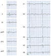
Electrocardiographic abnormalities in Case 1. A) Standard 12-lead ECG recorded at age 6 years showing marked QTc prolongation (579 ms) with atypical T wave morphology (late-peaking with notch on the descending limb). B) Atrial pacing at 100 bpm prolonged QTc from 596 ms to 610 ms. By contrast, mexiletine treatment shortened QT interval from 596 ms to 550 ms.
Figure 2
Figure 2
Electrocardiographic abnormalities in Case 2. A) Standard 12-lead ECG recorded at age 5 years showing QTc prolongation (478 ms). B) Epinephrine challenge test prolonged QTc at peak (466 ms to 558 ms) and at steady-state (535 ms). C) Propranolol or propranolol with mexieltine caused QTc shortening from 517 ms to 471 ms and 473 ms, respectively. Metoprolol normalized the QTc to 449 ms.
Figure 3
Figure 3
Electrocardiographic features of Case 3. A) Standard 12-lead ECG recorded at age 27 years showing QTc prolongation (567 ms). B) Polymorphic ventricular ectopy recorded (lead III) during exercise (step test) at age 28 years. During exercise, a progressive increase of heart rate was observed with no arrhythmias until 110 bpm was reached. A 3-beat episode of polymorphic ventricular tachycardia was recorded at 113 bpm. A representative 12-lead ECG during exercise is provided as Supplemental Fig. S1.
Figure 3
Figure 3
Electrocardiographic features of Case 3. A) Standard 12-lead ECG recorded at age 27 years showing QTc prolongation (567 ms). B) Polymorphic ventricular ectopy recorded (lead III) during exercise (step test) at age 28 years. During exercise, a progressive increase of heart rate was observed with no arrhythmias until 110 bpm was reached. A 3-beat episode of polymorphic ventricular tachycardia was recorded at 113 bpm. A representative 12-lead ECG during exercise is provided as Supplemental Fig. S1.
Figure 4
Figure 4
Electrocardiographic features of Case 5. Standard 12-lead ECG recorded at age 2 years showing QTc prolongation (555 ms) and bradycardia (HR = 55 bpm) during β-blocker treatment. Because of bradycardia, only one beat was recorded in the limb leads.
Figure 5
Figure 5
Novel de novo CALM2 mutations. A) Pedigrees and Sanger sequence electropherograms of each proband (marked by arrow). QTc values for each individual are shown underneath the corresponding pedigree symbol. B) Amino acid sequence alignments for calmodulins from different species with location of the five missense mutations. Amino acid sequence of EF-hands III and IV are underlined. C) Schematic model of Ca2+ binding loops in the N-terminal (I and II) and C-terminal (III and IV) domains of calmodulin showing the locations of mutations. Red circles represent the CALM2 mutations (p.N98S, p.N98I, p.D132E, p.D134H, p.Q136P) identified in our present study, green symbols represents previously reported mutations.
Figure 5
Figure 5
Novel de novo CALM2 mutations. A) Pedigrees and Sanger sequence electropherograms of each proband (marked by arrow). QTc values for each individual are shown underneath the corresponding pedigree symbol. B) Amino acid sequence alignments for calmodulins from different species with location of the five missense mutations. Amino acid sequence of EF-hands III and IV are underlined. C) Schematic model of Ca2+ binding loops in the N-terminal (I and II) and C-terminal (III and IV) domains of calmodulin showing the locations of mutations. Red circles represent the CALM2 mutations (p.N98S, p.N98I, p.D132E, p.D134H, p.Q136P) identified in our present study, green symbols represents previously reported mutations.
Figure 6
Figure 6
Impaired Ca2+ binding by mutant calmodulin C-domains. A) Representative Ca2+ titrations monitored by intrinsic fluorescence for recombinant WT (black circles) and mutant calmodulins (D132E, blue triangles; D134H, red squares). Kd values derived by fitting data from three replicates to a two-site model were (mean ± SEM) 2.1 ± 0.1, 48 ± 10, and 27 ± 5 μM for wildtype, D132E and D134H, respectively. B) Representative Ca2+ titrations for WT (black circles) and mutant (N98I, inverted orange triangles; Q136P, green diamonds) calmodulins. Kd values derived by fitting data from three replicates to a two-site model were (mean ± SEM) 2.3 ± 0.1, 19 ± 2, and 15 ± 1 μM for wildtype, N98I and Q136P, respectively.

References

    1. Schwartz PJ, Crotti L, Insolia R. Long-QT syndrome: from genetics to management. Circ Arrhythm Electrophysiol. 2012;5:868–877. - PMC - PubMed
    1. Moss AJ, Zareba W, Benhorin J, Locati EH, Hall WJ, Robinson JL, et al. ECG T-wave patterns in genetically distinct forms of the hereditary long QT syndrome. Circulation. 1995;92:2929–2934. - PubMed
    1. Zhang L, Timothy KW, Vincent GM, Lehmann MH, Fox J, Giuli LC, et al. Spectrum of ST-T-wave patterns and repolarization parameters in congenital long-QT syndrome: ECG findings identify genotypes. Circulation. 2000;102:2849–2855. - PubMed
    1. Schwartz PJ, Priori SG, Spazzolini C, Moss AJ, Vincent GM, Napolitano C, et al. Genotype-phenotype correlation in the long-QT syndrome: gene-specific triggers for life-threatening arrhythmias. Circulation. 2001;103:89–95. - PubMed
    1. Zareba W, Moss AJ, Schwartz PJ, Vincent GM, Robinson JL, Priori SG, et al. Influence of genotype on the clinical course of the long-QT syndrome. International Long-QT Syndrome Registry Research Group. N Engl J Med. 1998;339:960–965. - PubMed

Publication types

MeSH terms