Review
. 2014 Jun 3:5:130.
doi: 10.3389/fphar.2014.00130. eCollection 2014.

Dysregulation of cellular iron metabolism in Friedreich ataxia: from primary iron-sulfur cluster deficit to mitochondrial iron accumulation

Affiliations
Review

Dysregulation of cellular iron metabolism in Friedreich ataxia: from primary iron-sulfur cluster deficit to mitochondrial iron accumulation

Alain Martelli et al. Front Pharmacol. .

Abstract

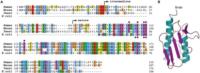
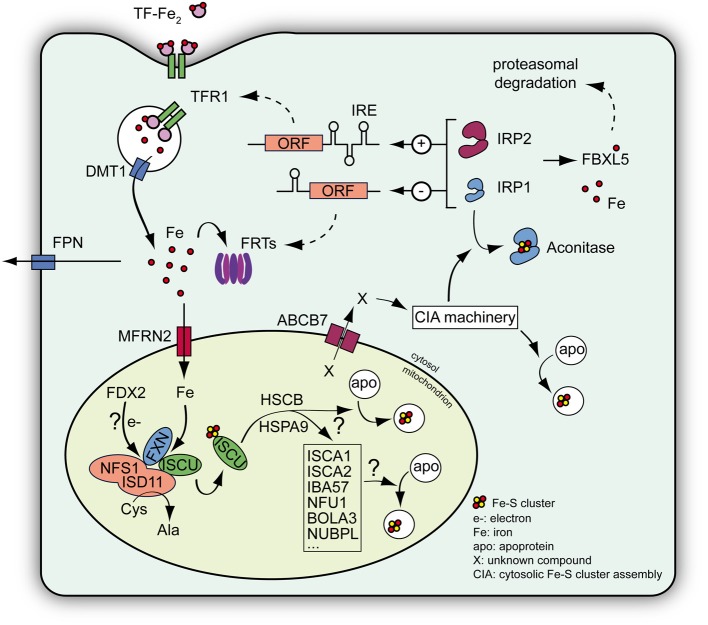
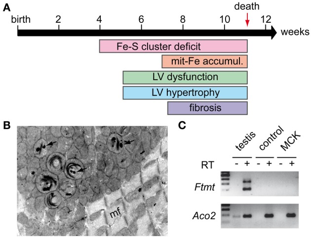
Friedreich ataxia (FRDA) is the most common recessive ataxia in the Caucasian population and is characterized by a mixed spinocerebellar and sensory ataxia frequently associating cardiomyopathy. The disease results from decreased expression of the FXN gene coding for the mitochondrial protein frataxin. Early histological and biochemical study of the pathophysiology in patient's samples revealed that dysregulation of iron metabolism is a key feature of the disease, mainly characterized by mitochondrial iron accumulation and by decreased activity of iron-sulfur cluster enzymes. In the recent past years, considerable progress in understanding the function of frataxin has been provided through cellular and biochemical approaches, pointing to the primary role of frataxin in iron-sulfur cluster biogenesis. However, why and how the impact of frataxin deficiency on this essential biosynthetic pathway leads to mitochondrial iron accumulation is still poorly understood. Herein, we review data on both the primary function of frataxin and the nature of the iron metabolism dysregulation in FRDA. To date, the pathophysiological implication of the mitochondrial iron overload in FRDA remains to be clarified.

Keywords: Friedreich ataxia; frataxin; iron metabolism; iron metabolism disorders; iron-sulfur cluster; mitochondria.


Figures

Figure 1
Figure 1
Sequences and structure of frataxin. (A) Sequence alignment of human, mouse, yeast, and bacterial frataxin. The non-conserved N-terminal parts of human, mouse, and yeast frataxins contain the mitochondrial targeting sequence of the protein. The arrows indicate the intermediate and mature forms of human FXN that are obtained during the two-step maturation process in vivo (Schmucker et al., 2008). The red dots indicate acidic residues of the N-terminal alpha helix of human and bacterial frataxins that are involved in the primary iron binding site of the monomeric protein. (B) Three dimensional structure of human frataxin obtained by NMR (1LY7) (Musco et al., 2000).
Figure 2
Figure 2
Schematic view of the Fe-S cluster machinery and the IRP-mediated cellular iron regulation. De novo Fe-S cluster biogenesis occurs within mitochondria and involves assembly of inorganic sulfur and iron on a scaffold protein ISCU. Iron is imported into the mitochondria by mitoferrins (MFRN). The process of Fe-S cluster assembly occurs within a complex consisting of NFS1-ISD11, the cysteine desulfurase providing the sulfur, ISCU and eventually frataxin (FXN), which regulates the NFS1 activity and the entry of iron within the complex (Colin et al., 2013). The process also needs electrons (e–) that may be provided by a mitochondrial ferredoxin (FDX2). Once assembled the cluster on ISCU is transferred to acceptor proteins with the help of additional proteins, such as the chaperones HSCB and HSPA9, ISCA proteins, IBA57, NFU1, BOLA3, and NUBPL (reviewed in Lill, ; Beilschmidt and Puccio, 2014). Alternatively, a still uncharacterized compound (X) provided by the mitochondrial machinery is exported to the cytosol via ABCB7 and is used by the cytosolic Fe-S cluster assembly machinery (CIA machinery) to assemble Fe-S clusters for cytosolic and nuclear acceptor proteins. Among the cytosolic Fe-S cluster acceptors, IRP1 is a regulator of cellular iron metabolism. In normal conditions, IRP1 binds a Fe-S cluster to become an aconitase devoid of regulatory capacity. IRP2 exists only as an apoprotein and is regulated through proteasomal degradation mediated by the iron sensor protein FBXL5 (Salahudeen et al., ; Vashisht et al., 2009). Both IRPs can regulate the expression of key genes involved in iron metabolism, such as transferrin receptor 1 (TFR1), ferritins (FRTs), and the iron exporter ferroportin (FPN), by binding a specific mRNA motif called IRE. Depending on the location of the IRE compared to the open reading frame (ORF), IRPs can increase (+) or decrease (−) protein expression, thus controlling cellular iron import and storage (reviewed in Anderson et al., 2012). DMT1: divalent metal transporter involved in iron import.
Figure 3
Figure 3
MCK mouse model. (A) Phenotypic characteristics of the MCK mouse model: MCK mice develop progressive hypertrophic cardiomyopathy characterized by progressive left ventricule (LV) dysfunction and hypertrophy starting around 5 weeks of age. The cardiomyopathy leads to cell death and fibrosis. MCK mice prematurely dye around 11 weeks. Fe-S cluster deficit is a primary feature in the mouse pathology with significant differences observed in 4 weeks old mice. Mitochondrial iron (mit-Fe) accumulation is observed in the later stage of the disease. (B) Electron microscopy picture obtained from a heart sample of a 7 weeks old MCK mouse showing mitochondrial abnormalities, in particular collapse cristae and electron-dense deposits (arrows) corresponding to mitochondrial iron deposits. mf, myofiber. (C) Semi-nested PCR on cDNA from heart samples of 8 weeks old control and deleted MCK mice was performed as described (Santambrogio et al., 2007) to assess mitochondrial ferritin (FTMT) expression. Total RNA from tissue was extracted using Trizol® reagent (Life Technologies) and submitted (+) or not (−) to reverse transcription (RT). Testis cDNA was used as positive control for Ftmt expression, and a classical PCR to amplify mitochondrial aconitase (Aco2) cDNA was carried out as a control for reverse transcription and loading. Samples without reverse transcription (RT–) were used as control for specific PCR amplification.

References

    1. Adamec J., Rusnak F., Owen W. G., Naylor S., Benson L. M., Gacy A. M., et al. (2000). Iron-dependent self-assembly of recombinant yeast frataxin: implications for Friedreich ataxia. Am. J. Hum. Genet. 67, 549–562 10.1086/303056 - DOI - PMC - PubMed
    1. Adinolfi S., Iannuzzi C., Prischi F., Pastore C., Iametti S., Martin S. R., et al. (2009). Bacterial frataxin CyaY is the gatekeeper of iron-sulfur cluster formation catalyzed by IscS. Nat. Struct. Mol. Biol. 16, 390–396 10.1038/nsmb.1579 - DOI - PubMed
    1. Adinolfi S., Trifuoggi M., Politou A. S., Martin S., Pastore A. (2002). A structural approach to understanding the iron-binding properties of phylogenetically different frataxins. Hum. Mol. Genet. 11, 1865–1877 10.1093/hmg/11.16.1865 - DOI - PubMed
    1. Allikmets R., Raskind W. H., Hutchinson A., Schueck N. D., Dean M., Koeller D. M. (1999). Mutation of a putative mitochondrial iron transporter gene (ABC7) in X-linked sideroblastic anemia and ataxia (XLSA/A). Hum. Mol. Genet. 8, 743–749 10.1093/hmg/8.5.743 - DOI - PubMed
    1. Al-Mahdawi S., Pinto R. M., Varshney D., Lawrence L., Lowrie M. B., Hughes S., et al. (2006). GAA repeat expansion mutation mouse models of Friedreich ataxia exhibit oxidative stress leading to progressive neuronal and cardiac pathology. Genomics 88, 580–590 10.1016/j.ygeno.2006.06.015 - DOI - PMC - PubMed