. 2014 Jun 3:5:194.
doi: 10.3389/fphys.2014.00194. eCollection 2014.

Effect of contraction intensity on sympathetic nerve activity to active human skeletal muscle

Affiliations

Effect of contraction intensity on sympathetic nerve activity to active human skeletal muscle

Daniel Boulton et al. Front Physiol. .

Abstract

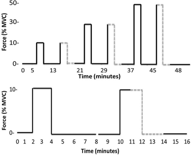
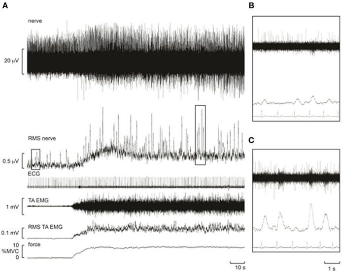
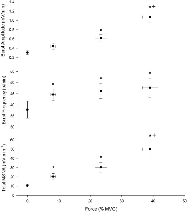
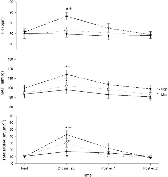
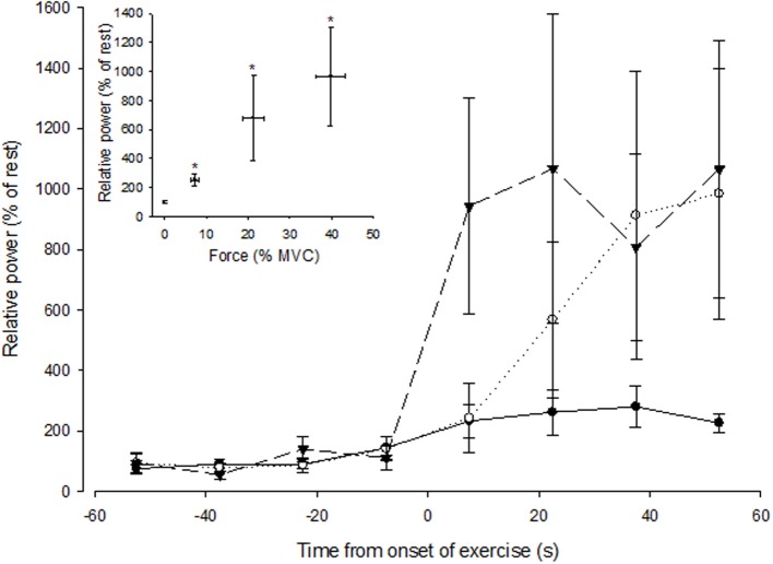
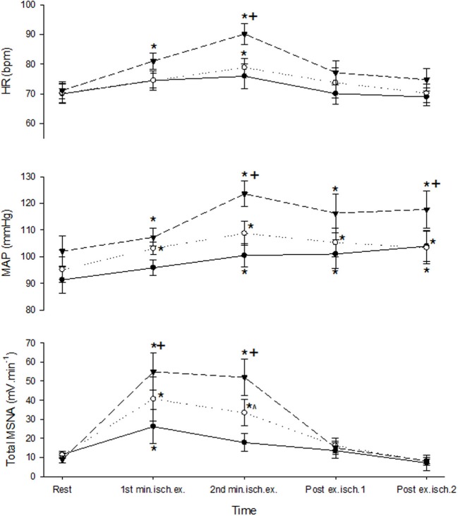
The effect of contraction intensity on muscle sympathetic nerve activity (MSNA) to active human limbs has not been established. To address this, MSNA was recorded from the left peroneal nerve during and after dorsiflexion contractions sustained for 2 min by the left leg at ~10, 25, and 40% MVC. To explore the involvement of the muscle metaboreflex, limb ischemia was imposed midway during three additional contractions and maintained during recovery. Compared with total MSNA at rest (11.5 ± 4.1 mv(.)min(-1)), MSNA in the active leg increased significantly at the low (21.9 ± 13.6 mv(.)min(-1)), medium (30.5 ± 20.8 mv(.)min(-1)), and high (50.0 ± 24.5 mv(.)min(-1)) intensities. This intensity-dependent effect was more strongly associated with increases in MSNA burst amplitude than burst frequency. Total MSNA then returned to resting levels within the first minute of recovery. Limb ischemia had no significant influence on the intensity-dependent rise in MSNA or its decline during recovery in the active leg. These findings reveal intensity-dependent increases in total MSNA and burst amplitude to contracting human skeletal muscle that do not appear to involve the muscle metaboreflex.

Keywords: contraction; ischemia; metaboreflex; muscle; sympathetic.


Figures

Figure 1
Figure 1
Timelines of the entire experimental protocol (top) and the initial pair of contractions plus rest periods (bottom). The latter timeline more clearly identifies the onset and offset of ischemia (gray dashed line) and that each contraction belongs to an 8-min “block” beginning with a 2 min rest and ending with a 4 min rest.
Figure 2
Figure 2
Experimental records from one subject during a control contraction at ~10% MVC. Note that an increase in muscle afferent activity occurs during the contraction, resulting in a baseline shift in the RMS-processed nerve signal. Despite this, bursts of MSNA remained clearly identifiable and increased in amplitude. (A) Raw recordings of the neurogram (top) and root mean square of this recording (“RMS Nerve”) which shows bursts of MSNA. (B) Zoom view of four bursts of MSNA (rms) at rest. (C) Zoom view of four bursts during contraction highlighting the increased amplitude of each burst compared with resting responses in (B).
Figure 3
Figure 3
Effect of contraction intensity on total MSNA, MSNA burst frequency, and MSNA burst amplitude during the first minute of contraction under control conditions. There was a significant main effect of intensity on total MSNA, burst frequency and burst amplitude. Asterisk (*) indicates significantly different (P < 0.05) from the resting responses. Cross (+) indicates significantly different (P < 0.05) from 10 to 30% MVC responses at the specified time. These contrasts were based on post-hoc, two-tailed pairwise tests.
Figure 4
Figure 4
Total MSNA, mean arterial pressure (MAP), and heart rate (HR) before, during, and after control contractions at low (closed circle), medium (open circle), and high intensities (triangle). Observations were made at rest, during the second minute of contraction, and during the first and second minute of recovery. Asterisk (*) indicates significantly different (P < 0.05) from the resting responses before contraction. Cross (+) indicates significantly different (P < 0.05) from the 10% MVC responses. These contrasts were based on post-hoc, two-tailed pairwise tests.
Figure 5
Figure 5
Spectral power of MSNA (normalized to the resting value) during 15 s intervals prior to and during the first minute of contraction at low (closed circle), medium (open circle), and high (triangle) forces. The onset of contraction is at t = 0 s. The inset shows spectral power of MSNA (1 min averaged values) as a function of force. The asterisk (*) indicates significantly different from rest at P < 0.05.
Figure 6
Figure 6
MSNA and cardiovascular responses before, during, and after ischemic contractions at low (closed circle), medium (open circle), and high intensities (triangle). Ischemia was imposed at the start of the second minute of contraction and maintained during recovery. Observations were made at rest and during the first and second minutes of contraction and recovery. Asterisk (*) indicates significantly different (P < 0.05) from the resting responses before contraction. Cross (+) indicates significantly different (P < 0.05) from the 10 to 30% MVC responses at the specified time. Hat (∧) indicates significantly different from 10% MVC responses at the specified time. These contrasts were based on post-hoc, two-tailed pairwise tests.

Similar articles

Cited by

References

    1. Alam M., Smirk F. H. (1937). Observations in man upon a blood pressure raising reflex arising from the voluntary muscles. J. Physiol. 89, 372–383 - PMC - PubMed
    1. Cui J., Blaha C., Moradhkan R., Gray K. S., Sinoway L. (2006). Muscle sympathetic nerve activity responses to dynamic passive muscle stretch in humans. J. Physiol. 576, 625–634 10.1113/jphysiol.2006.116640 - DOI - PMC - PubMed
    1. Green S., Langberg H., Skovgaard D., Bulow J., Kjaer M. (2000). Interstitial and arterial-venous [K+] in human calf muscle during dynamic exercise: effect of ischaemia and relation to muscle pain. J. Physiol. 529, 849–861 10.1111/j.1469-7793.2000.00849.x - DOI - PMC - PubMed
    1. Green S., Thorp R., Reeder E., Donnelly J., Fordy G. (2011). Venous occlusion plethysmography versus Doppler ultrasound in the assessment of leg blood flow during calf exercise. Eur. J. Appl. Physiol. 111, 1889–1900 10.1007/s00421-010-1819-6 - DOI - PubMed
    1. Hansen J., Thomas G. D., Jacobsen T. N., Victor R. G. (1994). Muscle metaboreflex triggers parallel sympathetic activation in exercising and resting human skeletal muscle. Am. J. Physiol. 266, H2508–H2514 - PubMed

LinkOut - more resources