Does antigen masking by ubiquitin chains protect from the development of autoimmune diseases?
- PMID: 24917867
- PMCID: PMC4042494
- DOI: 10.3389/fimmu.2014.00262
Does antigen masking by ubiquitin chains protect from the development of autoimmune diseases?
Abstract
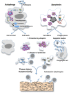
Autoimmune diseases are characterized by the production of antibodies against self-antigens and generally arise from a failure of central or peripheral tolerance. However, these diseases may develop when newly appearing antigens are not recognized as self by the immune system. The mechanism by which some antigens are "invisible" to the immune system is not completely understood. Apoptotic and complement system defects or autophagy imbalance can generate this antigenic autoreactivity. Under particular circumstances, cellular debris containing autoreactive antigens can be recognized by innate immune receptors or other sensors and can eventually lead to autoimmunity. Ubiquitination may be one of the mechanisms protecting autoreactive antigens from the immune system that, if disrupted, can lead to autoimmunity. Ubiquitination is an essential post-translational modification used by cells to target proteins for degradation or to regulate other intracellular processes. The level of ubiquitination is regulated during T cell tolerance and apoptosis and E3 ligases have emerged as a crucial signaling pathway for the regulation of T cell tolerance toward self-antigens. I propose here that an unrecognized role of ubiquitin and ubiquitin-like proteins could be to render intracellular or foreign antigens (present in cellular debris resulting from apoptosis, complement system, or autophagy defects) invisible to the immune system in order to prevent the development of autoimmunity.
Keywords: UBLs; apoptosis; autoimmunity; autophagy; immune tolerance; ubiquitin.
Figures

Similar articles
-
Pathogenesis and spectrum of autoimmunity.Methods Mol Biol. 2012;900:1-9. doi: 10.1007/978-1-60761-720-4_1. Methods Mol Biol. 2012. PMID: 22933062
-
Ubiquitination system and autoimmunity: the bridge towards the modulation of the immune response.Autoimmun Rev. 2008 Feb;7(4):284-90. doi: 10.1016/j.autrev.2007.11.026. Epub 2008 Jan 7. Autoimmun Rev. 2008. PMID: 18295731 Review.
-
E3 ubiquitin ligases in T-cell tolerance.Eur J Immunol. 2009 Sep;39(9):2337-44. doi: 10.1002/eji.200939662. Eur J Immunol. 2009. PMID: 19714573 Review.
-
Apoptosis and immune responses to self.Rheum Dis Clin North Am. 2004 Feb;30(1):193-212. doi: 10.1016/S0889-857X(03)00110-8. Rheum Dis Clin North Am. 2004. PMID: 15061575 Review.
-
The role of E3 ligases in autoimmunity and the regulation of autoreactive T cells.Curr Opin Immunol. 2007 Dec;19(6):665-73. doi: 10.1016/j.coi.2007.10.002. Epub 2007 Nov 26. Curr Opin Immunol. 2007. PMID: 18036806 Review.
Cited by
-
Ubiquitin-like Proteins in Autoimmune Diseases: Current Evidence and Therapeutic Opportunities.Immune Netw. 2025 May 1;25(3):e21. doi: 10.4110/in.2025.25.e21. eCollection 2025 Jun. Immune Netw. 2025. PMID: 40620421 Free PMC article. Review.
-
CSP ubiquitylation favours Plasmodium berghei survival during early liver stage infection.Sci Rep. 2025 Apr 25;15(1):14498. doi: 10.1038/s41598-025-98294-4. Sci Rep. 2025. PMID: 40281042 Free PMC article.
-
Antigenic mimicry of ubiquitin by the gut bacterium Bacteroides fragilis: a potential link with autoimmune disease.Clin Exp Immunol. 2018 Nov;194(2):153-165. doi: 10.1111/cei.13195. Epub 2018 Sep 17. Clin Exp Immunol. 2018. PMID: 30076785 Free PMC article.
-
Autophagy plays a protective role against Trypanosoma cruzi infection in mice.Virulence. 2019 Dec;10(1):151-165. doi: 10.1080/21505594.2019.1584027. Virulence. 2019. PMID: 30829115 Free PMC article.
-
Conformational changes in myeloperoxidase induced by ubiquitin and NETs containing free ISG15 from systemic lupus erythematosus patients promote a pro-inflammatory cytokine response in CD4+ T cells.J Transl Med. 2020 Nov 11;18(1):429. doi: 10.1186/s12967-020-02604-5. J Transl Med. 2020. PMID: 33176801 Free PMC article.
References
Publication types
LinkOut - more resources
Full Text Sources
Other Literature Sources

