. 2014 Dec;86(6):1150-60.
doi: 10.1038/ki.2014.196. Epub 2014 Jun 11.

Albumin-induced podocyte injury and protection are associated with regulation of COX-2

Affiliations

Albumin-induced podocyte injury and protection are associated with regulation of COX-2

Shipra Agrawal et al. Kidney Int. 2014 Dec.

Abstract

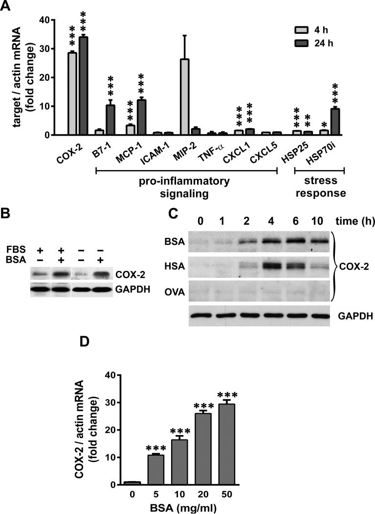
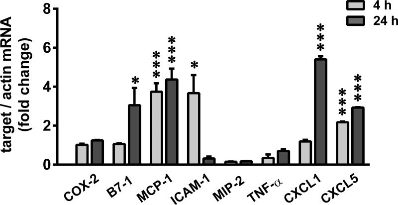
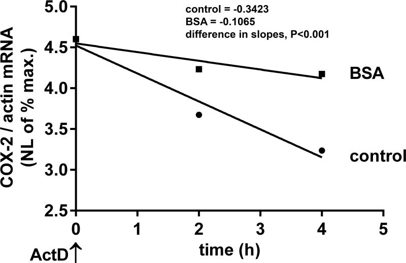
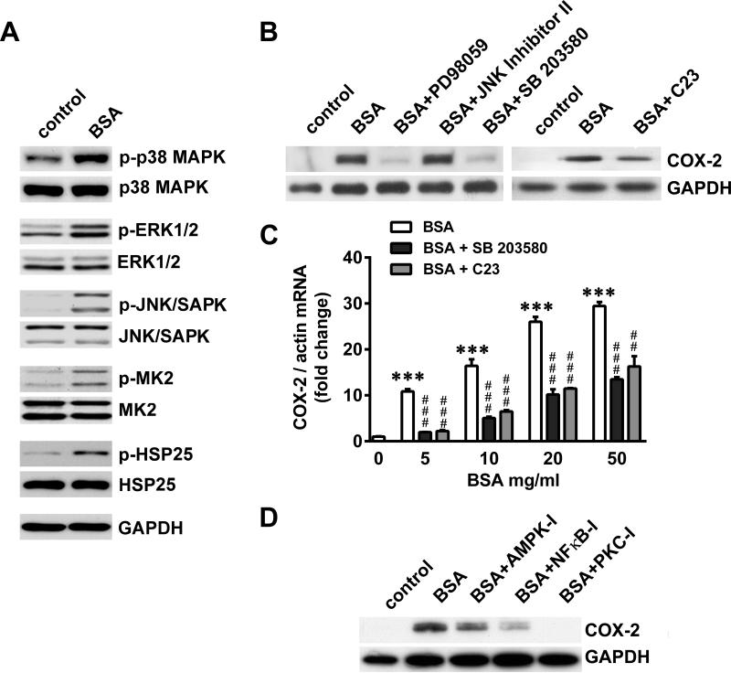
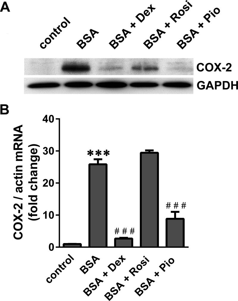
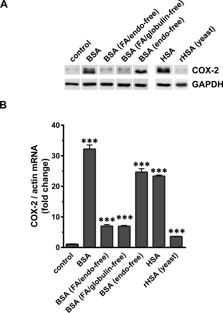
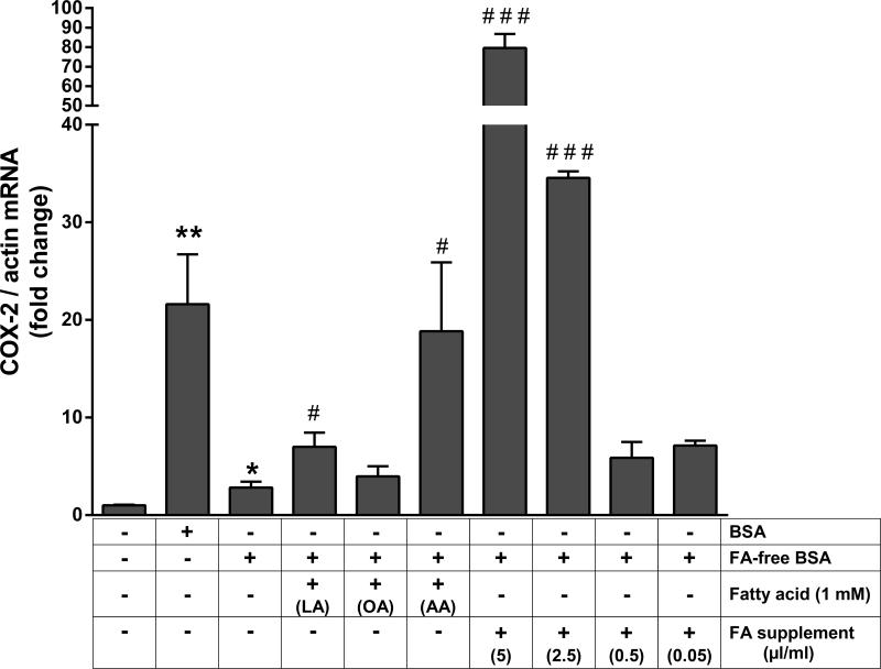
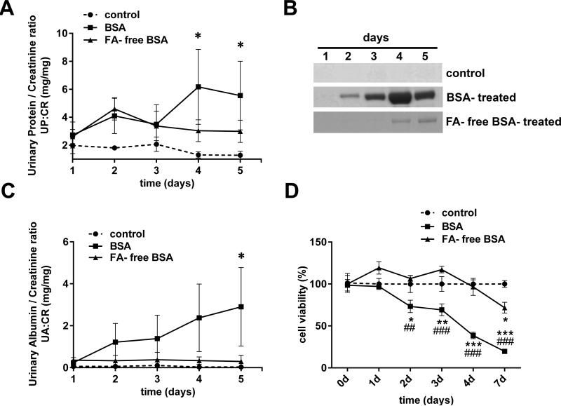
Albuminuria is both a hallmark and a risk factor for progressive glomerular disease, and results in increased exposure of podocytes to serum albumin with its associated factors. Here in vivo and in vitro models of serum albumin-overload were used to test the hypothesis that albumin-induced proteinuria and podocyte injury directly correlate with COX-2 induction. Albumin induced COX-2, MCP-1, CXCL1, and the stress protein HSP25 in both rat glomeruli and cultured podocytes, whereas B7-1 and HSP70i were also induced in podocytes. Podocyte exposure to albumin induced both mRNA and protein and enhanced the mRNA stability of COX-2, a key regulator of renal hemodynamics and inflammation, which renders podocytes susceptible to injury. Podocyte exposure to albumin also stimulated several kinases (p38 MAPK, MK2, JNK/SAPK, and ERK1/2), inhibitors of which (except JNK/SAPK) downregulated albumin-induced COX-2. Inhibition of AMPK, PKC, and NFκB also downregulated albumin-induced COX-2. Critically, albumin-induced COX-2 was also inhibited by glucocorticoids and thiazolidinediones, both of which directly protect podocytes against injury. Furthermore, specific albumin-associated fatty acids were identified as important contributors to COX-2 induction, podocyte injury, and proteinuria. Thus, COX-2 is associated with podocyte injury during albuminuria, as well as with the known podocyte protection imparted by glucocorticoids and thiazolidinediones. Moreover, COX-2 induction, podocyte damage, and albuminuria appear mediated largely by serum albumin-associated fatty acids.


Figures

Figure 1
Figure 1
SA-overload in rats induces proteinuria and glomerular expression of COX-2, pro-inflammatory and stress genes. A) Urinary protein/creatinine ratios (UP:CR) of the BSA-treated rats vs control rats (n=3 for each group; *P<0.05 versus control, determined by two way ANOVA Tukey's multiple comparisons test). B) Massive amounts of urinary albumin are excreted from BSA-treated rats but not control rats (representative gels are shown from each group). Urine was collected on five consecutive days and equal volumes (4 μl) analyzed by SDS-PAGE and Coomassie Brilliant Blue staining. C) Urinary albumin/creatinine ratio (UA:CR) of the BSA-treated rats vs control rats (n=3 for each group; *P<0.05 versus control, determined by two way ANOVA Tukey's multiple comparisons test). D) Total RNA was extracted from the isolated glomeruli of control and BSA-treated rats and relative mRNA levels of COX-2, pro-inflammatory and stress genes were measured by qRT-PCR and normalized to β-actin (*P<0.05, **P<0.01 and ***P<0.001 versus control, determined by t test).
Figure 2
Figure 2
SA induces COX-2, pro-inflammatory and stress genes in cultured podocytes. A) Serum-starved podocytes were exposed to 40 mg/ml BSA for 4 h and 24 h, and relative mRNA levels of COX-2, pro-inflammatory and stress response genes were measured by qRT-PCR and normalized to β-actin (*P<0.05, **P<0.01 and ***P<0.001 versus time matched control, determined by t test). B) Podocytes were maintained in culture medium containing FBS or serum-starved O/N, and exposed to 40 mg/ml of BSA for 4 h and subjected to immuno-blot analysis for COX-2 and GAPDH. C) Serum-starved podocytes were exposed to 40 mg/ml BSA, human serum albumin (HSA) and Ovalbumin (OVA), and harvested at indicated time points. Protein extracts were analyzed for COX-2 and GAPDH (shown as loading control for a representative blot). D) Serum-starved podocytes were exposed for 4 h to increasing concentrations of BSA as indicated, and mRNA levels of COX-2 were measured by qRT-PCR and normalized to β-actin (***P<0.001 versus control, determined by t test).
Figure 3
Figure 3
SA and LPS elicit disparate COX-2 and pro-inflammatory response in podocytes. Serum-starved podocytes were treated with 1 μg/ml LPS (026:B6) for 4 and 24 h and relative mRNA levels of COX-2 and pro-inflammatory signaling genes was measured by qRT-PCR and normalized to β-actin (*P<0.05 and ***P<0.001 versus time matched control, determined by t test).
Figure 4
Figure 4
SA stabilizes COX-2 mRNA. Podocytes were exposed to 20 mg/ml BSA or medium alone (control) for 2 h, transcription was inhibited with actinomycin D (ActD) and RNA was extracted after 2 and 4 h. COX-2 mRNA levels were quantified by qRT-PCR and normalized to β–actin. Natural logarithmic (NL) values of the maximum mRNA amount were plotted against time and half-lives calculated (control: t1/2=1.8h, BSA: t1/2=6h) from the slopes of linear regression analysis which were significantly different (P<0.001) between control and BSA groups (control: y = 0.3423*x + 4.523, BSA: y = 0.1065*x + 4.549).
Figure 5
Figure 5
Signaling pathways involving kinases mediate COX-2 induction by SA. A) Serum- starved podocytes were treated with 20 mg/ml BSA for 1 h and subjected to immuno-blot analysis for phosphorylated (p) and total forms of p38 MAPK, ERK1/2, JNK/SAPK, MK2, HSP25 and GAPDH. B) Serum-starved podocytes were treated with 20 mg/ml BSA for 4 h following 1 h pre-treatment with inhibitors at 20μM for ERK1/2 (PD98059), JNK/SAPK (JNK Inhibitor II), p38 MAPK (SB 203580) and MK2 (C23). Cells were subjected to immuno-blot analysis for COX-2 and GAPDH. C) Serum-starved podocytes were treated with increasing concentrations of BSA as indicated for 4 h following 24 h pre-treatment with 30 ZM SB203580 (p38 MAPK inhibitor) or 10 μM C23 (MK2 inhibitor), COX-2 mRNA levels were quantified by qRT-PCR and normalized to β-actin (***P<0.001 versus control; **P<0.01 and ***P<0.001 versus BSA, determined by one way ANOVA). D) Serum-starved podocytes were treated with 20 mg/ml BSA for 4 h following 1 h pre-treatment with inhibitors at 5μM for AMPK (Compound C), NFκB (NFκB-I) and PKC (PKC-I). Cells were subjected to immuno-blot analysis for COX-2 and GAPDH.
Figure 6
Figure 6
Glucocorticoids and thiazolidinediones inhibit SA-induced COX-2. Serum–starved podocytes were treated with 20 mg/ml BSA for 4 h following pre-treatment with 1 μM dexamethasone (Dex), 10 μM rosiglitazone (Rosi) and 0.1 μM pioglitazone (Pio) for 24 h. A) COX-2 and GAPDH protein were visualized by immuno-blot analysis, and B) COX-2 mRNA levels were quantified by qRT-PCR and normalized to β-actin (***P<0.001 versus control; ***P<0.001 versus BSA, determined by one way ANOVA).
Figure 7
Figure 7
SA-associated factors contribute to COX-2 induction in podocytes. Serum-starved podocytes were exposed to 40 mg/ml of BSA, charcoal-treated FA/endotoxin-free BSA, FA/globulin-free BSA, endotoxin-free BSA, HSA and recombinant HSA made in yeast (rHSA). (A) Cells were harvested after 4 h, processed for SDS-PAGE and western blotting and analyzed for COX-2 and GAPDH. (B) Total RNA was extracted and COX-2 mRNA was measured by RT-PCR and normalized to the β-actin mRNA (***P<0.001 versus control, determined by t test).
Figure 8
Figure 8
Fatty acids bound to SA contribute to COX-2 induction in podocytes. Serum-starved podocytes were exposed to 40 mg/ml of BSA, FA-free BSA, and FA-free BSA in combination with 1 mM Lauric acid (LA), 1 mM Oleic acid (OA), 1 mM Arachidonic acid (AA) and a combined fatty acid supplement at 5, 2.5, 0.5 and 0.05 μl/ml cell culture media. Cells were harvested after 4 h, total RNA was extracted and COX-2 mRNA was measured by RT-PCR and normalized to the β-actin mRNA (*P<0.05 and **P<0.01 versus control; *P<0.05 and ***P<0.001 versus FA-free BSA, determined by t test).
Figure 9
Figure 9
Delipidation of BSA attenuates proteinuria and podocyte damage. A) Urinary protein/creatinine ratio (UP:CR) of the control, BSA-treated and FA-free BSA-treated rats (n=3 for each group; *P<0.05 versus control, determined by two way ANOVA Tukey's multiple comparisons test). B) Representative gels from each group show that only small amounts of albumin are excreted from FA-free BSA-treated rats, while BSA treated rats excreted massive amounts of albumin in their urine compared to control rats. Urine was collected on five consecutive days and equal volumes (2.5 μl) analyzed by SDS PAGE and Coomassie Brilliant Blue staining. C) Urinary albumin/creatinine ratios (UA:CR) from control, BSA-treated and FA-free BSA-treated rats (n=3 for each group; *P<0.05 versus control, determined by two way ANOVA Tukey's multiple comparisons test). D) Cultured podocytes were exposed to media (control) or 40 mg/ml BSA and FA-free BSA for 0, 1, 2, 3, 4 and 7 days and assayed for cell viability using MTT (*P<0.05, **P<0.01 and ***P<0.001 versus control; ***P<0.01 and ***P<0.001 versus FA-free BSA, determined by two way ANOVA Tukey's multiple comparisons test).
Figure 10
Figure 10
Proposed schematic of COX-2 as a mediator of podocyte injury by SA and protection by GCs, TZDs and MAPK inhibitors. Serum Albumin (SA) exists as a complex entity and along with its associated factors (i.e. fatty acids, hormones, vitamins and bilirubin etc., depicted as ▲/ ■ / ● on SA molecule on left), it induces COX-2 in podocytes. This is coupled with an increase in phosphorylation (P) of kinases p38 MAPK, ERK1/2, MK2 and its downstream substrate HSP25. Inhibitors to p38 MAPK, MK2, ERK1/2, AMPK and PKC (depicted in rectangles) down-regulate SA-induced COX-2. Glucocorticoids (GCs) and thiazolidinediones (TZDs), which are known to protect podocytes against injury, also inhibit SA- induced COX-2. They have been previously shown to attenuate MAPK signaling and also impart their trans-repression activities via coupling of their respective ligand-bound receptors (GR and PPARγ) with NFκB to block COX-2 expression via the NFκB response element on COX-2. NFκB (p65 subunit) is over-expressed following SA-exposure and NFκB inhibition down-regulates SA-induced COX-2.

Similar articles

Cited by

References

    1. Iseki K, Ikemiya Y, Iseki C, et al. Proteinuria and the risk of developing end-stage renal disease. Kidney international. 2003;63:1468–1474. - PubMed
    1. Brunskill NJ. Albumin signals the coming of age of proteinuric nephropathy. J Am Soc Nephrol. 2004;15:504–505. - PubMed
    1. Ashworth CT, James JA. Glomerular excretion of macromolecular substances. Electron microscopic study of rat kidney after administration of human serum albumin. Am J Pathol. 1961;39:307–316. - PMC - PubMed
    1. Lawrence GM, Brewer DB. Effect of strain and sex on the induction of hyperalbuminaemic proteinuria in the rat. Clinical science. 1981;61:751–756. - PubMed
    1. Eddy AA, Kim H, Lopez-Guisa J, et al. Interstitial fibrosis in mice with overload proteinuria: deficiency of TIMP-1 is not protective. Kidney international. 2000;58:618–628. - PubMed

Publication types

MeSH terms

Substances