. 2014 Jul;21(7):579-84.
doi: 10.1038/nsmb.2849. Epub 2014 Jun 11.

Molecular basis for pseudokinase-dependent autoinhibition of JAK2 tyrosine kinase

Affiliations

Molecular basis for pseudokinase-dependent autoinhibition of JAK2 tyrosine kinase

Yibing Shan et al. Nat Struct Mol Biol. 2014 Jul.

Abstract

Janus kinase-2 (JAK2) mediates signaling by various cytokines, including erythropoietin and growth hormone. JAK2 possesses tandem pseudokinase and tyrosine-kinase domains. Mutations in the pseudokinase domain are causally linked to myeloproliferative neoplasms (MPNs) in humans. The structure of the JAK2 tandem kinase domains is unknown, and therefore the molecular bases for pseudokinase-mediated autoinhibition and pathogenic activation remain obscure. Using molecular dynamics simulations of protein-protein docking, we produced a structural model for the autoinhibitory interaction between the JAK2 pseudokinase and kinase domains. A striking feature of our model, which is supported by mutagenesis experiments, is that nearly all of the disease mutations map to the domain interface. The simulations indicate that the kinase domain is stabilized in an inactive state by the pseudokinase domain, and they offer a molecular rationale for the hyperactivity of V617F, the predominant JAK2 MPN mutation.


Figures
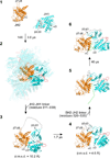
Figure 1
Figure 1
Steps of JAK2 JH2–JH1 model generation. (1) Starting positions of JAK2 JH2 (PDB code 4FVQ), residues 536–810, and JAK2 JH1 (PDB code 3KRR), residues 840–1131. The center-of-mass distance is 67 Å, with a minimum separation of 26 Å. JH2 is colored orange and JAK2 JH1 is colored cyan, with the activation loop (residues 994–1016) colored red. Structural elements that will converge in the final model of JH2–JH1 (αC (JH2) with αD (JH1) and β7–β8 (JH2) with β2–β3 (JH1)) are labeled at key steps. (2) JH2–JH1 interaction poses after 14 3-µs MD simulations (different initial random velocities for each simulation), superimposed on JH2. Pose 2, shown in solid coloring, was used in the subsequent modeling steps. (3) After adding the JH2–JH1 linker in an extended conformation. (4) After simulating JH2–JH1, residues 536–1131, for 1.7 µs. (5) After adding the SH2–JH2 linker in an extended conformation. (6) After simulating JH2–JH1, residues 520–1131, for 40 µs. RMSD values of JH1 and JH2 (Cα atoms) relative to the final model (state 6) are given in parenthesis for states 3 and 4.
Figure 2
Figure 2
Model of JAK2 JH2–JH1 derived from MD simulations. (a) Autoinhibitory pose of JAK2 JH2–JH1. The coloring scheme is the same as in Fig. 1. Residues that cause JAK2 activation upon mutation (to the indicated residues) are shown in sphere representation (side chains) and colored pink (carbon atoms). Phosphorylated Ser523 and Tyr570 are shown in stick representation and colored according to their location. Oxygen atoms are colored red, nitrogen atoms blue, sulfur atoms yellow, and phosphorus atoms black. A red superscript in a residue label indicates the figure part showing a zoom-in of that region. The N-terminus (residue 520) is labeled ‘N’, and the C-terminus (residue 1131) is labeled ‘C’. The JH2–JH1 interface (the SH2–JH2 and JH2–JH1 linkers excluded) buries 1670 Å2 of total surface area. (be) Regions of the JH2–JH1 interface near pTyr570 (b), near Arg683–Asp873 (c), near the hinge region of JH1 (d), and near the SH2–JH2 linker (e). Select residues are shown in stick representation, some with van der Waals surfaces. Black dashed lines represent salt bridges.
Figure 3
Figure 3
Experimental validation of the JAK2 JH2–JH1 model. (ad) Left: representative western blots of immunoprecipitated JAK2 from COS7 cells, wild type (WT) or the indicated JAK2 mutant, probed with anti-pTyr1007–1008 (pJAK2) (top) or anti-HA antibodies (bottom). The position of the 150-kDa molecular-weight marker is indicated. Middle: quantification of the pJAK2 signals normalized by JAK2 protein levels and plotted as fold-change relative to wild-type JAK2 (set to 1.0). Average values and standard deviations were derived from three independent experiments (N=3). Right: representative western blots of COS7 whole-cell lysates probed with anti-pTyr701 STAT1 antibodies (pSTAT1) (top) or anti-STAT1 antibodies (STAT1) (bottom) to detect endogenous STAT1 levels. The position of the 100-kDa molecular-weight marker is indicated. Original images of blots used in this study can be found in Supplementary Figure 6.
Figure 4
Figure 4
JH2-mediated autoinhibition of JH1 in JAK2. (a) Distance in JH1 between Lys882 (β3) and Glu898 (αC). The distance is plotted as a function of simulation time for simulations of JAK2 JH2–JH1 or JH1 alone (JH1 activation loop was unphosphorylated for both). To simplify the salt-bridge presentation (to account for both Oε1 and Oε2 of Glu898), the actual distance displayed is between Nζ of Lys882 and Cδ of Glu898, and the gray rectangle indicates the salt-bridging distance range. (b) DFG-in and -out states of the JH1 activation loop. Left: in the active state of JH1 (PDB code 3KRR), the Lys882–Glu898 salt bridge is formed, and Asp994 and Phe995 of the DFG motif in the activation loop adopt the DFG-in (active) conformation. Right: during the simulation of JH2–JH1, the Lys882–Glu898 salt bridge is disrupted and the DFG motif more readily adopts a DFG-out (inactive) conformation (shown is a snapshot taken after 12 µs of the simulation). Coloring is the same as in Fig. 1.
Figure 5
Figure 5
Model for JAK2 JH2-mediated autoinhibition of JH1. (Not shown are the FERM and SH2 domains of JAK2 and cytokine receptor.) A conformational equilibrium exists between JH1 in the JH2–JH1 autoinhibitory interaction (state I), in which JH1 is held in an inactive state (JH1, red), and configurations in which JH1 is disassociated from JH2 (orange) and is transiently active (state II; JH1, mixed red and green). The N and C lobes of JH2 and JH1 are labeled. Phosphorylated Ser523 and Tyr570 (magenta and mixed white and magenta spheres, respectively) stabilize the autoinhibited state by binding to positively charged residues in JH1 and JH2 (blue patches; see Fig. 2b,e). Ser523 is constitutively phosphorylated, whereas Tyr570 is sub-stoichiometrically phosphorylated in the basal state, and its phosphorylation level increases upon JAK2 activation, which probably serves as a negative feedback mechanism (to stabilize the autoinhibited state). In the basal state (no cytokine), the two JAK2 molecules (only one JH2–JH1 shown) associated with a cytokine-receptor dimer are maintained in positions that limit trans-phosphorylation of the JH1 activation loop (Tyr1007–1008). Cytokine binding and receptor rearrangement juxtapose the two JAK2 molecules to facilitate JH1 trans-phosphorylation of the activation loop, which activates JAK2 (state III; JH1, green). Activating mutations such as D873N, R683S, or V617F destabilize the autoinhibited state, permitting trans-phosphorylation of the JH1 activation loop in the basal state.

References

    1. O'Shea JJ, Holland SM, Staudt LM. JAKs and STATs in immunity, immunodeficiency, and cancer. N. Engl. J. Med. 2013;368:161–170. - PMC - PubMed
    1. Wallweber HJ, Tam C, Franke Y, Starovasnik MA, Lupardus PJ. Structural basis of recognition of interferon-alpha receptor by tyrosine kinase 2. Nat. Struct. Mol. Biol. 2014;21:443–448. - PMC - PubMed
    1. Vainchenker W, Delhommeau F, Constantinescu SN, Bernard OA. New mutations and pathogenesis of myeloproliferative neoplasms. Blood. 2011;118:1723–1735. - PubMed
    1. Haan C, Behrmann I, Haan S. Perspectives for the use of structural information and chemical genetics to develop inhibitors of Janus kinases. J. Cell. Mol. Med. 2010;14:504–527. - PMC - PubMed
    1. Kralovics R, et al. A gain-of-function mutation of JAK2 in myeloproliferative disorders. N. Engl. J. Med. 2005;352:1779–1790. - PubMed

Publication types