. 2014 Jun 10;7(6):723-53.
doi: 10.3390/ph7060723.

Antimicrobial peptides in reptiles

Affiliations

Antimicrobial peptides in reptiles

Monique L van Hoek. Pharmaceuticals (Basel). .

Abstract

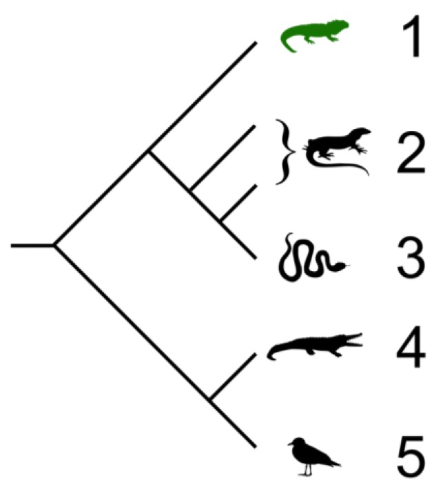
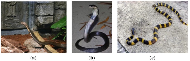
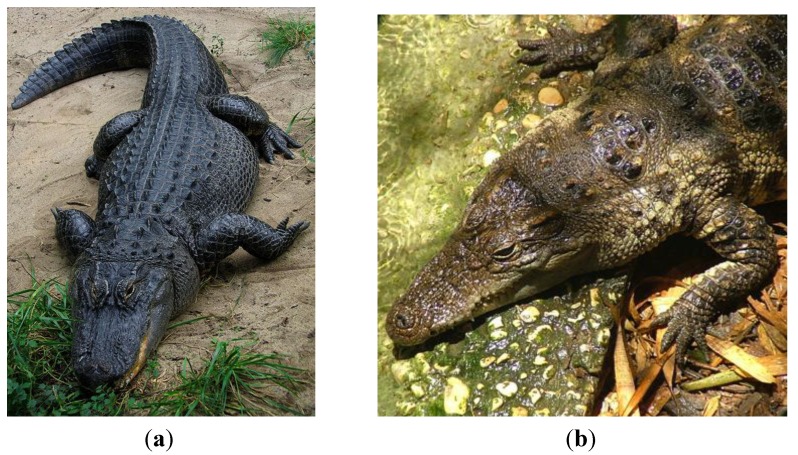
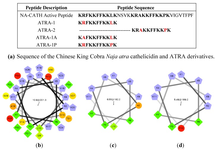
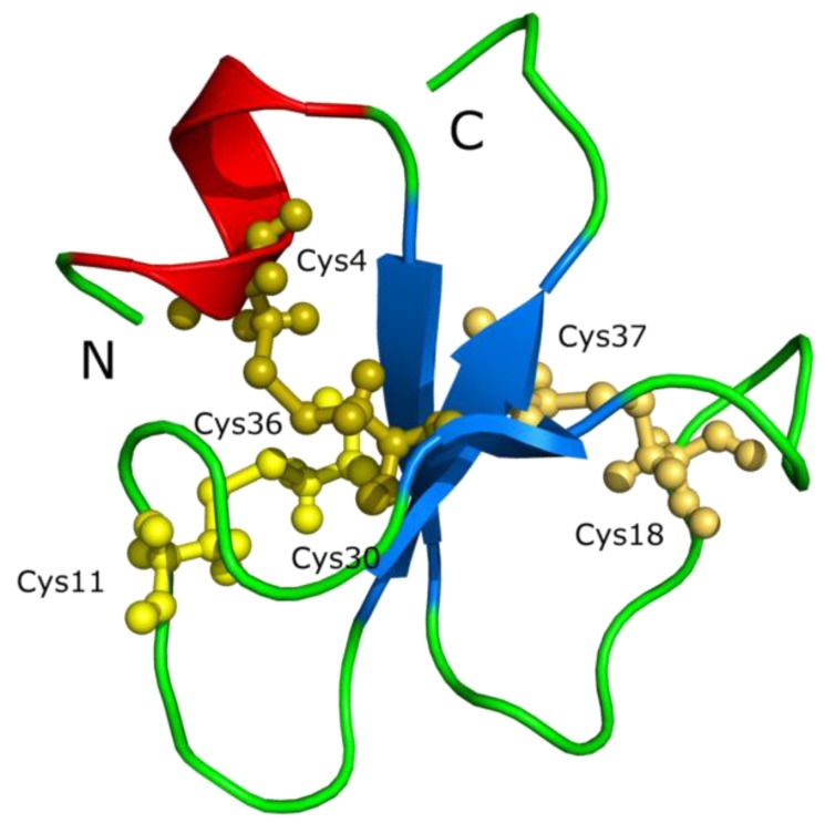
Reptiles are among the oldest known amniotes and are highly diverse in their morphology and ecological niches. These animals have an evolutionarily ancient innate-immune system that is of great interest to scientists trying to identify new and useful antimicrobial peptides. Significant work in the last decade in the fields of biochemistry, proteomics and genomics has begun to reveal the complexity of reptilian antimicrobial peptides. Here, the current knowledge about antimicrobial peptides in reptiles is reviewed, with specific examples in each of the four orders: Testudines (turtles and tortosises), Sphenodontia (tuataras), Squamata (snakes and lizards), and Crocodilia (crocodilans). Examples are presented of the major classes of antimicrobial peptides expressed by reptiles including defensins, cathelicidins, liver-expressed peptides (hepcidin and LEAP-2), lysozyme, crotamine, and others. Some of these peptides have been identified and tested for their antibacterial or antiviral activity; others are only predicted as possible genes from genomic sequencing. Bioinformatic analysis of the reptile genomes is presented, revealing many predicted candidate antimicrobial peptides genes across this diverse class. The study of how these ancient creatures use antimicrobial peptides within their innate immune systems may reveal new understandings of our mammalian innate immune system and may also provide new and powerful antimicrobial peptides as scaffolds for potential therapeutic development.


Figures

Figure 1
Figure 1
Cladogram showing the relationships of extant members of the Sauria (Sauropsida) which includes birds and reptiles. Branch lengths are not representative of divergence time. 1. Tuataras; 2. Lizards; 3. Snakes; 4. Crocodiles; 5. Birds. Cladogram by Benchill, licensed under the Creative Commons Attribution 3.0 Unported license [24].
Figure 2
Figure 2
Western Painted Turtle Chrysemys picta bellii. (a) Western painted turtle. Photo by Gary M. Stolz, U.S. Fish and Wildlife Service in the Public domain [26]. (b) Underside of a Western Painted Turtle. Photo by Matt Young [27].
Figure 3
Figure 3
Sphenodon punctatus, Tuatara, Nga Manu, Waikanae, New Zealand. Photo by PhillipC [39].
Figure 4
Figure 4
Elapid snakes (a) The King cobra (O. hannah) [44] (b) A juvenile Chinese cobra (N. atra) [45] (c) Banded Krait (B. fasciatus) [46].
Figure 5
Figure 5
Male Carolina Anole with partially expanded dewlap [47].
Figure 6
Figure 6
Crocodilians. (a) The American alligator, Alligator mississippiensis [52]. (b) The Siamese crocodile, Crocodylus siamesnsis [53].
Figure 7
Figure 7
Naja atra cathelicidin peptide analysis. (a) Sequences of the NA-CATH active peptide and derivatives [102,103,104,105]. (b) Helical wheel projection of NA-CATH. (c) Analysis of the active ATRA-1A peptide (d) Analysis of the inactive ATRA-1P peptide. From Rzlab.ucr.edu/scripts/wheel/wheel.cgi: “The hydrophilic residues are presented as circles, hydrophobic residues as diamonds, potentially negatively charged as triangles, and potentially positively charged as pentagons. Hydrophobicity is color coded as well: the most hydrophobic residue is green, and the amount of green is decreasing proportionally to the hydrophobicity, with zero hydrophobicitycoded as yellow. Hydrophilic residues are coded red with pure red being the most hydrophilic (uncharged) residue, and the amount of red decreasing proportionally to the hydrophilicity. The potentially charged residues are light blue.”
Figure 8
Figure 8
The crotamine chemical structure. Crotamine, a Na+ channel-affecting toxin from Crotalus durissus terrificus venom (PDB 1H5O) [125].

References

    1. Wang G. Antimicrobial Peptide Database. [(accessed on 9 May 2014)]. Available online: http://aps.unmc.edu/AP.
    1. Wang G., Li X., Wang Z. APD2: The updated antimicrobial peptide database and its application in peptide design. Nucleic Acids Res. 2009;37:D933–D937. doi: 10.1093/nar/gkn823. - DOI - PMC - PubMed
    1. Zhao H., Gan T.X., Liu X.D., Jin Y., Lee W.H., Shen J.H., Zhang Y. Identification and characterization of novel reptile cathelicidins from elapid snakes. Peptides. 2008;29:1685–1691. doi: 10.1016/j.peptides.2008.06.008. - DOI - PubMed
    1. Li S.A., Lee W.H., Zhang Y. Efficacy of OH-CATH30 and its analogs against drug-resistant bacteria in vitro and in mouse models. Antimicrob. Agents Chemother. 2012;56:3309–3317. doi: 10.1128/AAC.06304-11. - DOI - PMC - PubMed
    1. Zhang B.Y., Li S.M., Gao Z.H., Shen J.H. Protective effects of snake venom antimicrobial peptide OH-CATH on E. coli induced rabbit urinary tract infection models. Dong Wu Xue Yan Jiu. 2013;34:27–32. - PubMed