. 2014 Aug 21;512(7514):328-32.
doi: 10.1038/nature13428. Epub 2014 Jun 11.

Dynamic pathways of -1 translational frameshifting

Affiliations

Dynamic pathways of -1 translational frameshifting

Jin Chen et al. Nature. .

Abstract

Spontaneous changes in the reading frame of translation are rare (frequency of 10(-3) to 10(-4) per codon), but can be induced by specific features in the messenger RNA (mRNA). In the presence of mRNA secondary structures, a heptanucleotide 'slippery sequence' usually defined by the motif X XXY YYZ, and (in some prokaryotic cases) mRNA sequences that base pair with the 3' end of the 16S ribosomal rRNA (internal Shine-Dalgarno sequences), there is an increased probability that a specific programmed change of frame occurs, wherein the ribosome shifts one nucleotide backwards into an overlapping reading frame (-1 frame) and continues by translating a new sequence of amino acids. Despite extensive biochemical and genetic studies, there is no clear mechanistic description for frameshifting. Here we apply single-molecule fluorescence to track the compositional and conformational dynamics of individual ribosomes at each codon during translation of a frameshift-inducing mRNA from the dnaX gene in Escherichia coli. Ribosomes that frameshift into the -1 frame are characterized by a tenfold longer pause in elongation compared to non-frameshifted ribosomes, which translate through unperturbed. During the pause, interactions of the ribosome with the mRNA stimulatory elements uncouple EF-G catalysed translocation from normal ribosomal subunit reverse-rotation, leaving the ribosome in a non-canonical intersubunit rotated state with an exposed codon in the aminoacyl-tRNA site (A site). tRNA(Lys) sampling and accommodation to the empty A site and EF-G action either leads to the slippage of the tRNAs into the -1 frame or maintains the ribosome into the 0 frame. Our results provide a general mechanistic and conformational framework for -1 frameshifting, highlighting multiple kinetic branchpoints during elongation.


Figures
Extended Data Figure 1
Extended Data Figure 1. Explanation and schematics of the experimental signals
(a) Different stages of the elongation cycle during translation. Frameshifting have been proposed to occur at each of the steps: (1) during accommodation of the A-site tRNA, (2) subsequent to accommodation, but prior to peptide bond formation, (3) during tRNA hybrid-state intermediates, (4) during translocation, and (5) at the start of the next found of elongation. (b) The ribosome starts each round of elongation in the nonrotated state. In this “locked” state, the P-site tRNA is stably bound in the classical state, preserving the reading frame of the mRNA. Upon A-site tRNA selection and peptide bond formation, the 30S subunit rotates 3~10° counterclockwise with respect to the 50S subunit to the rotated state (pre-translocation),,. This “unlocked” state permits tRNA motions and the tRNA can fluctuate freely between the classical state and hybrid state, thus facilitating translocation of tRNA and movement of ribosome by one codon over mRNA,. Peptide-bond formation also triggers spontaneous fluctuations of the L1 stalk between open and closed conformations as well as spontaneous rotations in ribosome conformations,. EF-G then catalyzes translocation, and the ribosome returns to the nonrotated state (post-translocation). To monitor the rotational state of the ribosome in real time, we employed FRET between the small (30S) and large (50S) subunits. The 30S subunit was site-specifically labeled with Cy3B on helix 44, and a nonfluorescent quencher, BHQ-2, was placed on helix 101 of 50S subunit,,. Reagent delivery of BHQ-50S, tRNA ternary complex, and EF-G to surface immobilized Cy3B-30S pre-initiation complexes in ZMWs results in IF2-guided 70S assembly during initiation and establishment of FRET between the two ribosomal subunits: upon subunit joining, the green (Cy3B) intensity drops, which is followed by alternating low-high-low intensities Each alternating cycle corresponds to the ribosome translating a single codon, with the two intensity states consistent with the two rotational states of the ribosome: the low intensity state (high FRET) defining the nonrotated (locked) ribosome conformation and the high intensity state (low FRET) the rotated (unlocked) conformation. The rotated- and nonrotated-state lifetimes at each codon can be statistically analyzed. (c) During each cycle of elongation, the ribosome selects the aminoacyl-tRNA in a ternary complex with EF-Tu-GTP, and positions the tRNA in the A site. Upon A-site tRNA accommodation, the ribosome rapidly catalyzes peptide bond formation with the P-site tRNA,. Translocation moves A- and P-site tRNA-mRNA complexes to the E and P site respectively, catalyzed by the EF-G. The compositional dynamics of tRNA and EF-G on the ribosome, here defined as the relative timing of their arrival and departure during elongation, can be observed by labeling the tRNA or EF-G with Cy3 or Cy5. Cy5/Cy3-tRNA arrival to the surface immobilized ribosomes is marked by a red/green fluorescent pulse. Translation can be monitored by the arrival and departure of dye-labeled tRNA. Each productive tRNA binding event results in a fluorescence pulse that lasts as a ribosome translates 2 codons – beginning with arrival of tRNA in the A-site, continuing through tRNA translocation to the P-site, arrival of A-site tRNA to the next codon, a second round of translocation, and ending with spontaneous dissociation of tRNA from the E site. To track tRNA and EF-G dynamics on translating ribosomes at near-physiological concentrations of fluorescent factors (0.1 – 1 µM), we used ZMWs to detect hundreds of individual ribosomes (d) Substituting the traditional FRET acceptor, Cy5, with BHQ-2 allowed the use of Cy5 to label other translation components for correlation studies. The Cy3B intensity reports on the conformational state of the ribosome, while Cy5 pulses indicate arrival, occupancy, and departure of ribosomal ligands.
Extended Data Figure 2
Extended Data Figure 2. Phe codon in the −1 frame confirms the characteristic long pause during frameshifting
(a) Histogram of the fraction of ribosomes translating to a particular codon for the dnaX −1 frameshift mRNA, with a schematic. Many of the ribosomes translate up to 12 codons where the 0 frame stop codon is, though a large percentage of ribosomes translate up to 9 codons, where the −1 frame stop codon is. There are also ribosomes that stall at codon 7, limited by Cy3B photobleaching or end of movie (8 minutes) from the long rotated state pause. Interestingly, there are also a significant number of ribosomes that stall at codon 8 (see Extended Data Fig. 10 for discussion). By parsing the number of ribosomes that translate beyond codon 9 and up to codon 9, the frameshifting percentage can be calculated (75%). However, since non-frameshifted ribosomes may terminate early, this would lead to a slight over-estimate of the frameshifting percentage (3~10%). The frameshifting efficiency has been independently confirmed using a Cy5-tRNAPhe score, as described below. Number of molecules analyzed n = 256. (b) A UUC(Phe) codon is introduced in the −1 frame downstream of the slippery site. Frameshifting can be scored by an appearance of a Cy5 (red) pulse with Cy5-tRNAPhe in addition to the Cy3B/BHQ conformational FRET signal. This allows us to independently score for frameshifting. (c) Using the Cy5-tRNAPhe as a score to confirm frameshifting, we get the same dynamics and lifetimes: the nonrotated state lifetimes remain constant at each codon, and the rotated state lifetime increases 10-fold at codon Lys7 at the slippery sequence. This confirms and justifies our results in Figure 1. Number of molecules analyzed n = 474. Error bars, s.e. (d) By using the Cy5-tRNAPhe as a score, we can parse the rotated state lifetimes into ribosomes that frameshifted and ribosomes that did not frameshift. We also get the same results as Figure 1: non-frameshifted ribosomes translate through the frameshift sequence seemingly unaffected; frameshifted ribosomes exhibit the characteristic long-rotated state pause at codon Lys7. Number of molecules analyzed n = 474. Error bars, s.e.
Extended Data Figure 3
Extended Data Figure 3. Hairpin and the internal Shine-Dalgarno sequence are important for frameshifting
(a) mRNA sequence of the no hairpin (no HP) mutant. The mRNA consists of the same sequence as the wild-type dnaX frameshift sequence, but with the sequence after the UGA stop codon in the 0 frame in the hairpin deleted. (b) Nonrotated and rotated state lifetimes in the presence of 80 nM EF-G and 1 µM tRNAtot. The nonrotated state lifetimes are constant at each codon. There is an increase in rotated state lifetime at codon Lys7. Number of molecules n = 124. Error bars, s.e. (c) mRNA sequence of the no Shine-Dalgarno (no SD) mutant. The mRNA consists of the same sequence as the wild-type dnaX frameshift sequence, but with the original internal Shine-Dalgarno sequence GGGAGC mutated to AGGCGC, decreasing the rRNA-mRNA interaction energy from −3.00 kJ/mol to −0.06 kJ/mol. (d) Nonrotated and rotated state lifetimes in the presence of 80 nM EF-G and 1 µM tRNAtot. The nonrotated state life is constant. There is an increase in rotated state lifetime at codon Lys7. Number of molecules n = 225. Error bars, s.e. (e) Frameshifting percentages of the no SD and no HP mutant. Without the Shine-Dalgarno sequence, frameshifting percentage drops by half. Without the hairpin, frameshifting percentage drops to a quarter of the wild-type sequence. This indicates that both the internal Shine-Dalgarno sequence and the hairpin are required for efficient frameshifting, confirming that the hairpin and internal SD are stimulatory elements for frameshifting. These stimulatory elements may present a barrier and tension to translocation that is a prerequisite for efficient frameshifting.
Extended Data Figure 4
Extended Data Figure 4. Hairpin and internal Shine-Dalgarno sequences increases the energy barrier to translocation
(a) Translation of a short linear mRNA, 6(FK), in the presence of 80 nM EF-G and 1 µM tRNAtot, with an example trace. (b) Histogram of fraction of ribosomes translating to a particular codon. Most of the ribosomes translate up to 12 codons. Ribosomes translate <12 codons are due to photobleaching of the Cy3B dye, or non-processive ribosomes. This gives us a background level of 3~10% for our frameshifting efficiency analysis. The small number of ribosomes that translate beyond codon 12 are likely errors in our statistical analysis or read-through of the stop codon. Number of molecules analyzed n = 462. (c) Rotated and nonrotated state lifetimes are fairly constant. Number of molecules analyzed n = 462. Error bars, s.e. (d) Translation of a Phe-Lys sequence preceded by an internal Shine-Dalgarno sequence (same SD sequence used in the dnaX frameshift mRNA of this study) in the presence of 80 nM EF-G and 1 µM tRNAtot, with an example trace. (e) Histogram of fraction of ribosomes translating to a particular codon. Number of molecules analyzed n = 60. (f) There is an increase in rotated state lifetime at codon 5 ~ 7. There is an increase in the rotated state lifetimes 3~4 fold over 3~5 codons downstream of the SD-like sequence, while the non-rotated state lifetime remains unaffected. The internal SD-like sequences may base pair with the 3’ end of the 16S rRNA and slow down ribosomes in the pre-translocation state, echoing several work done previously by tracking ribosome movement and ribosome profiling,. Number of molecules analyzed n = 60. Error bars, s.e. (g) Translation of a Phe-Lys sequence followed by a hairpin (same hairpin used in the dnaX frameshift mRNA of this study) in the presence of 80 nM EF-G and 1 µM tRNAtot, with an example trace. (h) Histogram of fraction of ribosomes translating to a particular codon. Number of molecules analyzed n = 332. (i) Nonrotated state lifetimes are fairly constant. There is an increase in rotated state lifetime at codon 5, exactly 3 codons before the start of the hairpin, placing the ribosome directly at the first encounter of the hairpin. The relative position also matches where we see the long-rotated state pause during frameshift. This echoes the work done by Tinoco et al. where they showed the ribosome is capable of translating through the secondary structure through two mechanisms: ribosome translocating when encountering an open-state junction, occurring naturally or induced by the ribosome, or mechanically unwinding by the ribosome when encountering a closed-state junction. When the ribosome encounters an open-state junction, translation proceeds at a constant rate; however, when a closed-state junction is encountered, the ribosome actively unwinds the secondary structure, resulting in a slight waiting time for translocation, after which the hairpin is biased by the ribosome into an open-state and translation occurs normally. The shunt to either pausing in the rotated state (which leads to uncoupled translocation) or normal translation during frameshifting is likely due to this mechanism. Number of molecules analyzed n = 332. Error bars, s.e.
Extended Data Figure 5
Extended Data Figure 5. Dynamics of frameshifting at different factor concentrations
(a) Example trace and schematic of a ribosome translating the dnaX frameshift mRNA at much higher factor concentrations (6 µM tRNAtot and 480 nM EF-G). (b) Frameshifting efficiency does not depend on EF-G and tRNAtot concentrations. (c) Increasing the tRNAtot and EF-G concentrations two-fold (from 1 µM tRNAtot and 80 nM EF-G to 2 µM tRNAtot and 160 nM EF-G) decreases both the rotated state lifetime and nonrotated state lifetimes. This confirms that our conformational FRET signal depends correctly on factor concentration. The lifetime of the long rotated state pause at codon Lys7 is also decreased by half, indicating that EF-G and/or tRNA interacts with the ribosome in that state. Number of molecules analyzed n = 256, n = 234. Error bars, s.e. (d) Increasing the tRNAtot concentration (from 1 µM to 3 µM) while keeping EF-G concentrations constant decrease the non-rotated state lifetimes 3-fold as expected. The rotated state lifetimes remain the same except for codon Lys7; this is expected because the rotated state lifetime depends only on concentration of EF-G. Unexpectedly, the rotated state lifetime at codon Lys7 is also slightly decreased, suggesting a linkage between tRNA and EF-G dynamics at that long rotated-state stall. This echoes our results in Figure 2 that tRNA (tRNALys in this case) samples the uncoupled rotated-state after translocation, and the tRNA sampling and accommodation may help to re-establish the ribosome’s reading frame and reverse-rotate subsequently. Thus, increasing tRNA concentrations (especially tRNALys in this case) will decrease the long rotated-state lifetime. Number of molecules analyzed n = 526. Error bars, s.e. (e) Increasing the EF-G concentration (from 80 nM to 240 nM) while keeping tRNAtot concentration constant decreases the rotated state lifetime 3-fold. The nonrotated state lifetime, which depends on the tRNA concentration, remains the same. However, the decrease in the rotated lifetime at codon Lys7 is only around 2-fold, rather than 3-fold as expected. This echoes our result in Figure 2 as well as part (b) above, suggesting that tRNA sampling also plays a role at this codon. Number of molecules analyzed n = 314. Error bars, s.e. (f) Increasing the EF-G and tRNAtot concentrations further to 6 µM tRNAtot and 480 nM EF-G further decreases the rotated and nonrotated state lifetimes. However, the rotated state lifetime at codon Lys7 remains the same when compared with 2 µM tRNAtot and 160 nM EF-G. The long tRNALys sampling events observed in Figure 2 may be contributing to the long rotated state lifetime at codon Lys7.
Extended Data Figure 6
Extended Data Figure 6. Slippery sequence mutation (A21GA24G) decreases frameshifting percentage
(a) Example trace of a ribosome translating the A21GA24G mutant mRNA in the presence of 80 nM EF-G and 1 µM tRNAtot. There seems to be a slightly longer pause at codon Lys7. (b) Histogram of the fraction of ribosomes translating to a particular codon for the dnaX −1 frameshift A21GA24G mRNA. Most of the ribosomes translate up to 12 codons where the 0 frame stop codon is. The buildup of ribosomes stalled at codon 9 present during frameshifting disappears. By parsing the number of ribosomes that translate beyond codon 9 and up to codon 9, the frameshifting percentage can be calculated (12%). (c) The rotated-state lifetime. The long stall at Lys7 is decreased with the slippery site mutant, suggesting that the extra-long pause is indeed a result of frameshifting. The slight increase in lifetime at Lys7 is due to the effects of the hairpin and internal Shine-Dalgarno sequence. Number of molecules analyzed n = 230. Error bars, s.e. (d) A UUC(Phe) is introduced in the −1 frame downstream of the slippery site of the A21GA24G mutant, similar to above. The A21GA24G mutation is known to decrease frameshifting efficiency down to background levels. (e) The nonrotated state lifetime and rotated-state lifetime match with our results using codon counting (see above). In the absence of frameshifting, there is still an increase in rotated state lifetime at codon Lys7, due to the increased energy barrier to translocation by the hairpin and internal Shine-Dalgarno sequence, though this increased lifetime is still much less than the Lys7 rotated state lifetime during frameshifting. Number of molecules analyzed n = 538. Error bars, s.e. (f) Using Cy5-tRNAPhe as a score for frameshifting, frameshifting percentage matches with our previous results. The slippery sequence A21GA24G mutant decreases frameshifting percentage down to background levels. Number of molecules analyzed n = 474, n = 538.
Extended Data Figure 7
Extended Data Figure 7. tRNA dynamics during frameshifting with the dnaX GCA(Ala) to GUA(Val) mutant mRNA
(a) The 3 nucleotides upstream of the slippery sequence (GCA(Ala)) is mutated to GUA(Val) (named the C20U mutant) so that E-site tRNA dynamics can be observed during frameshifting since tRNAVal can be labeled with Cy3-maleimide (see Figure 2). This allows us to estimate the time to translocation during the long rotated-state pause at codon Lys7, since translocation of the Cy3-tRNAVal from the P-site to the E-site leads to rapid departure of the tRNAVal and disappearance of the Cy3 signal. We want to make sure that the C20U mutation does not affect frameshifting dynamics. The nonrotated state and rotated state lifetimes, as well as frameshifting percentages, are consistent with what we have observed before for the wild-type sequence. Number of molecules analyzed n = 266. Error bars, s.e. (b) tRNA-tRNA FRET between the Cy3-tRNAVal in the P site and the incoming Cy5-tRNALys at Lys7 in the A site at the slippery sequence. The tRNAs are in a hybrid state upon encountering of hairpin and engagement with the internal Shine-Dalgarno sequence. After translocation, the Cy3-tRNAVal departs from the ribosome, resulting in a disappearance of FRET. After translocation and uncoupling with ribosome reverse-rotation, the now P-site tRNALys is likely in a hybrid, or “distorted” conformation, according to the structure by Namy et al. Number of molecules analyzed, top n = 227, bottom n = 337.
Extended Data Figure 8
Extended Data Figure 8. tRNALys transit and sampling dynamics
(a) Example trace of Cy5-tRNALys transit during translation of the dnaX frameshift mRNA, indicating the definition of pulse lifetime and time between pulse. (b) The time between tRNALys pulse and lifetime of each pulse for the first three Lys codons are consistent with what is expected, and decrease expectedly with the increase of EF-G concentration and tRNAtotal(ΔLys) concentration ([Cy5-tRNALys]= 200 nM). The time between pulse for the first Lys pulse corresponds to the decoding of the first codon Lys1 of the mRNA after 50S subunit joining to the immobilized 30S, which is short as expected and does not depend on factor concentration. The second Lys pulse has a longer time between pulse, corresponding to the ribosome translating four codons from Lys1 to Lys5. The third pulse has a slightly shorter time between pulse, corresponding to the ribosome translating from codon Lys5 to Lys7. The lifetimes for the first two pulses are short, since the ribosomes decode and translocate the corresponding codons normally. The lifetime for the third pulse at codon Lys7 is long, corresponding to the ribosome at the long rotated-state pause during frameshifting. Number of molecules analyzed, n = 179 and n = 212. Error bars, s.e. (c) Mean number of additional tRNALys sampling pulses to the long rotated-state pause at codon Lys7, sampling lifetimes, and sampling arrival times, at various concentrations of EF-G and tRNAtot ([Cy5-tRNALys] = 200 nM) for ribosomes translating the dnaX −1 wild-type frameshifting sequence. There is a mean number of ~2.3 sampling tRNALys pulses, which remains constant at the various factor concentrations. There seems to be an interplay (competition) between EF-G and the other tRNAs on tRNALys sampling. Increasing the concentration of other factors increase the arrival time for tRNALys, probably because they are all competing for the ribosomal A site. Number of molecules analyzed, from left to right, n = 179, n = 212, n = 180, n = 162. Error bars, s.e. (d) The time between tRNALys pulse and lifetime of each pulse for the first three Lys codons are the same for the wild-type AAG sequence and the AAG(AAA) mutant sequence. Number of molecules analyzed n = 212 and n = 454. Error bars, s.e. (e) By translating the AAG(AAA) mutant in the presence of Cy5-tRNALys, we see similar dynamics as the dnaX wild-type sequence. Cy5-tRNALys samples the A site at codon Lys8 after uncoupled translocation at the frameshift site. The fraction of ribosomes exhibiting >4 tRNALys pulses are the same for the wild-type sequence and the AAG(AAA) mutant. The mean number of tRNALys sampling pulses, the mean arrival time, and the mean lifetimes of the sampling pulses to the long rotated-state stall are the same for the AAG(AAA) mutant and the dnaX wild-type sequence. Number of molecules analyzed, n = 212, n = 454. Error bars, s.e.
Extended Data Figure 9
Extended Data Figure 9. tRNA sampling dynamics and slippage during frameshifting
(a) Example traces of Cy5-tRNAPhe (red) sampling to the A-site Phe8 codon during the long rotated-state pause correlated with Cy3B/BHQ conformational FRET signal (green) for the AAG(UUU) mutant. (b) By translating the AAG(UUU) mutant in the presence of Cy5-tRNAPhe (red) and correlating with the Cy3B/BHQ conformational FRET signal (green), we can observe the fraction of ribosomes exhibiting only 1 Cy5-tRNAPhe pulse or >1 Cy5-tRNAPhe pulse sampling to the long rotated state pause at codon Lys7. There is a significant number of ribosomes exhibiting >1 Cy5-tRNAPhe pulse even when there is only one Phe codon, suggesting that even without frameshifting, many of the ribosomes still pause in an uncoupled rotated state at Lys7, where tRNAPhe samples the exposed UUU codon in the A site. Number of molecules analyzed n = 106. (c) The arrival time of the first tRNA sampling to the long stalled codon for wild-type mRNA (with Cy5-tRNALys) and AAG(UUU) with Cy5-tRNAPhe are the same. Although frameshifting in principle could occur through an incomplete +2 translocation, with weakened codon-anticodon-ribosome interactions and the final reading-frame determined through the Lys8 tRNALys sampling in the −1 frame, our data support +3 translocation that weakens codon-anticodon-ribosome interactions followed by tRNA sampling and accommodation that defines the reading frame and shifts −1 through effects of the hairpin and internal-SD interaction. For +2 translocation, we would expect to see tRNA sampling to both −1 frame and the 0 frame of A-site codon. For the AAG(UUU) mutant, tRNAPhe will sample the 0 frame U25U26U27, while tRNAIle will sample the −1 frame A24U25U26. Since, Cy5-tRNAPhe arrival times and lifetimes for sampling to the AAG(UUU) mutant match Cy5-tRNALys arrival times and lifetimes for the wild-type sequence, there is likely no competition between tRNAPhe and tRNAIle suggesting that the AUU codon is not initially exposed for tRNAIle sampling. Furthermore, for the +2 model, we would not expect a AAG(UUU) mutation to lead to a decrease in frameshifting efficiency. Thus, our results favor a +3 translocation followed by a −1 shift driven by sampling, accommodation and base-pairing stability. Unfortunately, our single-molecule assay is blind to the actual movement of the ribosome on the mRNA, so the details of this mechanism will require further exploration. See Extended Data Fig. 10 for possible implications for heterogeneous frameshift products observed previously. Error bars, s.e. Number of molecules analyzed n = 212, n = 106. (d) Mean sampling lifetime and mean sampling arrival time for Cy5-tRNAPhe to the Phe8 codon for the AAG(UUU) mutant. The arrival time and lifetime are the same as Cy5-tRNALys sampling to the Lys8 codon for the dnaX wild-type sequence. Number of molecules analyzed n = 106. (e) Example trace for Cy5-tRNALys transit through the dnaX frameshift mRNA AAG(UUU) mutant. For the AAG(UUU) mutant, we see only three Lys pulses, as expected, since the fourth Lys codon (Lys8) is mutated to a Phe codon. Most of the ribosomes (~80%) exhibit only three Cy5-tRNALys pulses, indicating that the additional Cy5-tRNALys sampling pulses we saw characteristic of frameshifting are indeed sampling to the Lys8 codon. Sampling now is by tRNAPhe, which are dark and invisible to our observations. The time between pulses are consistent with both the wild-type sequence and AAG(AAA) mutant. The lifetime of the third pulse (at Lys7) is long, consistent with the long pause at that codon. Number of molecules analyzed n = 318. Error bars, s.e. (f) Example trace for Cy5-tRNALys transit through the dnaX frameshift mRNA AAG(AAC) mutant. For the AAG(AAC) mutant, we see mostly only three Lys pulses (~75%) since the fourth Lys codon (Lys8) is mutated to a Asn codon. Number of molecules analyzed n = 406. This further argues against the +2 translocation model (see part (c)). For +2 translocation, we would expect to see long Lys-tRNA sampling to the −1 frame AAA codon, which is not observed significantly. The additional Cy5-tRNALys pulses have a shorter lifetime and longer arrival time when compared with the translation of the wild-type mRNA, suggesting that these pulses are non-cognate sampling to the AAC codon in the 0 frame or sampling unstably to the AAA codon in the −1 frame. Even though our data supports a +3 translocation followed by a −1 slippage, multiple frameshifting pathways probably occurs. The details of this mechanism will require further exploration.
Extended Data Figure 10
Extended Data Figure 10. Hetereogeneous frameshift products
(a) Two different protein products are possible after −1 frameshifting, dependent on whether peptide bond formation occurs during sampling in the −1 or 0 frame. For the first scenario, tRNA sampling to the last three nucleotides of the slippery sequence (YYZ) redefines the ribosome in the −1 frame (YYY), after which the tRNA dissociates to leave an empty A-site codon. After the long rotated state is reverse-rotated by EF-G, tRNAYYY decodes that codon normally, creating a frameshift product denoted by XXY-YYY. For the second scenario, peptide bond formation occurs after slippage of tRNAYYZ into the −1 frame; peptide-bond formation occurs slowly, since the P- and A-site tRNAs would likely not be positioned correctly in the rotated ribosomal conformation. EF-G would then normally and rapidly resolve the newly-created A/P hybrid state and the ribosome reverse-rotates. In this case, the frameshift product will be denoted by XXY-YYZ. (b) Histogram of the fraction of ribosomes translating to a particular codon for the dnaX −1 frameshift AAG(UUU) mRNA, with a schematic. Since the frameshifting percentage for the AAG(UUU) sequence is low, we see that most of the ribosomes translate up to 12 codons where the 0 frame stop codon is. Though, there is a significant number of ribosomes that translate to 11 codons (~25%), compared to ~5% for our previous experiments. There are two possible scenarios for tRNAPhe sampling to the long rotated-state pause to codon Phe8. In the first case, the tRNAPhe defines the reading frame and falls off, after which the ribosome resolves itself through the action of EF-G, followed by the normal decoding of Phe codon at codon 8. In this case, we get 12 cycles of low-high-low FRET intensity, and hence 12 codons translated by our signal. In the second case, the tRNAPhe defines the reading frame, followed by slow peptide bond formation. After peptide bond formation, the ribosome returns to the canonical hybrid and rotated state, for which EF-G then catalyzes reverse rotation. In this case, one low-high-low FRET cycle is missed, so we count 11 codons translated by our signal. This explains the heterogeneity in frameshift products observed in many frameshift systems. Number of molecules analyzed n = 353. (c) Example traces of Cy5-tRNAPhe (red) sampling to the long rotated-state pause at codon Lys7 correlated with Cy3B/BHQ conformational FRET signal (green), showing the two possible scenarios for tRNA sampling. Case 1 (as described in part (a)) leads to correlation of tRNA arrival and ribosome rotation after the long rotated state pause whereas case 2 leads to overlap of a tRNAPhe pulse with the reverse-rotation of the long pause. Both scenarios occur when translating the AAG(UUU) mutant, with ~58% of ribosomes for case 1 and 42% for case 2. For the 2nd case, the time between the last Cy5-tRNAPhe arrival and the ribosome reverse-rotation is 27.2 s, much longer than the 7.7 s during normal decoding and translocation, suggesting a slow peptidyltransfer reaction. Our results provide a possible explanation for why heterogeneous frameshifting products are observed in many frameshifting systems. Number of molecules analyzed n = 55.
Figure 1
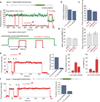
Figure 1. Frameshifting is characterized by a long rotated-state pause
(a) Schematic of the mRNA used in this study, modified from the dnaX gene. (b) Schematic of the Cy3B/BHQ conformational FRET signal, with each low-high-low Cy3B intensity cycle representing a ribosome elongating one codon. (c) Example traces of Cy3B (green) fluorescent intensity for frameshifted and non-frameshifted ribosomes translating with 80 nM EF-G and 1 µM tRNAtot TC. Codon Lys7 of the frameshift site is shaded yellow. (d) The mean rotated-state lifetime and nonrotated state lifetime. The nonrotated state lifetime is constant. There is a 10-fold increase in rotated state lifetime at codon Lys7. n = 256. Error bars, s.e. (e) By parsing the rotated state lifetimes in (c) into ribosomes that frameshift (75%) and ribosomes that do not frameshift (25%), we see only the rotated state pause at codon Lys7 for frameshifted ribosomes.
Figure 2
Figure 2. tRNA samples the rotated state after uncoupled translocation and defines the reading frame
(a) Sample trace and time to translocation at the frameshift site, which is estimated by Cy3-tRNAVal (green) departure from the E-site during frameshifting relative to the arrival of Cy5-tRNALys (red) at codon Lys7 (shaded in yellow) on a GCA21 (Ala) to GUA21 (Val) mRNA mutant. From left to right, n = 337, 449, 455. Error bars, s.e. (b) Example trace of correlation of the Cy3B/BHQ ribosome conformational signal (green) with Cy5-tRNALys (red), confirming the long pause at codon Lys7 (shaded yellow). Upon reaching codon Lys7, additional tRNALys pulses sample codon Lys8 in the A site (shaded red), which results in a buildup of Cy5 intensity from the two Cy5-tRNALys in the A and P sites of the ribosome. (c) Fraction of elongating ribosomes exhibiting >4 Lys pulses (additional sampling pulses) for the frameshift wild-type mRNA and the A21GA24G mutant. From left to right, n = 179 and n = 147. (d) Two-dimensional density plot of Cy5-tRNALys sampling to codon Lys8 in the A site post-synchronized to the time of translocation (indicated by the green line). The sampling of tRNALys at Lys8 only begins after translocation. (e) Rotated and nonrotated state lifetimes for the slippery sequence mutant (A25A26G27 codon to AAA). There is now an extra subpopulation of ribosomes with a long rotated-state pause but does not lead to frameshifting. n = 310. Error bars, s.e. (f) Mutation of the last A25A26G27 codon to UUU (Phe). Similar to (e), there are two subpopulations within the non-frameshifted ribosomes. n = 353. Error bars, s.e. (g) Pathways of frameshifting for the various slippery sequence mutants, indicating how tRNA sampling defines the final reading frame.
Figure 3
Figure 3. EF-G samples and resolves the uncoupled rotated state after frameshifting
(a) Sample trace and schematic of the correlation of Cy3B/BHQ ribosome conformational signal (green) with Cy5-EF-G binding (red). Cy5-EF-G pulses are correlated with ribosome reverse-rotation at each codon. At the rotated state pause at codon Lys7 (shaded in yellow), multiple EF-G sampling events with long dwell times can be observed. (b) EF-G sampling and EF-G lifetimes for each codon for the wild-type frameshift mRNA. There is an increased number of sampling as well as increased mean EF-G lifetime at codon Lys7. n = 122. Error bars, s.e. (c) For the A21GA24G mutant mRNA, there is a slight increase in number of sampling at Lys7 codon due to the increase in energy barrier from the hairpin and internal-SD interaction, but the long EF-G lifetime disappears. n = 157. Error bars, s.e. (d) Two-dimensional histogram plotting time at the stalled rotated state at codon Lys7 vs. lifetime of EF-G. Longer EF-G lifetimes only appear after uncoupled translocation, as roughly indicated by the red line. For the A21GA24G mutant, no long EF-G lifetimes are observed. n = 122, n = 157. (e) Postsynchronization plot correlating ribosome reverse-rotation at the Lys7 codon with EF-G. n = 436.
Figure 4
Figure 4. Branchpoint of pathways and mechanism of dnaX −1 frameshifting
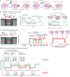
The first branchpoint during frameshifting is likely due to the stochastic interaction of the ribosome with the hairpin in an open or closed state, and/or formation of the SD-antiSD pairing, that represent the shunt to either pausing or normal translation. Translocation of the paused ribosomes under the slippery sequence with the tension caused by the hairpin and SD leads to +3 translocation, but uncoupled from reverse ribosomal rotation, creating a non-canonical intermediate in translation (denoted Rotated*). The uncoupled translocation exposes the A site, to which tRNALys and EF-G sample. tRNALys sampling and accommodation to the AAG codon stimulates the ribosome to slip into the −1 frame. Finally, EF-G catalyzes the final reverse-rotation, after which the ribosome resumes normal translation.

References

    1. Jenner LB, Demeshkina N, Yusupova G, Yusupov M. Structural aspects of messenger RNA reading frame maintenance by the ribosome. Nature structural & molecular biology. 2010;17:555–560. - PubMed
    1. Tinoco I, Jr, Kim HK, Yan S. Frameshifting dynamics. Biopolymers. 2013;99:1147–1166. - PMC - PubMed
    1. Tsuchihashi Z, Kornberg A. Translational frameshifting generates the gamma subunit of DNA polymerase III holoenzyme. Proceedings of the National Academy of Sciences of the United States of America. 1990;87:2516–2520. - PMC - PubMed
    1. Plant EP, et al. The 9-A solution: how mRNA pseudoknots promote efficient programmed −1 ribosomal frameshifting. RNA. 2003;9:168–174. - PMC - PubMed
    1. Baranov PV, Gesteland RF, Atkins JF. P-site tRNA is a crucial initiator of ribosomal frameshifting. RNA. 2004;10:221–230. - PMC - PubMed

Publication types