. 2014 Aug;20(8):1337-48.
doi: 10.1261/rna.044602.114. Epub 2014 Jun 11.

A homolog of lariat-debranching enzyme modulates turnover of branched RNA

Affiliations

A homolog of lariat-debranching enzyme modulates turnover of branched RNA

Stephen M Garrey et al. RNA. 2014 Aug.

Abstract

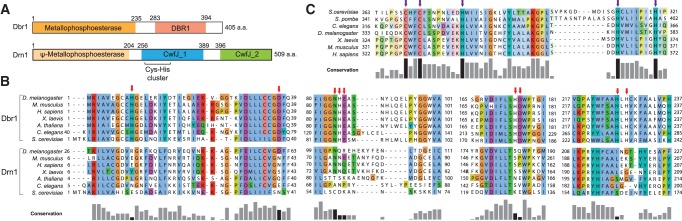
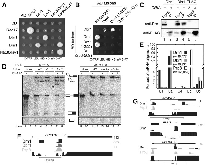
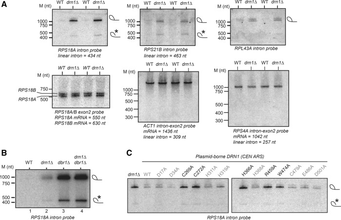
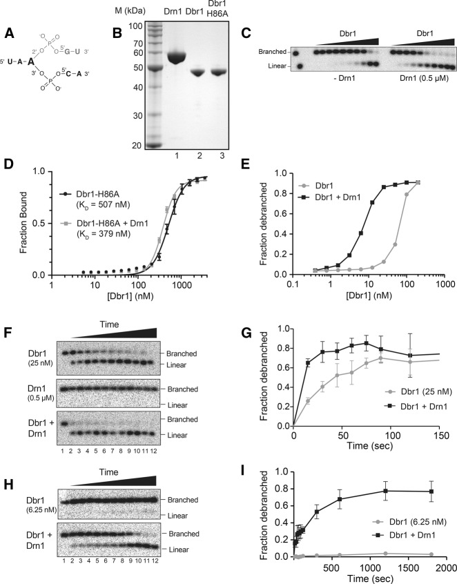
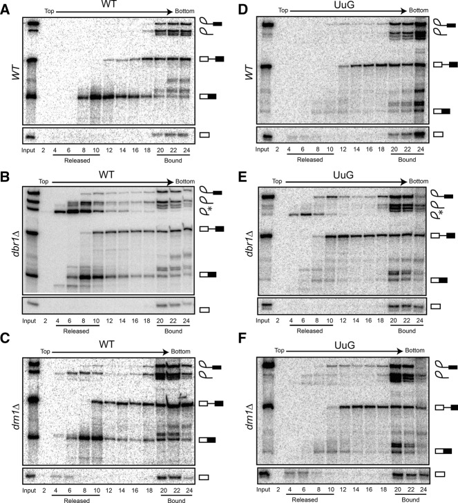
Turnover of the branched RNA intermediates and products of pre-mRNA splicing is mediated by the lariat-debranching enzyme Dbr1. We characterized a homolog of Dbr1 from Saccharomyces cerevisiae, Drn1/Ygr093w, that has a pseudo-metallophosphodiesterase domain with primary sequence homology to Dbr1 but lacks essential active site residues found in Dbr1. Whereas loss of Dbr1 results in lariat-introns failing broadly to turnover, loss of Drn1 causes low levels of lariat-intron accumulation. Conserved residues in the Drn1 C-terminal CwfJ domains, which are not present in Dbr1, are required for efficient intron turnover. Drn1 interacts with Dbr1, components of the Nineteen Complex, U2 snRNA, branched intermediates, and products of splicing. Drn1 enhances debranching catalyzed by Dbr1 in vitro, but does so without significantly improving the affinity of Dbr1 for branched RNA. Splicing carried out in in vitro extracts in the absence of Drn1 results in an accumulation of branched splicing intermediates and products released from the spliceosome, likely due to less active debranching, as well as the promiscuous release of cleaved 5'-exon. Drn1 enhances Dbr1-mediated turnover of lariat-intermediates and lariat-intron products, indicating that branched RNA turnover is regulated at multiple steps during splicing.

Keywords: Dbr1; Nineteen Complex; branched RNA; splicing.


Figures

FIGURE 1.
FIGURE 1.
Drn1 is a homolog of the lariat-debranching enzyme Dbr1. (A) Protein domains in Drn1 and Dbr1. The pseudo-metallophosphoesterase domain in Drn1 is homologous to the metallophosphoesterase domain of Dbr1. (B) Multiple sequence alignment showing N-terminal homology between the metallophosphoesterase regions of Dbr1 and Drn1. Essential catalytic residues in Dbr1 (Khalid et al. 2005) are marked with red arrows (note regions are discontinuous). (C) Multiple sequence alignment of a portion of the Drn1 CwfJ_1 domain showing evolutionary conservation of a series of Cys and His residues (purple arrows).
FIGURE 2.
FIGURE 2.
Drn1 interacts with Dbr1, components of the Nineteen Complex, U2 snRNA, and branched intermediates and products of pre-mRNA splicing. (A) Yeast two-hybrid analysis of Drn1 interactions. Proteins were tested as Gal4 activation (AD) and binding (BD) domain fusions on selective media. Mec3/Rad17 is a positive control, and Drn1 and Dbr1 interact as reciprocal AD/BD fusions. Drn1 interacts with Ntc90; the Ntc90-Ntc30 (Syf1-Isy1) interaction was reported previously (Dix et al. 1999). (B) Yeast two-hybrid analysis of Drn1 domain interactions. The Drn1 N-terminal domain (residues 1–255) and C-terminal domain (256–509) were tested as Gal4-AD and BD fusions against Dbr1-BD and Ntc90/Syf1-AD fusions. Strong interactions were observed between Drn1, Drn1-1-255, and Ntc90/Syf1, and weaker interactions we observed with Drn1, Drn1-1-255, and Dbr1. (C) Drn1 and Dbr1 interact in splicing extracts. Splicing extracts were prepared from wild-type (WT; lanes 1,2), Dbr1-Flag (lanes 3,4), and Dbr1-Flag drn1Δ (lanes 5,6) yeast strains. Inputs (5%; lanes 1,3,5) and anti-Flag immunoprecipitations (lanes 2,4,6) were analyzed by Western blotting with anti-Drn1 antibodies (top) and anti-Flag antibodies (bottom). Drn1 is specifically isolated in the Dbr1-Flag splicing extracts (lane 4). A nonspecific protein isolated by anti-Flag antibodies is marked with an asterisk. (D) Copurification of Drn1 and branched RNA intermediates. Anti-Drn1 antibodies were used to immunoprecipitate Drn1 from in vitro splicing reactions with WT, drn1Δ, and dbr1Δ extracts. IP − indicates 10% input; IP + indicates RNA recovered by immunoprecipitation with anti-Drn1 antibodies of remaining 90%. WT ACT1 pre-mRNA and a 3′-splice site mutant (UAG-to-UuG) were used in the assays. A black arrow indicates a faster-migrating RNA species likely derived from processing of lariat-intermediate or lariat-intron. (E) Summary of snRNA interactions observed in Drn1, Dbr1, and Prp8 CLIP-seq experiments (read numbers indicated in the legend; y-axis is discontinuous). Prp8 is crosslinked mainly to U5 snRNA, whereas Dbr1 and Drn1 crosslink to U2 snRNA. (F) CLIP-seq signals in the RPS17B intron. Sequencing coverage (i.e., reads per base) is indicated to the right of each track. Conservation among six sensu stricto yeast strains (Siepel et al. 2005) is indicated at the bottom in black. The peak of conservation indicated by the gray arrow is the branch site. Drn1, Dbr1, and Prp8 (Li et al. 2013) crosslink near the branch point sequence; Drn1 also crosslinks downstream from the 5′-splice site. (G) Drn1 and Prp8 CLIP-seq signals over the RPL43A, RPP1B, and RPS16A pre-mRNAs; annotations are as in F. No Dbr1 CLIP-seq signals were observed for any of these pre-mRNAs.
FIGURE 3.
FIGURE 3.
Loss of Drn1 causes accumulation of pre-mRNA splicing intermediates and products in vivo. (A) Pre-mRNA intermediate accumulation was measured by acrylamide Northern blot. Predicted positions of lariat-intron are indicated in the top panels. The indicated probes are specific for intron, exon2, or intron-exon2 junctions. A band comigrating with linear intron is marked with an asterisk, consistent with nicked lariat-intron. Sizes were estimated by staining of an RNA molecular weight standard excised prior to blotting. (B) Northern blot for RPS18A intron in cells lacking combinations of DRN1 and DBR1. Loss of drn1Δ causes mild accumulation of RPS18A lariat-intron, whereas dbr1Δ causes high levels of RPS18A lariat-intron accumulation independent of DRN1. A presumed nicked lariat-intron is marked with an asterisk. (C) Complementation of RPS18A intron accumulation in drn1Δ cells by plasmid-encoded DRN1 variants. WT DRN1 rescues the intron accumulation phenotype of drn1Δ. Alanine substitutions in the N-terminal Dbr1 domain (D17A, D43A) fail to disrupt Drn1 activity, whereas alanine substitutions at conserved positions in the CwfJ domains (C269A, C272A, H366A, R459A, W747A, black) are unable to complement RPS18A intron accumulation caused by drn1Δ.
FIGURE 4.
FIGURE 4.
Drn1 enhances Dbr1 debranching activity in vitro. (A) Structure of the 7-nt branched RNA substrate used in debranching assays. The 5-nt linear product of debranching is in black. (B) Coomassie-stained SDS-PAGE gel of purified recombinant Drn1 (lane 1), Dbr1 (lane 2), and Dbr1-H86A (lane 3). (C) Increasing amounts of Dbr1 (0, 0.4, 0.8, 1.6, 3.1, 6.3, 12.5, 25, 50, 100, and 200 nM) were incubated with 1 nM 32P-end-labeled substrate for 30 min at 22°C in the presence (right) or absence (left) of 0.5 µM Drn1 and analyzed by acrylamide gel electrophoresis. (Lanes 1,2) Standards for the branched and linear substrates. (D) Binding isotherms for complexes of Dbr1-H86A and a 7-nt branched RNA substrate were determined by filter binding in the absence of Drn1 (black; KD = 507 nM) and in the presence of 4 µM Drn1 (gray; KD = 379 nM). Error bars, SD of three independent experiments. (E) Quantitation of RNA debranching shown in C. (F) Time course of RNA debranching. End-labeled substrate (1 nM) was incubated at 22°C in the presence of 25 nM Dbr1 (top), 0.5 µM Drn1 (middle), or 25 nM Dbr1 and 0.5 µM Drn1 (bottom). Aliquots were removed at 15, 30, 45, 60, 75, 90, 120, 300, 600, 1200, and 1800 sec and analyzed by denaturing acrylamide gel electrophoresis. (G) Quantitation of cleavage shown in top and bottom panels of F; error bars, SD for three independent assays. (H) Time course of RNA debranching. End-labeled substrate (1 nM) was incubated in the presence of 6.25 nM Dbr1 (top) or 6.25 nM Dbr1 and 0.5 µM Drn1 (bottom). Aliquots were removed at time points as in F and analyzed by denaturing acrylamide gel electrophoresis. (I) Quantitation of cleavage shown in H; error bars, SD for three independent assays.
FIGURE 5.
FIGURE 5.
Dbr1 and Drn1 modulate branched RNA turnover during in vitro splicing. Splicing of ACT1 pre-mRNA was analyzed by glycerol gradient sedimentation. RNA in even-numbered fractions collected from the top (“released”) to the bottom (“bound”) of the gradient was analyzed on denaturing polyacrylamide gels. Positions of lariat-intermediate, lariat-intron, a faster-migrating intron form (marked with an asterisk, possibly a lariat lacking a tail), pre-mRNA, spliced mRNA, and 5′-exon are shown for each panel. Splicing of a WT ACT1 pre-mRNA was analyzed in WT (A), dbr1Δ (B), and drn1Δ (C) extracts. Splicing of WT ACT1 pre-mRNA in dbr1Δ and drn1Δ extracts causes accumulation of branched intermediates in released fractions (B,C, fractions 4–10). A faster-migrating intron (i.e., a lariat without a tail, marked with an asterisk) migrates just below lariat-intron (B, top panel, fractions 4–10). Splicing of WT pre-mRNA in drn1Δ extract causes modest accumulation of 5′-exon in released fractions (C, bottom panel, fractions 4–6). Splicing of a pre-mRNA with a 3′-splice site mutation (UAG to UuG) was analyzed in WT (D), dbr1Δ (E), and drn1Δ (F) extracts. Splicing of UuG substrates in drn1Δ extract enhances accumulation of released 5′-exon relative to WT extract (F, bottom panel, fractions 4–6).

Similar articles

Cited by

References

    1. Alberti S, Gitler AD, Lindquist S 2007. A suite of Gateway cloning vectors for high-throughput genetic analysis in Saccharomyces cerevisiae. Yeast 24: 913–919 - PMC - PubMed
    1. Arenas J, Hurwitz J 1987. Purification of a RNA debranching activity from HeLa cells. J Biol Chem 262: 4274–4279 - PubMed
    1. Barrett T, Troup DB, Wilhite SE, Ledoux P, Evangelista C, Kim IF, Tomashevsky M, Marshall KA, Phillippy KH, Sherman PM, et al.2010. NCBI GEO: archive for functional genomics data sets: 10 years on. Nucleic Acids Res 39: D1005–D1010 - PMC - PubMed
    1. Braich RS, Damha MJ 1997. Regiospecific solid-phase synthesis of branched oligonucleotides: effect of vicinal 2′,5′- (or 2′,3′-) and 3′,5′-phosphodiester linkages on the formation of hairpin DNA. Bioconjug Chem 8: 370–377 - PubMed
    1. Chang KJ, Chen HC, Cheng SC 2009. Ntc90 is required for recruiting first step factor Yju2 but not for spliceosome activation. RNA 15: 1729–1739 - PMC - PubMed

Publication types