. 2014 Aug;137(Pt 8):2193-209.
doi: 10.1093/brain/awu140. Epub 2014 Jun 11.

Connexin-43 induces chemokine release from spinal cord astrocytes to maintain late-phase neuropathic pain in mice

Affiliations

Connexin-43 induces chemokine release from spinal cord astrocytes to maintain late-phase neuropathic pain in mice

Gang Chen et al. Brain. 2014 Aug.

Abstract

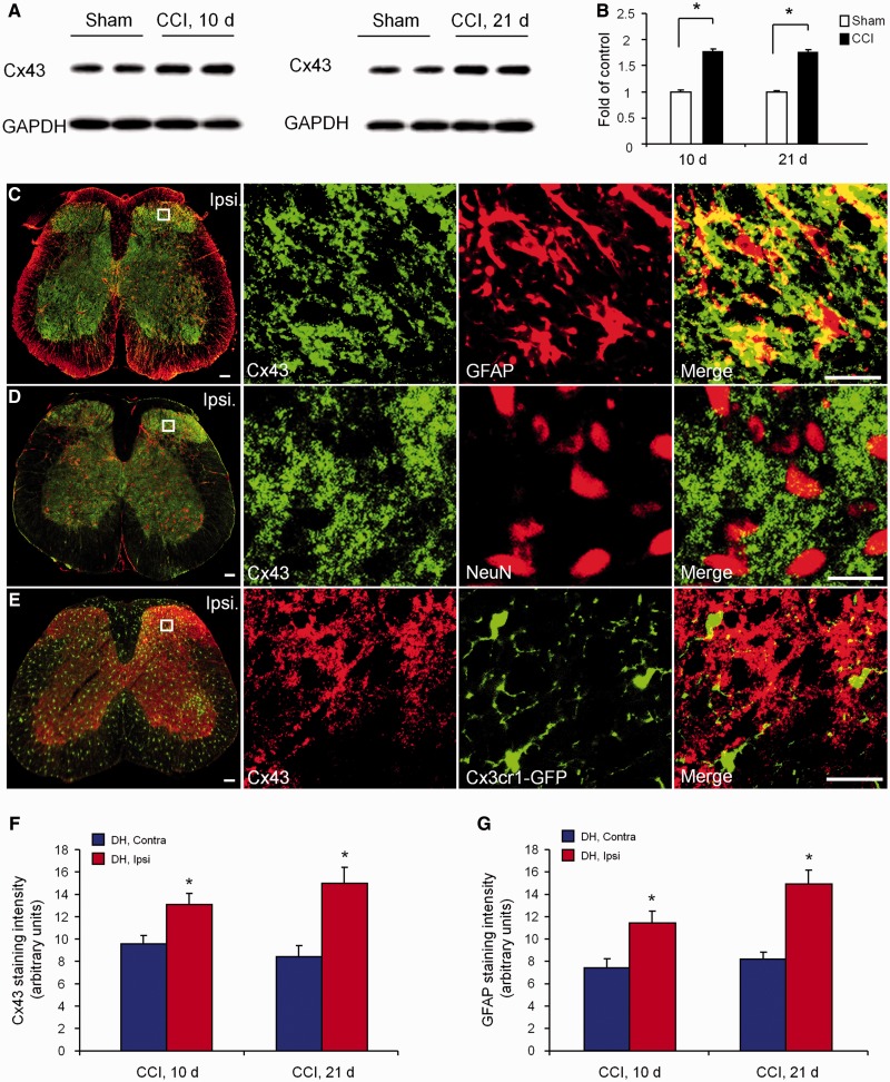
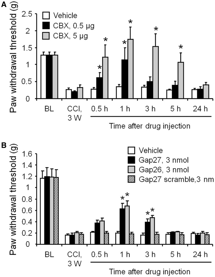
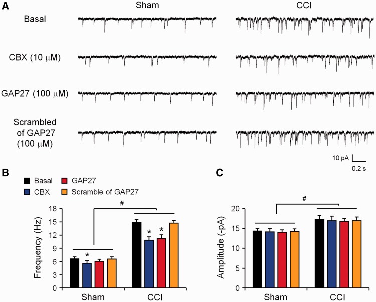
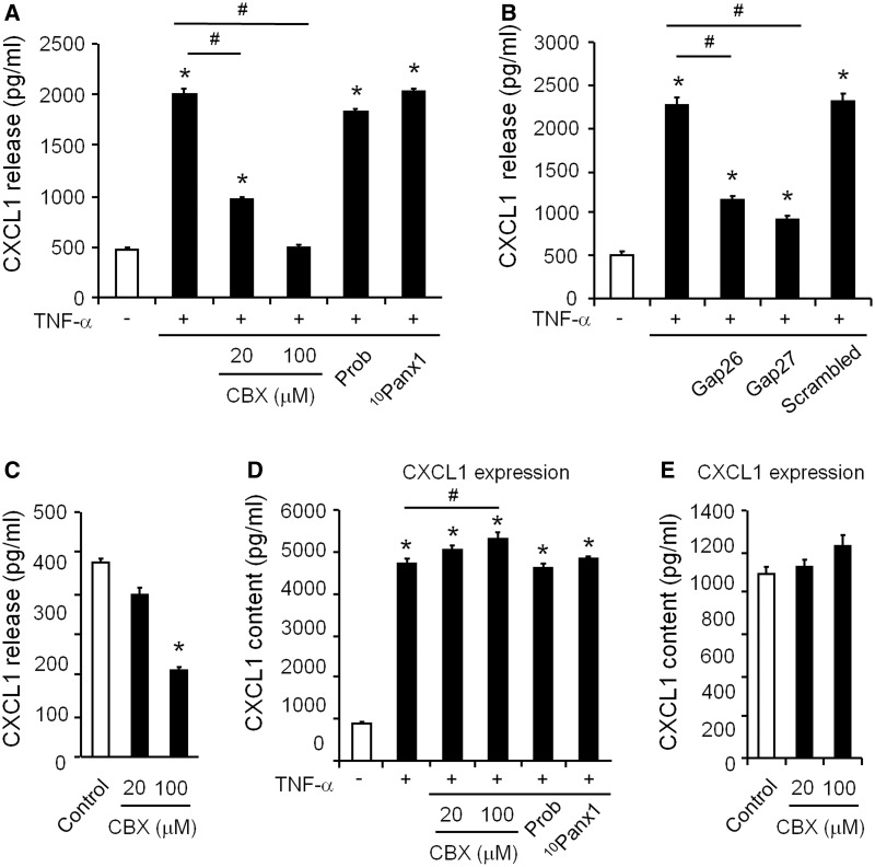
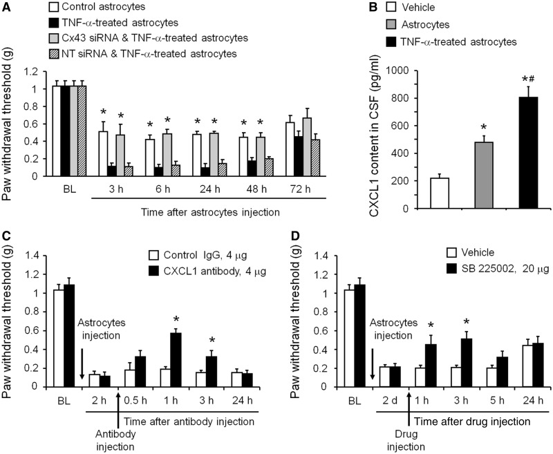
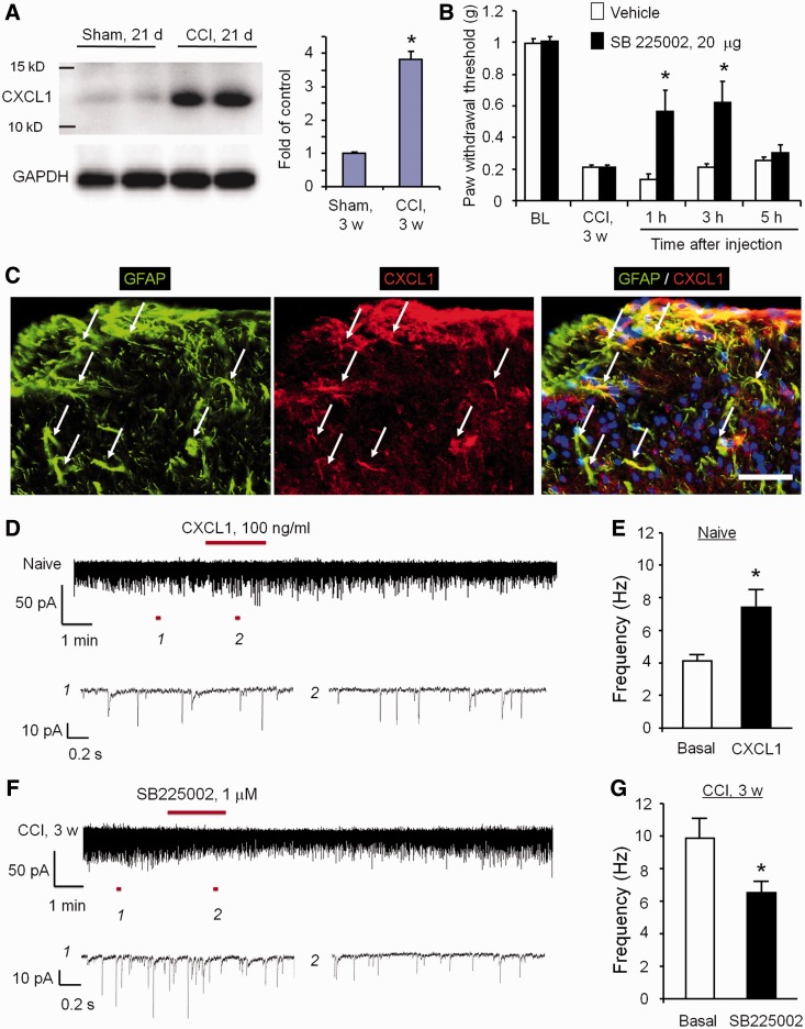
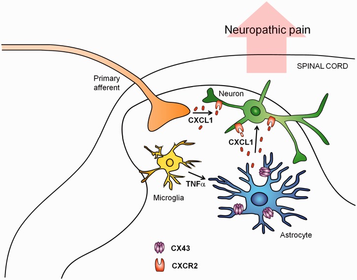
Accumulating evidence suggests that spinal cord astrocytes play an important role in neuropathic pain sensitization by releasing astrocytic mediators (e.g. cytokines, chemokines and growth factors). However, it remains unclear how astrocytes control the release of astrocytic mediators and sustain late-phase neuropathic pain. Astrocytic connexin-43 (now known as GJ1) has been implicated in gap junction and hemichannel communication of cytosolic contents through the glial syncytia and to the extracellular space, respectively. Connexin-43 also plays an essential role in facilitating the development of neuropathic pain, yet the mechanism for this contribution remains unknown. In this study, we investigated whether nerve injury could upregulate connexin-43 to sustain late-phase neuropathic pain by releasing chemokine from spinal astrocytes. Chronic constriction injury elicited a persistent upregulation of connexin-43 in spinal astrocytes for >3 weeks. Spinal (intrathecal) injection of carbenoxolone (a non-selective hemichannel blocker) and selective connexin-43 blockers (connexin-43 mimetic peptides (43)Gap26 and (37,43)Gap27), as well as astroglial toxin but not microglial inhibitors, given 3 weeks after nerve injury, effectively reduced mechanical allodynia, a cardinal feature of late-phase neuropathic pain. In cultured astrocytes, TNF-α elicited marked release of the chemokine CXCL1, and the release was blocked by carbenoxolone, Gap26/Gap27, and connexin-43 small interfering RNA. TNF-α also increased connexin-43 expression and hemichannel activity, but not gap junction communication in astrocyte cultures prepared from cortices and spinal cords. Spinal injection of TNF-α-activated astrocytes was sufficient to induce persistent mechanical allodynia, and this allodynia was suppressed by CXCL1 neutralization, CXCL1 receptor (CXCR2) antagonist, and pretreatment of astrocytes with connexin-43 small interfering RNA. Furthermore, nerve injury persistently increased excitatory synaptic transmission (spontaneous excitatory postsynaptic currents) in spinal lamina IIo nociceptive synapses in the late phase, and this increase was suppressed by carbenoxolone and Gap27, and recapitulated by CXCL1. Together, our findings demonstrate a novel mechanism of astrocytic connexin-43 to enhance spinal cord synaptic transmission and maintain neuropathic pain in the late-phase via releasing chemokines.

Keywords: CXCL1; CXCR2; carbenoxolone (CBX); hemichannels; neuro-glial interaction.


Figures

Figure 1
Figure 1
Nerve injury induces persistent upregulation of Cx43 in astrocytes of the spinal cord dorsal horn. (A) Cx43 expression in the spinal cord dorsal horn, as shown by western blotting, at 10 and 21 days after CCI and sham surgery. (B) Quantification of Cx43 levels in the dorsal horn. The western blot results are presented as a fold of sham control. *P < 0.05, compared with sham group, Student’s t-test, n = 4 mice/group. (C–E) Confocal images in the dorsal horn 21 days after CCI show co-localization of Cx43 with GFAP (red, C) but not with the neuronal marker NeuN (red, D) and microglial marker CX3CR1 (green, E). Scale bar = 100 µm. (F and G) Quantification of Cx43 (F) and GFAP (G) immunoflurorescence intensity in the ipsilateral (Ipsi) and contralateral (Contra) superficial dorsal horn (DH) 10 and 21 days after CCI. *P < 0.05, compared with the contralateral group, Student’s t-test, n = 4 mice/group. All data are mean ± SEM.
Figure 2
Figure 2
Spinal injection of CBX and Cx43 mimetic peptides 21 days after nerve injury reduces CCI-induced mechanical allodynia in the late phase. (A) Intrathecal injection of CBX (0.5 or 5 µg) rapidly (<0.5 h) and completely reversed mechanical allodynia for 5 h, in a dose-dependent manner. This inhibitory effect recovered after 24 h. (B) Intrathecal injection of Cx43 mimetic peptides (43Gap26, or 37,43Gap27) also reduced mechanical allodynia for 3 h. However, the scrambled peptide (Gap27 scrambled) had no effect. All data are mean ± SEM. The differences between groups were analysed by ANOVA. *P < 0.05, compared with vehicle, n = 6–7 mice/group.
Figure 3
Figure 3
Cx43 inhibition 21 days after nerve injury reverses CCI-induced increase in spontaneous EPSCs in lamina II neurons of spinal cord slices. (A) Traces of spontaneous EPSCs. (B) Frequency of spontaneous EPSCs. CCI induced profound increases in spontaneous EPSC frequency in the late-phase, which is suppressed by CBX (10 µM) and Gap27 (100 µM). (C) spontaneous EPSC amplitude was not affected by CBX and Gap27. Note that superfusion of the scrambled peptide has no effects on the frequency and amplitudes of spontaneous EPSCs. *P < 0.05, compared with corresponding baseline (basal); #P < 0.05, compared with sham surgery, ANOVA followed by Newman–Keuls test, n = 5 neurons/group.
Figure 4
Figure 4
Cx43 is required for TNF-α-evoked and basal release of CXCL1 in astrocyte cultures. (A and B) CXCL1 release in astrocytes following TNF-α stimulation (10 ng/ml, 60 min). Note the TNF-α-induced CXCL1 release is suppressed by pretreatment (60 min) of CBX (20 and 100 µM, A) and Gap26 and Gap27 (100 µM, B) but not by the inhibitors of pannexin hemichannels probenecid (Prob, 500 µM, A) and PANX1 mimetic peptide 10Panx1 (100 µM, A) and the scrambled peptide (Gap27 scrambled, 100 µM, B). *P < 0.05, compared with control; #P < 0.05, compared with TNF-α. (C) Inhibition of basal release of CXCL1 by CBX in astrocytes. *P < 0.05, compared with control. (D) Evoked expression (content) of CXCL1 in astrocytes following TNF-α stimulation (10 ng/ml, 60 min). Note the TNF-α-induced CXCL1 expression is not suppressed by pretreatment (60 min) of CBX (20 and 100 µM) and inhibitors of pannexin hemichannels probenecid (Prob, 500 µM) and PANX1 mimetic peptide 10Panx1 (100 µM). In contrast, a high dose of CBX (100 µM) increases CXCL1 expression. *P < 0.05, compared with control; #P < 0.05, compared with TNF-α. (E) Effects of CBX on the basal expression (content) of CXCL1 in astrocytes. All data are mean ± SEM. n = 8 cultures/group. The differences between groups were analysed by ANOVA followed by Newman–Keuls test.
Figure 5
Figure 5
TNF-α increases Cx43 expression and hemichannel activity in cultured astrocytes. (A) TNF-α (10 ng/ml, 3 h) induces Cx43 expression (western blotting) in astrocyte cultures. *P < 0.05, compared with control, Student’s t-test, n = 8 cultures/group. (B) Double staining showing increased Cx43 expression in GFAP-expressing astrocytes following the TNF-α treatment. Scale bar = 10 µm. (C) TNF-α treatment (10 ng/ml, 60 min) does not alter gap junction communication in astrocytes, revealed by diffusion of Lucifer yellow following microinjection into an astrocyte. Scale bars = 10 µm. (D) Number of Lucifer yellow-labelled astrocytes following dye injection into a single astrocyte. P > 0.05, compared with control, Student’s t-test, n = 8 cultures/group. (E) TNF-α treatment (10 ng/ml, 60 min) increases hemichannel function revealed by ethidium bromide (EthrBr) uptake in astrocytes. Note this increases is suppressed by Gap27 (100 µM). Scale bar = 20 µm. (F) Number of ethidium bromide-positive astrocytes and the effects of TNF-α, Gap27 and its control peptide (scrambled Gap27, 100 µM). The differences between groups were analysed by ANOVA followed by Newman–Keuls test. n = 9 cultures/group. *P < 0.05, compared with control without treatment; #P < 0.05, compared with TNF-α group.
Figure 6
Figure 6
Spinal injection of TNF-α-activated astrocytes induces mechanical allodynia via Cx43-mediated CXCL1 release. (A) Intrathecal injection of TNF-α-activated astrocytes elicited persistent mechanical allodynia for >48 h. Note this allodynia is reduced by pretreatment of astrocytes with Cx43 small interfering RNA (1 µg/ml, 18 h). *P < 0.05, compared with TNF-α or TNF-α + non-targeting control small interfering RNA treated group; n = 6 mice/group. (B) ELISA analysis shows increased CXCL1 release in the CSF at 3 h after the intrathecal injection of TNF-α-activated astrocytes. *P < 0.05, compared with vehcile group; #P < 0.05, compared with non-activated astrocytes; n = 4 mice/group. (C) Intrathecal injection of a CXCL1 neutralizing antibody (4 µg) transiently and partially reversed mechanical allodynia, induced by TNF-α-treated astrocytes. *P < 0.05, compared with control IgG group; n = 6 mice/group. (D) Intrathecal injection of the CXCR2 antagonist SB225002 (20 µg = 57 nmol) transiently and partially reversed mechanical allodynia, induced by TNF-α-activated astrocytes. *P < 0.05, compared with vehicle (PBS); n = 5–6 mice/group. All data are mean ± SEM. The differences between groups were analysed by ANOVA followed by Newman–Keuls test.
Figure 7
Figure 7
CXCL1, upregulated in spinal cord astrocytes after nerve injury, enhances excitatory synaptic transmission in spinal cord neurons and maintains neuropathic pain via CXCR2. (A) Western blotting shows CXCL1 upregulation in the spinal cord dorsal horn 21 days after CCI. Right, quantification of Cx43 levels in the dorsal horn. The western blot results are presented as a fold of sham control. *P < 0.05, compared to sham control, Student’s t-test, n = 4 mice/group. (B) Intrathecal injection of SB 225002 (20 µg), 21 days after CCI, reduced CCI-induced mechanical allodynia in the late phase. *P < 0.05, compared with vehicle (saline), Student’s t-test, n = 6 mice/group. (C) Double immunostaining of CXCL1 and GFAP in the dorsal horn 21 days after CCI. Note CXCL1 is primarily colocalized with GFAP. Arrows indicate doubled-labelled cells. Scale bar = 50 µm. (D and E) CXCL1 superfusion (100 ng/ml) increases spontaneous EPSC frequency (revealed by patch clamp recordings) in lamina IIo neurons of spinal cord slices. (E) Spontaneous EPSC frequency. *P < 0.05, Student’s t-test, n = 5 neurons/group. (F and G) CCI (21 d) increases spontaneous EPSC frequency, which is reversed by the CXCR2 antagonist SB225002 (1 µM). *P < 0.05, Student’s t-test, n = 5 neurons/group.
Figure 8
Figure 8
Schematic of working hypothesis for astrocytic Cx43-mediated late-phase neuropathic pain. CCI induces a persistent upregulation of Cx43 in spinal cord astrocytes. Cx43 expression and activity is also upregulated by TNF-α, secreted from microglia. Upregulation of Cx43 hemichannel activities results in CXCL1 release. Astrocytic CXCL1 secretion activates CXCR2 on neurons (central terminals of primary sensory neurons and spinal cord neurons), leading to enhanced excitatory synaptic transmission in nociceptive neurons (e.g. lamina IIo excitatory interneurons) and sustained neuropathic pain in the late-phase. Additionally, CXCL1 can also be secreted from intact or injured primary afferents in the spinal cord especially in the early phase of CCI.

References

    1. Anand P, Shenoy R, Palmer JE, Baines AJ, Lai RY, Robertson J, et al. Clinical trial of the p38 MAP kinase inhibitor dilmapimod in neuropathic pain following nerve injury. Eur J Pain. 2011;15:1040–8. - PubMed
    1. Basbaum AI, Bautista DM, Scherrer G, Julius D. Cellular and molecular mechanisms of pain. Cell. 2009;139:267–84. - PMC - PubMed
    1. Bennett GJ, Xie YK. A peripheral mononeuropathy in rat that produces disorders of pain sensation like those seen in man. Pain. 1988;33:87–107. - PubMed
    1. Bennett MV, Garre JM, Orellana JA, Bukauskas FF, Nedergaard M, Saez JC. Connexin and pannexin hemichannels in inflammatory responses of glia and neurons. Brain Res. 2012;1487:3–15. - PMC - PubMed
    1. Berta T, Park CK, Xu ZZ, Xie RG, Liu T, Lü N. Extracellular caspase-6 drives murine inflammatory pain via microglial TNF-α secretion. J Clin Invest. 2014;124:1173–86. - PMC - PubMed

Publication types

MeSH terms