. 2014 Aug;91(2):34.
doi: 10.1095/biolreprod.113.115717. Epub 2014 Jun 11.

Ovarian membrane-type matrix metalloproteinases: induction of MMP14 and MMP16 during the periovulatory period in the rat, macaque, and human

Affiliations

Ovarian membrane-type matrix metalloproteinases: induction of MMP14 and MMP16 during the periovulatory period in the rat, macaque, and human

Muraly Puttabyatappa et al. Biol Reprod. 2014 Aug.

Abstract

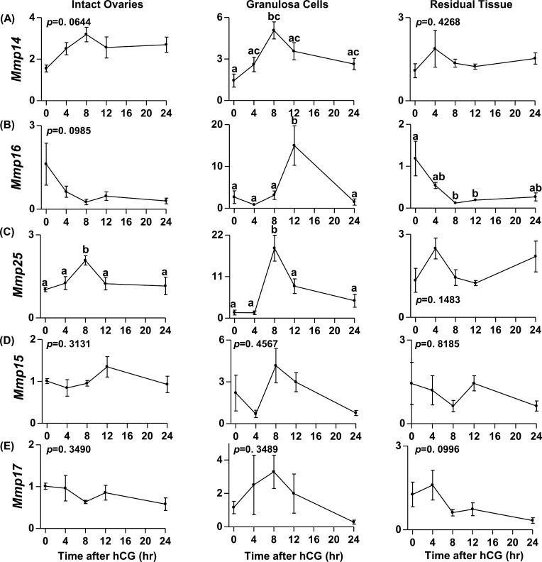
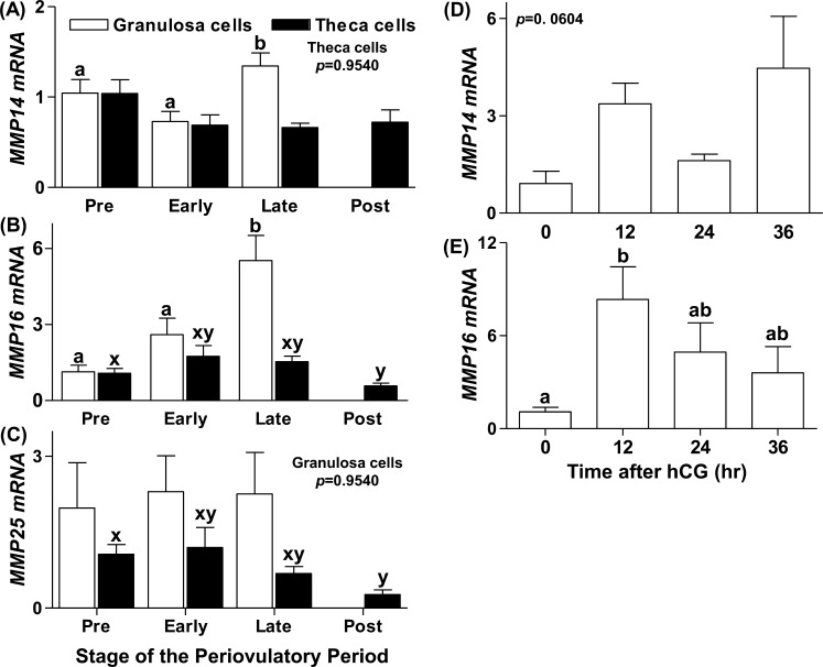
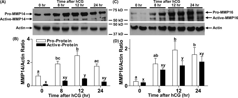
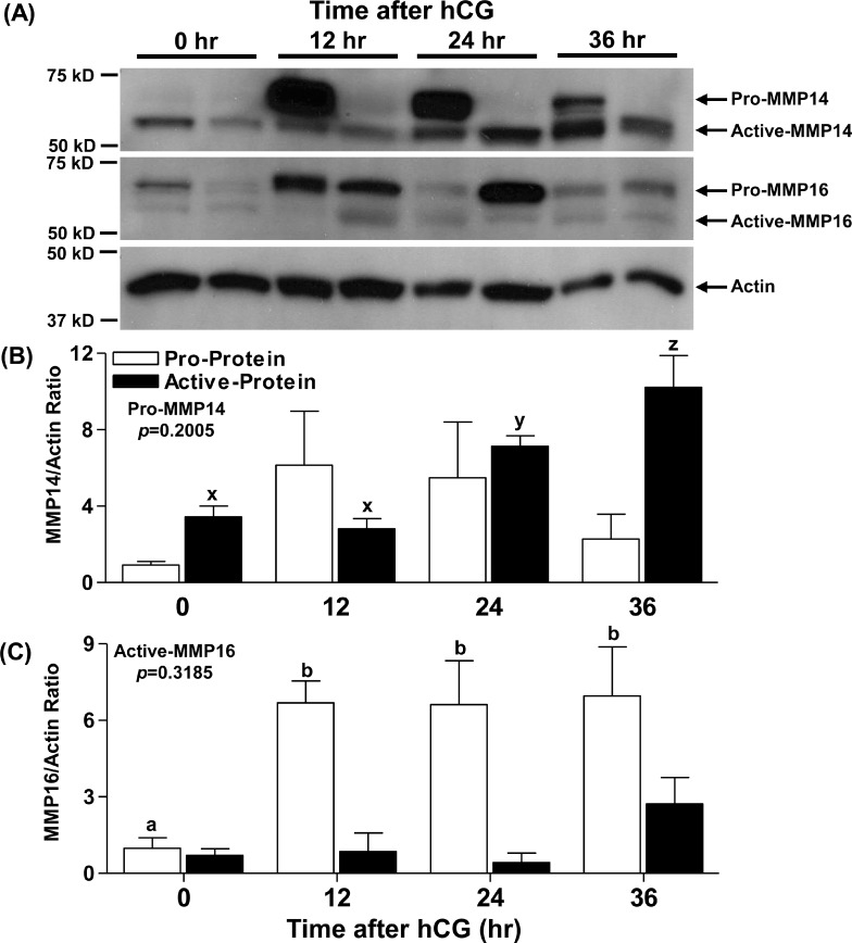
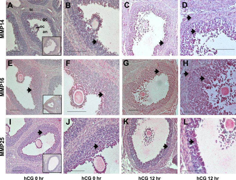
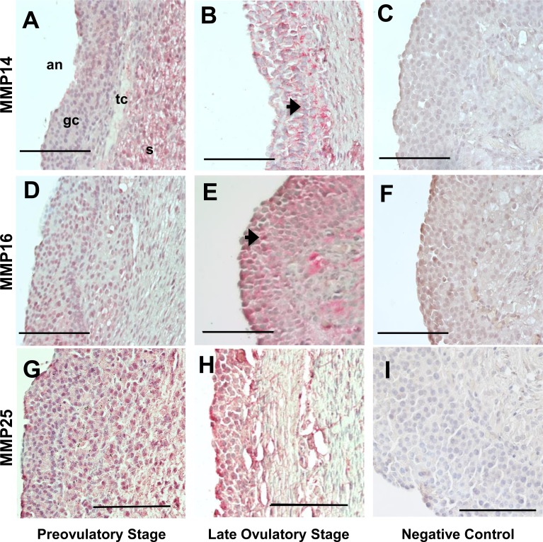
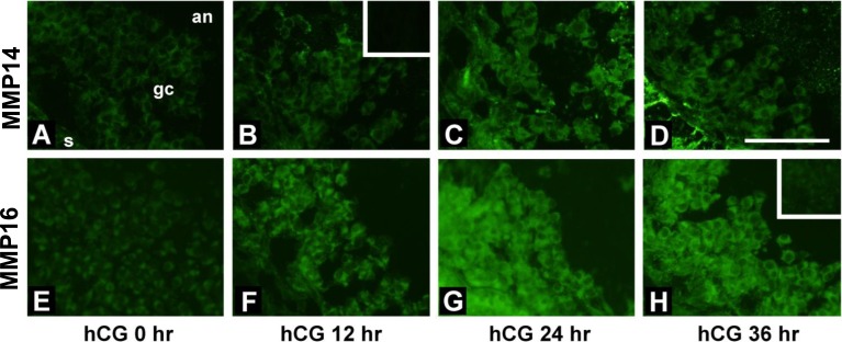
An intrafollicular increase in proteolytic activity drives ovulatory events. Surprisingly, the periovulatory expression profile of the membrane-type matrix metalloproteinases (MT-MMPs), unique proteases anchored to the cell surface, has not been extensively examined. Expression profiles of the MT-MMPs were investigated in ovarian tissue from well-characterized rat and macaque periovulatory models and naturally cycling women across the periovulatory period. Among the six known MT-MMPs, mRNA expression of Mmp14, Mmp16, and Mmp25 was increased after human chorionic gonadotropin (hCG) administration in rats. In human granulosa cells, mRNA expression of MMP14 and MMP16 increased following hCG treatment. In contrast, mRNA levels of MMP16 and MMP25 in human theca cells were unchanged before ovulation but declined by the postovulatory stage. In macaque granulosa cells, hCG increased mRNA for MMP16 but not MMP14. Immunoblotting showed that protein levels of MMP14 and MMP16 in rats increased, similar to their mRNA expression. In macaque granulosa cells, only the active form of the MMP14 protein increased after hCG, unlike its mRNA or the proprotein. By immunohistochemistry, both MMP14 and MMP16 localized to the different ovarian cell types in rats and humans. Treatment with hCG resulted in intense immunoreactivity of MMP14 and MMP16 proteins in the granulosa and theca cells. The present study shows that MMP14 and MMP16 are increased by hCG administration in the ovulating follicle, demonstrating that these MMPs are conserved among rats, macaques, and humans. These findings suggest that MT-MMPs could have an important role in promoting ovulation and remodeling of the ovulated follicle into the corpus luteum.

Keywords: extracellular matrix; granulosa cells; matrix metalloproteinase; ovary; ovulation; theca cell.


Figures

FIG. 1
FIG. 1
Messenger RNA expression profiles of the MT-MMPs during the rat periovulatory period. The mRNA expression profiles of the MT-MMPs Mmp14 (A), Mmp16 (B), Mmp25 (C), Mmp15 (D), and Mmp17 (E) in the intact ovary, granulosa cells, and residual ovarian tissue collected from eCG-primed immature rats at 0 h (48 h after eCG) or at 4, 8, 12, and 24 h after administration of hCG are shown. Relative levels of mRNA were normalized to L32 in each sample and expressed as the fold-change relative to 0 h. Data are presented as the mean ± SEM (n = 3–4 per time point) of the fold-change in mRNA levels, and different superscripts indicate significant changes (P < 0.05) in mRNA levels within an MT-MMP panel.
FIG. 2
FIG. 2
Messenger RNA expression profiles of the MT-MMPs in human and macaque ovarian cells during the periovulatory period in vivo. Human granulosa and theca cells were collected from women undergoing elective surgery for tubal sterilization at different stages of the periovulatory period. The mRNA expression profiles of MMP14 (A), MMP16 (B), and MMP25 (C) as determined by real-time RT-PCR in the granulosa (open bars) and theca (closed bars) cells are shown. Macaque granulosa cells were obtained 0, 12, 24, and 36 h after hCG administration from monkeys undergoing controlled ovarian stimulation and analyzed for MMP14 (D) and MMP16 (E). Relative levels of mRNA were normalized to GAPDH or L32 (for analysis of human or macaque samples, respectively) in each sample and expressed as the fold-change relative to the preovulatory stage or 0 h. Data are presented as the mean ± SEM (n = 3–4 per time point) of the fold-change in mRNA levels, and different superscripts indicate significant changes (P < 0.05) in mRNA levels.
FIG. 3
FIG. 3
Changes in MMP14 and MMP16 protein levels in rat ovaries during the periovulatory period. Rat ovaries were obtained 0, 8, 12, and 24 h after hCG administration from eCG-primed rats and processed for protein analysis. Representative immunoblots of MMP14 and MMP16 in the rat ovarian lysates at different times after hCG administration (top blots in A and C, respectively) are shown. The beta-actin immunoblots are from the respective blots obtained after stripping of the primary MMP antibodies (bottom blots in A and C) are also shown. Densitometric analysis was used to semiquantitate the changes in pro-MMP14 and active MMP14 (B) and in pro-MMP16 and active MMP16 (D). Arbitrary optical density units measured for proform and active form using ImageJ software were normalized to those measured for the internal control beta-actin. The graph depicts the mean ± SEM (n = 4 animals/group). Groups with no common letters are significantly different (P < 0.05).
FIG. 4
FIG. 4
Changes in MMP14 and MMP16 protein levels in macaque granulosa cells during the periovulatory period. Monkey granulosa cells were obtained 0, 12, 24, and 36 h after hCG administration from monkeys undergoing controlled ovarian stimulation and processed for protein analysis. A representative immunoblot of MMP14 and MMP16 along with pan-actin (A) in granulosa cell lysates at different times after hCG administration is shown. Densitometry was used to semiquantitate changes in pro-MMP14 and active MMP14 (B) as well as pro-MMP16 and active MMP16 (C) protein in granulosa cell lysates. Arbitrary optical density units measured for proform and active form were normalized to those measured for the internal control actin. The graph depicts the mean ± SEM (n = 4 animals/group). Groups with no common letters are significantly different (P < 0.05).
FIG. 5
FIG. 5
Immunohistochemical detection of MMP14, MMP16, and MMP25 in the rat ovary during the periovulatory period. Ovaries collected from eCG-primed rats at different times after administration of an ovulatory dose of hCG were processed for immunolocalization of MMP14 (AD), MMP16 (EH), and MMP25 (IL). In ovaries collected at 0 h (A, B, E, F, I, and J) and 12 h (C, D, G, H, K, and L) h after hCG administration, immunoreactive MMP14, MMP16, and MMP25 protein are identified as a red reaction product (arrows). B, D, F, H, J, and L are higher-magnification views (×40) of the images in A, C, E, G, I, and K (×20), respectively. Insets in A, E, and I are ovary sections in which the primary antibodies against MMP14, MMP16, and MMP25, respectively, were omitted. The full preovulatory (0 h) or periovulatory (12 h) ovarian follicle is shown (gc, granulosa cell layer; tc, theca cell layer; an, follicular antrum; oc, oocyte). Representative photomicrographs are shown for each time point. Bar = 50 μm (×20 panels) and 100 μm (×40 panels).
FIG. 6
FIG. 6
Immunohistochemical detection of MMP14, MMP16, and MMP25 in human periovulatory follicles. Human follicles collected from women at different stages of the periovulatory period were processed for immunolocalization of MMP14, MMP16, and MMP25. Immunoreactive MMP14, MMP16, and MMP25 in follicles from preovulatory (A, D, and G) and late ovulatory (B, E, and H) stages, respectively, are identified as a red reaction product (arrow). C, F, and I represent follicle sections in which the primary antibodies against MMP14, MMP16, and MMP25, respectively, were omitted. All images are oriented as in A, with ovarian stroma (s), theca cells (tc), granulosa cells (gc), and follicle antrum (an) from right to left. Representative photomicrographs are shown for the different periovulatory stages. Bar =100 μm.
FIG. 7
FIG. 7
Immunofluorescent detection of MMP14 and MMP16 in monkey periovulatory follicles. Monkeys received gonadotropins to stimulate the development of multiple follicles. Ovarian tissue from these animals was obtained before (0 h; A and E) or at 12 h (B and F), 24 h (C and G), or 36 h (D and H) after administration of hCG to initiate periovulatory events. Immunolocalization was performed for MMP14 (AD) and MMP16 (EH). All images are oriented as in A, with ovarian stroma (s) in the lower left, granulosa cells (gc) central, and follicle antrum (an) in the upper right. Insets in B and H show that faint immunofluorescence was present when the primary antibody was omitted. Bar = 50 μm.

References

    1. Curry TE, Jr, , Smith MF. Impact of extracellular matrix remodeling on ovulation and the folliculo-luteal transition. Semin Reprod Med. 2006;24:228–241. - PubMed
    1. Chaffin CL, Stouffer RL. Expression of matrix metalloproteinases and their tissue inhibitor messenger ribonucleic acids in macaque periovulatory granulosa cells: time course and steroid regulation. Biol Reprod. 1999;61:14–21. - PubMed
    1. Curry TE, Jr, , Osteen KG. The matrix metalloproteinase system: changes, regulation, and impact throughout the ovarian and uterine reproductive cycle. Endocr Rev. 2003;24:428–465. - PubMed
    1. Liu YX. Plasminogen activator/plasminogen activator inhibitors in ovarian physiology. Front Biosci. 2004;9:3356–3373. - PubMed
    1. Mittaz L, Russell DL, Wilson T, Brasted M, Tkalcevic J, Salamonsen LA, Hertzog PJ, Pritchard MA. Adamts-1 is essential for the development and function of the urogenital system. Biol Reprod. 2004;70:1096–1105. - PubMed

MeSH terms

Substances