Review
. 2014 Sep;52(9):771-92.
doi: 10.1002/dvg.22798. Epub 2014 Jun 25.

Using zebrafish to study podocyte genesis during kidney development and regeneration

Affiliations
Review

Using zebrafish to study podocyte genesis during kidney development and regeneration

Paul T Kroeger Jr et al. Genesis. 2014 Sep.

Abstract

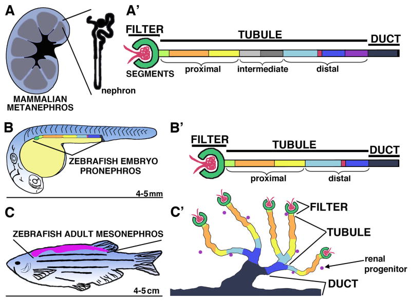
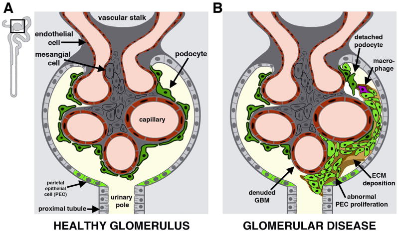
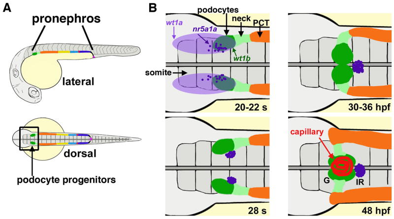
During development, vertebrates form a progression of up to three different kidneys that are comprised of functional units termed nephrons. Nephron composition is highly conserved across species, and an increasing appreciation of the similarities between zebrafish and mammalian nephron cell types has positioned the zebrafish as a relevant genetic system for nephrogenesis studies. A key component of the nephron blood filter is a specialized epithelial cell known as the podocyte. Podocyte research is of the utmost importance as a vast majority of renal diseases initiate with the dysfunction or loss of podocytes, resulting in a condition known as proteinuria that causes nephron degeneration and eventually leads to kidney failure. Understanding how podocytes develop during organogenesis may elucidate new ways to promote nephron health by stimulating podocyte replacement in kidney disease patients. In this review, we discuss how the zebrafish model can be used to study kidney development, and how zebrafish research has provided new insights into podocyte lineage specification and differentiation. Further, we discuss the recent discovery of podocyte regeneration in adult zebrafish, and explore how continued basic research using zebrafish can provide important knowledge about podocyte genesis in embryonic and adult environments. genesis 52:771-792, 2014. © 2014 Wiley Periodicals, Inc.

Keywords: development; glomerulus; kidney; podocyte; regeneration; renal corpuscle; zebrafish.


Figures

FIGURE 1
FIGURE 1. Nephron segmental anatomy is conserved among vertebrates
(A) Schematic of a mammalian metanephros with (A′) a linear diagram of nephron segments. (B) Schematic of a zebrafish embryo with lateral location of the pronephros indicated, and with (B′) a linear diagram of the nephron segments. The zebrafish embryo is approximately 4 mm long (tip to tail) by 5 dpf, and continues to utilize the pronephros as the fish grows. (C) Schematic of a zebrafish adult with dorsal location of the mesonephros, and with (C′) a diagram depicting the arborized arrangements of nephrons with common duct exitways. The adult zebrafish is typically 4–5 cm in length (tip to tail). Analogous nephron segments are color-coded, with the vasculature (red ball), podocytes (dark green), neck (light green), proximal tubule segments (orange, yellow), intermediate tubule segments (gray), distal tubule segments (light blue, dark blue, purple) with intervening macula densa (mammals) or corpuscle of Stannius (fish) (red), and finally the duct (black). [Reprinted from Transl Res, 163(2), McCampbell K, Wingert RA, New Tides: using zebrafish to study renal regeneration, Pages No. 109–122, Copyright 2014, with permission from Elsevier.]
FIGURE 2
FIGURE 2. Composition of the nephron blood filter
(A) Healthy blood filter with intact glomerular components. Inset depicts nephron outline and boxed area enlarged in panel. (B) Maladaptive response to podocyte attrition, in which activated parietal epithelial cells proliferate and fill the capsule, impeding flow to the nephron tubule and leading to nephron atrophy. [Figure adapted from Clin Transl Med, 2(1), Li Y, Wingert RA, Regenerative medicine for the kidney: stem cell prospects and challenges, Pages 1–16, doi: 10.1186/2001-1326-2-11, Copyright 2013, permission through the creative commons license.]
FIGURE 3
FIGURE 3. Zebrafish podocyte lineage specification and glomerular development
(A) The zebrafish pronephros contains podocytes (dark green) at the rostral-most position. (B) Developmental timecourse of the cell populations that develop in proximity to podocytes. Gene expression of wt1a (light purple) is broad, while wt1b transcripts (dark green) are restricted next to somite (s) three, and interrenal precursors marked by nr5a1a transcripts (dark purple) are interspersed in this region. The neck (light green) is located caudal to the podocytes, followed by the proximal convoluted tubule (PCT, orange). Cell movements between the 20 somite stage to 48 hours post fertilization (hpf) lead to formation of a single glomerulus (G) with central capillary nexus (red). The interrenal gland (IR) (dark purple) is situated just caudal to the glomerulus. [Figure adapted from Wiley Interdiscip Rev Dev Biol, 2, Gerlach G, Wingert RA, Kidney organogenesis in the zebrafish: Insights into vertebrate nephrogenesis and regeneration, Pages 559–585, Copyright 2013, with author permission].

Similar articles

Cited by

References

    1. Amacher SL, Draper BW, Summers BR, Kimmel CB. The zebrafish T-box genes no tail and spadetail are required for development of trunk and tail mesoderm and medial floor plate. Development. 2002;129:3311–3323. - PubMed
    1. Amsterdam A, Hopkins N. Mutagenesis strategies in zebrafish for identifying genes involved in development and disease. Trends Genet. 2006;22:473–478. - PubMed
    1. Appel D, Kershaw DB, Smeets B, Yuan G, Fuss A, Frye B, Elger M, Kriz W, Floege J, Moeller MJ. Recruitment of podocytes from glomerular parietal epithelial cells. J Am Soc Nephrol. 2009;20:333–343. - PMC - PubMed
    1. Arif E, Kumari B, Wagner MC, Zhou W, Holzman LB, Nihalani D. Myo1c is an unconventional myosin required for zebrafish glomerular development. Kid Int. 2013;84:1154–1165. - PMC - PubMed
    1. Arif E, Rathore YS, Kumari B, Ashish F, Wong HN, Holzman LB, Nihalani D. Slit diaphragm protein Neph1 and its signaling: a novel therapeutic target for protection of podocytes against glomerular injury. J Biol Chem. 2014;289:9502–9518. - PMC - PubMed

Publication types

LinkOut - more resources