. 2014 Jun 11;34(24):8210-8.
doi: 10.1523/JNEUROSCI.5456-13.2014.

Is Parkinson's disease a vesicular dopamine storage disorder? Evidence from a study in isolated synaptic vesicles of human and nonhuman primate striatum

Affiliations

Is Parkinson's disease a vesicular dopamine storage disorder? Evidence from a study in isolated synaptic vesicles of human and nonhuman primate striatum

Christian Pifl et al. J Neurosci. .

Erratum in

  • J Neurosci. 2015 Nov 25;35(47):15767

Abstract

The cause of degeneration of nigrostriatal dopamine (DA) neurons in idiopathic Parkinson's disease (PD) is still unknown. Intraneuronally, DA is largely confined to synaptic vesicles where it is protected from metabolic breakdown. In the cytoplasm, however, free DA can give rise to formation of cytotoxic free radicals. Normally, the concentration of cytoplasmic DA is kept at a minimum by continuous pumping activity of the vesicular monoamine transporter (VMAT)2. Defects in handling of cytosolic DA by VMAT2 increase levels of DA-generated oxy radicals ultimately resulting in degeneration of DAergic neurons. Here, we isolated for the first time, DA storage vesicles from the striatum of six autopsied brains of PD patients and four controls and measured several indices of vesicular DA storage mechanisms. We found that (1) vesicular uptake of DA and binding of the VMAT2-selective label [(3)H]dihydrotetrabenazine were profoundly reduced in PD by 87-90% and 71-80%, respectively; (2) after correcting for DA nerve terminal loss, DA uptake per VMAT2 transport site was significantly reduced in PD caudate and putamen by 53 and 55%, respectively; (3) the VMAT2 transport defect appeared specific for PD as it was not present in Macaca fascicularis (7 MPTP and 8 controls) with similar degree of MPTP-induced nigrostriatal neurodegeneration; and (4) DA efflux studies and measurements of acidification in the vesicular preparations suggest that the DA storage impairment was localized at the VMAT2 protein itself. We propose that this VMAT2 defect may be an early abnormality promoting mechanisms leading to nigrostriatal DA neuron death in PD.

Keywords: Parkinson's disease; VMAT2; dopamine; striatum; synaptic vesicles.


Figures

Figure 1.
Figure 1.
DA uptake by synaptic vesicles prepared from human control striatal tissue. A, Time-dependence of DA uptake. Synaptic vesicle preparations from striatum were incubated in the absence or presence of 1 μm reserpine with 0.3 μm [3H]DA at 30°C for the times indicated. Specific uptake (filled symbols, solid line) was calculated by subtraction of unspecific from total uptake. Data shown are mean values of triplicates. B, Protein-dependence of DA uptake. Synaptic vesicle preparations from caudate/putamen at the protein amounts indicated were incubated with 0.1 μm [3H]DA at 30°C for 4 min in the absence or presence of 1 μm reserpine. Data shown are mean values of triplicates. C, Concentration-dependence of DA uptake. Synaptic vesicle preparations from striatum were incubated with [3H]DA at the concentration indicated at 30°C for 4 min in the absence or presence of 1 μm reserpine. Inset, Scatchard transformation of specific uptake. Data shown are mean values ± SE of three independent experiments, each done in triplicates. D, ATP-dependence and striatal specifity of DA uptake. Synaptic vesicle preparations (33–35 μg protein) of putamen or thalamus were incubated with 0.3 μm [3H]DA at 30°C for 4 min in the absence or presence of 1 μm reserpine and 2 mm MgATP. Data shown are mean values of triplicate.
Figure 2.
Figure 2.
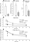
Synaptic vesicles prepared from caudate of patients with PD and controls. A, Specific DA uptake. Synaptic vesicle preparations (16 ± 4 and 19 ± 3 μg protein for controls and PD, respectively) were incubated in the absence or presence of 1 μm reserpine with 0.1 μm [3H]DA at 30°C for 4 min; **p < 0.002 specific uptake in PD versus control by Student's t test. B, DTBZ-binding. Synaptic vesicle preparations (3.7 ± 1.6 and 4.2 ± 0.7 μg protein for controls and PD, respectively) were incubated in the absence or presence of 1 μm tetrabenazine with 4 nm [3H]DTBZ at 30°C for 90 min; **p < 0.004 specific binding in PD versus control by Student's t test. C, Uptake per DTBZ-binding site. For each synaptic vesicle preparation specific DA uptake was divided by specific DTBZ-binding; *p < 0.03 uptake rate per transport site in PD versus control by Student's t test. D, DA efflux. Synaptic vesicle preparations were loaded with DA by incubation with 0.1 μm [3H]DA at 30°C for 4 min, centrifuged and the pellet resuspended at 4°C and finally incubated at 30°C for 2 (in the presence and absence of 0.1 mg/L nigericin), 10, and 30 min. DA retained was determined by filtration. E, DA efflux presented in percentage of loaded DA. *p < 0.05 efflux in PD versus control by Student's t test. Data shown are mean values ± SE of four controls and six PD cases, each done in triplicate.
Figure 3.
Figure 3.
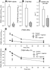
Synaptic vesicles prepared from putamen of patients with PD and controls. A, Specific DA uptake. Synaptic vesicle preparations (28 ± 5 and 31 ± 6 μg protein for controls and PD, respectively) were incubated in the absence or presence of 1 μm reserpine with 0.1 μm [3H]DA at 30°C for 4 min; **p < 0.00001 specific uptake in PD versus control by Student's t test. B, DTBZ-binding. Synaptic vesicle preparations (6.5 ± 0.71 and 8.0 ± 1.3 μg protein for controls and PD, respectively) were incubated in the absence or presence of 1 μm tetrabenazine with 4 nm [3H]DTBZ at 30°C for 90 min; **p < 0.0005 specific binding in PD versus control by Student's t test. C, Uptake per DTBZ-binding site. For each synaptic vesicle preparation specific DA uptake was divided by specific DTBZ-binding; **p < 0.05 uptake rate per transport site in PD versus control by Student's t test. D, DA efflux. Synaptic vesicle preparations were loaded with DA by incubation with 0.1 μm [3H]DA at 30°C for 4 min, centrifuged and the pellet resuspended at 4°C and finally incubated at 30°C for 2 (in the presence and absence of 0.1 mg/L nigericin), 10, and 30 min. DA retained was determined by filtration. E, DA efflux presented in percentage of loaded DA. *p < 0.05 of release in PD versus control by Student's t test. Data shown are mean values ± SE of four controls and six PD cases, each done in triplicate.
Figure 4.
Figure 4.
Synaptic vesicles prepared from putamen of MPTP monkeys and controls. A, Specific DA uptake. Synaptic vesicle preparations (25 ± 3 and 26 ± 2 μg protein for controls and MPTP, respectively) were incubated in the absence or presence of 1 μm reserpine with 0.1 μm [3H]DA at 30°C for 4 min; **p < 0.00001 specific uptake in MPTP versus control by Student's t test. B, DTBZ-binding. Synaptic vesicle preparations (16 ± 2 and 16 ± 1 μg protein for controls and MPTP, respectively) were incubated in the absence or presence of 1 μm tetrabenazine with 4 nm [3H]DTBZ at 30°C for 90 min; **p < 0.00001 specific binding in MPTP versus control by Student's t test. C, Uptake per DTBZ-binding site. For each synaptic vesicle preparation, specific DA uptake was divided by specific DTBZ-binding. Data shown are mean values ± SE of eight control and seven MPTP monkeys, each done in triplicate.
Figure 5.
Figure 5.
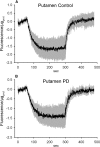
Protein gradient measurement on synaptic vesicles prepared from putamen of patients with PD and controls. Synaptic vesicle preparations (47 ± 4 and 48 ± 7 μg protein for controls and PD, respectively) were incubated with 2 μm acridine orange and acidification was measured by quenching of fluorescence at 493 nm (excitation) and 530 nm (emission) after addition of 0.5 mm MgATP at 60 s and proton gradient abolition by addition of 1 μm carbonyl cyanide FCCP at 300 s. Shown are mean values of fluorescent traces shifted to 0 fluorescence at the time interval 0–58 s ± SE (gray area) of four controls (A) and six PD patients (B).

References

    1. Betarbet R, Sherer TB, MacKenzie G, Garcia-Osuna M, Panov AV, Greenamyre JT. Chronic systemic pesticide exposure reproduces features of Parkinson's disease. Nat Neurosci. 2000;3:1301–1306. doi: 10.1038/81834. - DOI - PubMed
    1. Bilska A, Dubiel M, Sokołowska-Jezewicz M, Lorenc-Koci E, Włodek L. Alpha-lipoic acid differently affects the reserpine-induced oxidative stress in the striatum and prefrontal cortex of rat brain. Neuroscience. 2007;146:1758–1771. doi: 10.1016/j.neuroscience.2007.04.002. - DOI - PubMed
    1. Blesa J, Pifl C, Sánchez-González MA, Juri C, García-Cabezas MA, Adanez R, Iglesias E, Collantes M, Peñuelas I, Sánchez-Hernández JJ, Rodríguez-Oroz MC, Avendaño C, Hornykiewicz O, Cavada C, Obeso JA. The nigrostriatal system in the presymptomatic and symptomatic stages in the MPTP monkey model: a PET, histological and biochemical study. Neurobiol Dis. 2012;48:79–91. doi: 10.1016/j.nbd.2012.05.018. - DOI - PubMed
    1. Caudle WM, Richardson JR, Wang MZ, Taylor TN, Guillot TS, McCormack AL, Colebrooke RE, Di Monte DA, Emson PC, Miller GW. Reduced vesicular storage of dopamine causes progressive nigrostriatal neurodegeneration. J Neurosci. 2007;27:8138–8148. doi: 10.1523/JNEUROSCI.0319-07.2007. - DOI - PMC - PubMed
    1. Caudle WM, Colebrooke RE, Emson PC, Miller GW. Altered vesicular dopamine storage in Parkinson's disease: a premature demise. Trends Neurosci. 2008;31:303–308. doi: 10.1016/j.tins.2008.02.010. - DOI - PubMed

MeSH terms