. 2014 Jun 11;6(240):240ra73.
doi: 10.1126/scitranslmed.3008659.

Macrophage models of Gaucher disease for evaluating disease pathogenesis and candidate drugs

Affiliations

Macrophage models of Gaucher disease for evaluating disease pathogenesis and candidate drugs

Elma Aflaki et al. Sci Transl Med. .

Abstract

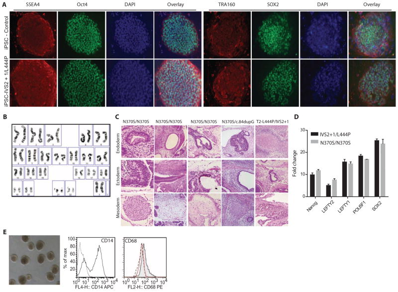
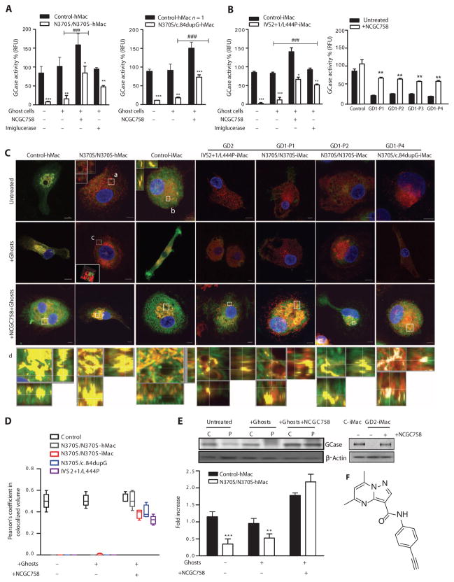
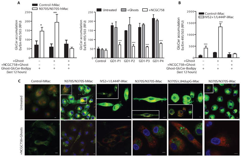
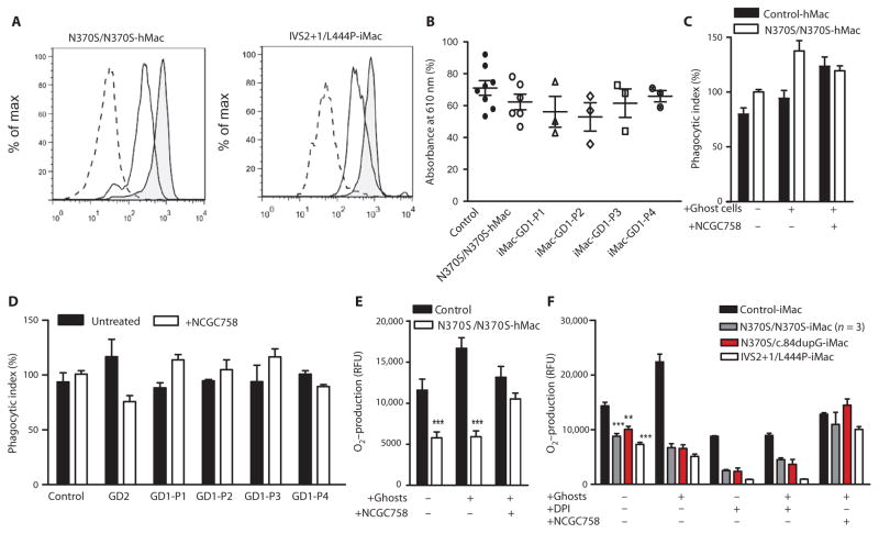
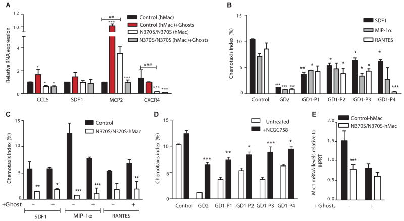
Gaucher disease is caused by an inherited deficiency of glucocerebrosidase that manifests with storage of glycolipids in lysosomes, particularly in macrophages. Available cell lines modeling Gaucher disease do not demonstrate lysosomal storage of glycolipids; therefore, we set out to develop two macrophage models of Gaucher disease that exhibit appropriate substrate accumulation. We used these cellular models both to investigate altered macrophage biology in Gaucher disease and to evaluate candidate drugs for its treatment. We generated and characterized monocyte-derived macrophages from 20 patients carrying different Gaucher disease mutations. In addition, we created induced pluripotent stem cell (iPSC)-derived macrophages from five fibroblast lines taken from patients with type 1 or type 2 Gaucher disease. Macrophages derived from patient monocytes or iPSCs showed reduced glucocerebrosidase activity and increased storage of glucocerebroside and glucosylsphingosine in lysosomes. These macrophages showed efficient phagocytosis of bacteria but reduced production of intracellular reactive oxygen species and impaired chemotaxis. The disease phenotype was reversed with a noninhibitory small-molecule chaperone drug that enhanced glucocerebrosidase activity in the macrophages, reduced glycolipid storage, and normalized chemotaxis and production of reactive oxygen species. Macrophages differentiated from patient monocytes or patient-derived iPSCs provide cellular models that can be used to investigate disease pathogenesis and facilitate drug development.


Conflict of interest statement

Competing interests: The molecule described in this work has been patented by the NIH under the filing “Substituted pyrazolopyrimidines as glucocerebrosidase activators” (authors: J.M., N.S., E.G., W.Z., S.P., E.S., O. Motabar, and W. Westbroek; U.S. Application 61/420946; International Patent Application PCT/US2011/63928). Please refer to the NIH Office of Transfer Technology for further information. The other authors declare that they have no competing interests.

Figures

Fig. 1
Fig. 1. Generation and differentiation of iPSCs into monocytes and macrophages
(A) Immunostaining for common embryonic markers (Oct4, SOX2, SSEA4, TRA160, TRA180, and Nanog) in control and type 2 Gaucher disease (IVS2+1/L444P) iPSC lines using a Zeiss fluorescence microscope. (B) iPSCs derived from a patient with type 2 Gaucher disease have a normal 46XX karyotype. (C) Teratomas were formed from iPSCs generated from four patients with type 1 Gaucher disease (three with genotype N370S/N370S and one with genotype N370S/c.84dupG) and from one patient with type 2 Gaucher disease (genotype IVS2+1/L444P). (D) Total RNA from two skin fibroblast lines from patients with type 1 Gaucher disease (genotype N370S/N370S), and the corresponding two iPSC lines derived from them were characterized in triplicate using a PCR array. The fold changes refer to the expression levels for each gene compared to that in the fibroblast line. (E) iPSCs were differentiated into macrophages using a three-step method. Embryoid bodies were made and differentiated into iPSC monocytes. The monocytes harvested from the supernatant stained positive for CD14 with flow cytometry. The monocytes were then differentiated into macrophages, which stained positive for CD68 expression with flow cytometry.
Fig. 2
Fig. 2. Increased CD163 expression in Gaucher disease macrophages
(A to C) Flow cytometry analysis of control iMacs and type 1 Gaucher disease hMacs (genotype N370S/N370S) and iMacs (genotypes N370S/N370S and N370S/c.84dupG) and type 2 Gaucher disease iMacs (genotype IVS2+1/L444P) stained for (A) CD33 and CD11b, (B) CD64 and CD163, and (C) CD15 and CD105 markers. (D) hMacs and iMacs were also stained with CD163 alone. The flow cytometry images of hMacs shown are representative of 7 patients with type 1 Gaucher disease (all with genotype N370S/N370S) and 10 healthy controls in independent experiments. The antibody isotype controls are shown in fig. S3.
Fig. 3
Fig. 3. NCGC00188758 increases mutant glucocerebrosidase activity
(A) Glucocerebrosidase activity is shown as percent of control glucocerebrosidase activity in type 1 Gaucher disease hMacs in the presence and absence of Gaucher erythrocyte ghosts, 8 μM NCGC00188758 (referred to in figures as NCGC758), or imiglucerase. The left panel reflects independent experiments using N370S/N370S hMacs from 17 different patients; the right panel shows data for hMacs from one patient with genotype N370S/c.84dupG. Four different hMacs with genotype N370S/N370S were treated with imiglucerase (20 μM) (left panel). Error bars indicate SD. ***P < 0.0016, **P < 0.01. RFU, relative fluorescence units. (B) Effect of NCGC00188758 on iMacs in the presence and absence of Gaucher erythrocyte ghosts. The left panel shows glucocerebrosidase activity in iMacs from an infant with type 2 Gaucher disease (genotype IVS2+1/L444P). The right panel shows iMacs from four patients with type 1 Gaucher disease (GD1-P1, GD1-P2, and GD1-P3 each have the genotype N370S/N370S, and GD1-P4 has the genotype N370S/c.84dupG). Glucocerebrosidase activity in iMacs was measured in two independent experiments. Error bars indicate SD. *P = 0.018, **P = 0.003, ***P < 0.0017, ###P < 0.001. (C) Gaucher disease and control hMacs and iMacs were treated with 8 μM NCGC00188758 for 6 days and then stained for glucocerebrosidase (Alexa Fluor 488, green), the lysosomal marker LAMP2 (Alexa Fluor 555, red), and 4′,6-diamidino-2-phenylindole (DAPI) (nuclear stain, blue). Z-stack images were acquired using a Zeiss 510 confocal microscope (×63 magnification). Single images are presented in fig. S6. Insets correspond to the regions marked a, b, and c. Figures on lower panel (d) show higher magnifications of the areas outlined in the images of the cells treated with NCGC00188758. The location of cross section (Z stack) is shown in x-y, x-z, and y-z planes (corresponding movies are shown in the Supplementary Materials). iMacs from four different patients with Gaucher disease with genotypes N370S/N370S, N370S/c.84dupG, and IVS2+1/L444P are shown. Images are taken at the same laser settings and are representative of 12 independent experiments. Scale bars, 5 μm. (D) Pearson’s coefficient was calculated for the degree of colocalization between glucocerebrosidase (green), LAMP2, and nuclear stain (blue). Results represent >20 cells per condition. (E) Left: Western blot showing glucocerebrosidase in lysed hMacs treated with Gaucher erythrocyte ghosts in the presence and absence of 8 μM NCGC00188758 for 6 days. Patient (P) (genotype N370S/N370S) and control (C) hMacs are shown. Right: Western blot showing glucocerebrosidase concentrations in lysed iMacs from a patient with type 2 Gaucher disease (genotype IVS2+1/L444P) treated with Gaucher erythrocyte ghosts in the presence and absence of 8 μM NCGC00188758 for 6 days. The graph represents Western blots performed on hMacs from six different patients with type 1 Gaucher disease with genotype N370S/N370S. Groups were compared using one-way analysis of variance (ANOVA) (n = 6). **P = 0.03; ***P = 0.009, Bonferroni correction. (F) Chemical structure of NCGC00188758.
Fig. 4
Fig. 4. NCGC00188758 reduces glucosylceramide accumulation in Gaucher disease hMacs and iMacs
(A and B) Gaucher disease and control hMacs and iMacs from four patients with type 1 Gaucher disease (GD1-P1, GD1-P2, and GD1-P3 each with genotype N370S/N370S, and GD1-P4 with genotype N370S/c.84dupG) and one with type 2 Gaucher disease (IVS2+1/L444P) were treated with 8 μM NCGC00188758 in the presence and absence of erythrocyte ghosts. On day 6, the macrophages were loaded with glucosylceramide-Bodipy (GlcCer-Bodipy)–labeled erythrocyte ghosts, and fluorescence was measured at 495/503 nm. (A) Left panel: Pooled data for hMacs from 15 different patients with genotype N370S/N370S; right panel: iMacs from four different type 1 Gaucher disease iPSC lines (GD1-P1, GD1-P2, and GD1-P3 have genotype N370S/N370S; GD1-P4 has genotype N370S/c.84dupG). Each experiment was performed in triplicate. (B) Data for type 2 Gaucher disease iMacs, with measurements performed in triplicate. Error bars indicate SD. *P = 0.018; **P = 0.007; ***P ≤ 0.0012, two-way ANOVA with Bonferroni post test. (C) N370S/N370S hMacs and four different Gaucher disease iMacs (genotypes N370S/N370S for two patients, N370S/c.84dupG, and IVS2+1/L444P). Cells were treated with labeled erythrocyte ghosts (green) for 12 hours, fixed, and stained with LAMP2 (Alexa Fluor 555, red). They were imaged using a confocal microscope with a 488-nm argon laser. Images were acquired using a Plan Neofluar 63× 1.3 oil DIC (differential interference contrast) objective. Scale bars, 5 μm. Insets show specific regions with a lower intensity of the green channel.
Fig. 5
Fig. 5. NCGC00188758 corrects ROS production in Gaucher disease macrophages
(A) Expression of CD32 measured by flow cytometry. The first peak (dashed) is unstained cells; the second peak is control hMacs stained with CD32; the third peak (shaded) is N370S/N370S hMacs (left) or IVS2+1/L444P iMacs (right) stained with CD32. Data represent six independent experiments. (B) Phagocytosis of IgG-opsonized erythrocytes was evaluated in hMacs (genotype N370S/N370S) and in iMacs from four different iPSC lines (GD1-P1, GD1-P2, and GD1-P3 with genotype N370S/N370S, and GD1-P4 with genotype N370S/c.84dupG). Experiments were performed five times in hMacs and three times in iMacs for each sample. Results of hMacs from six different patients and eight different controls are shown. (C and D) Phagocytosis in Gaucher disease macrophages treated with erythrocyte ghosts in the presence and absence of 8 μM NCGC00188758 for 6 days. On day 6, fluorescein-labeled E. coli bacteria were added for 2 hours, and fluorescence was measured at 494/518 nm. Phagocytosis of control hMacs was arbitrarily set at 100%. (C) N370S/N370S and control hMacs. Data for N370S/N370S and control hMacs reflect 12 independent experiments. (D) Phagocytic index of iMacs from four different iPSC lines (GD1-P1, GD1-P2, and GD1-P3 with genotype N370S/N370S, and GD1-P4 with genotype N370S/c.84dupG). Data reflect two independent experiments performed in triplicate. Significance was determined using one-way ANOVA. (E and F) ROS production was measured in macrophages treated with NCGC00188758 in the presence and absence of IgG-opsonized erythrocytes. (E) Ten independent experiments were performed with hMacs. ***P = 0.0009. (F) iMacs from five different patients were treated with diphenyleneiodonium (DPI) (5 μM), a potent inhibitor of NADPH oxidase, for 30 min in the presence or absence of IgG-opsonized erythrocyte ghosts. Two independent experiments were performed in quadruplicate. N370S/N370S iMacs represent data from three different iPSC lines. Groups were compared using one-way ANOVA with Bonferroni correction. ***P = 0.0021, **P = 0.012.
Fig. 6
Fig. 6. NCGC00188758 improves chemotaxis in Gaucher disease macrophages
(A) mRNA expression of chemokines CCL5, SDF1, MCP2, and CXCR4 measured in control hMacs and hMacs from six patients with genotype N370S/N370S in the presence and absence of Gaucher erythrocyte ghosts. *P = 0.015, ***P < 0.001, ##P = 0.012, ###P = 0.0018. (B) Chemotaxis of iMacs from four different patients with type 1 Gaucher disease (GD1-P1, GD1-P2, and GD1-P3 with genotype N370S/N370S, and GD1-P4 with genotype N370S/c.84dupG) and from one patient with type 2 Gaucher disease (genotype IVS2+1/L444P) was evaluated using Transwell chambers. *P < 0.062, **P < 0.0048, ***P < 0.0002. (C) Chemotaxis of type 1 Gaucher disease hMacs (genotype N370S/N370S). Experiments were performed on hMacs from seven patients in independent experiments. *P = 0.026, **P = 0.0051, ***P ≤ 0.00044. (D) Chemotaxis toward SDF1 by iMacs from five different patients with Gaucher disease was measured in the presence and absence of NCGC00188758. Data represent two independent experiments, and groups were compared using two-way ANOVA. *P = 0.02, **P = 0.007, ***P < 0.003. (E) Mannose receptor 1 (Mrc1) expression was measured in N370S/N370S hMacs using real-time PCR. Independent experiments were performed on hMacs from 10 patients with genotype N370S/N370S, and groups were compared using one-way ANOVA with Bonferroni correction. ***P = 0.0021.

References

    1. Grabowski GA, Beutler E. The Metabolic and Molecular Bases of Inherited Disease. McGraw-Hill; New York: 2001. pp. 3635–3668.
    1. Orvisky E, Park JK, LaMarca ME, Ginns EI, Martin BM, Tayebi N, Sidransky E. Glucosylsphingosine accumulation in tissues from patients with Gaucher disease: Correlation with phenotype and genotype. Mol Genet Metab. 2002;76:262–270. - PubMed
    1. Lee RE. The fine structure of the cerebroside occurring in Gaucher’s disease. Proc Natl Acad Sci USA. 1968;61:484–489. - PMC - PubMed
    1. Sidransky E, Nalls MA, Aasly JO, Aharon-Peretz J, Annesi G, Barbosa ER, Bar-Shira A, Berg D, Bras J, Brice A, Chen CM, Clark LN, Condroyer C, De Marco EV, Dürr A, Eblan MJ, Fahn S, Farrer MJ, Fung HC, Gan-Or Z, Gasser T, Gershoni-Baruch R, Giladi N, Griffith A, Gurevich T, Januario C, Kropp P, Lang AE, Lee-Chen GJ, Lesage S, Marder K, Mata IF, Mirelman A, Mitsui J, Mizuta I, Nicoletti G, Oliveira C, Ottman R, Orr-Urtreger A, Pereira LV, Quattrone A, Rogaeva E, Rolfs A, Rosenbaum H, Rozenberg R, Samii A, Samaddar T, Schulte C, Sharma M, Singleton A, Spitz M, Tan EK, Tayebi N, Toda T, Troiano AR, Tsuji S, Wittstock M, Wolfsberg TG, Wu YR, Zabetian CP, Zhao Y, Ziegler SG. Multicenter analysis of glucocerebrosidase mutations in Parkinson’s disease. N Engl J Med. 2009;361:1651–1661. - PMC - PubMed
    1. Gardai SJ, McPhillips KA, Frasch SC, Janssen WJ, Starefeldt A, Murphy-Ullrich JE, Bratton DL, Oldenborg PA, Michalak M, Henson PM. Cell-surface calreticulin initiates clearance of viable or apoptotic cells through trans-activation of LRP on the phagocyte. Cell. 2005;123:321–334. - PubMed

Publication types