. 2014 Aug 15;289(33):22915-22925.
doi: 10.1074/jbc.M114.574731. Epub 2014 Jun 11.

Unstable reaction intermediates and hysteresis during the catalytic cycle of 5-aminolevulinate synthase: implications from using pseudo and alternate substrates and a promiscuous enzyme variant

Affiliations

Unstable reaction intermediates and hysteresis during the catalytic cycle of 5-aminolevulinate synthase: implications from using pseudo and alternate substrates and a promiscuous enzyme variant

Bosko M Stojanovski et al. J Biol Chem. .

Abstract

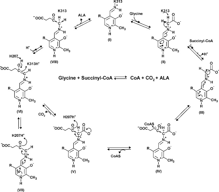
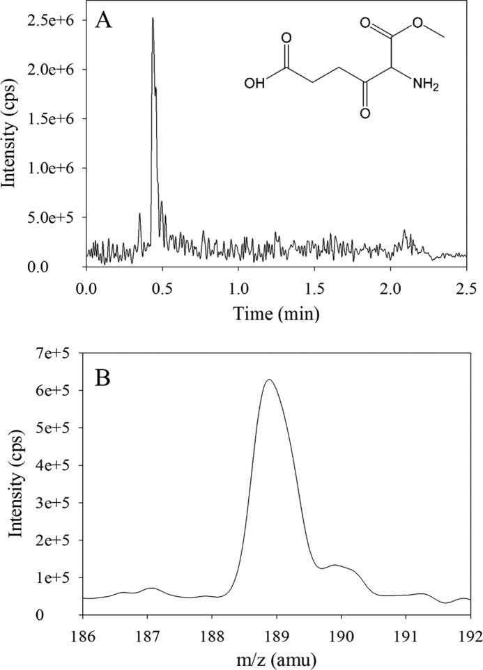
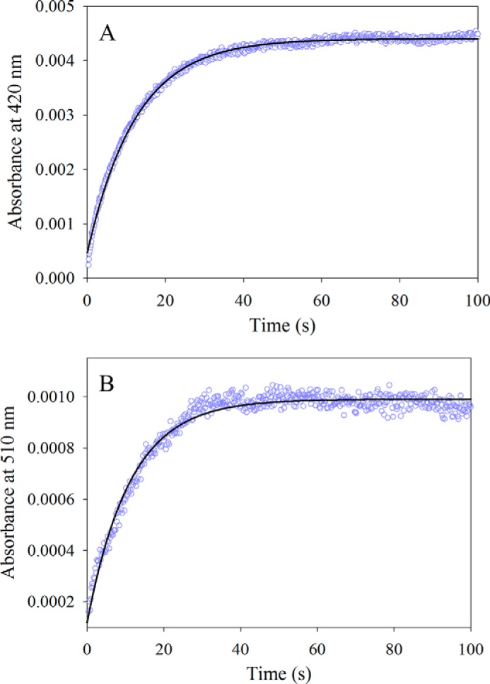
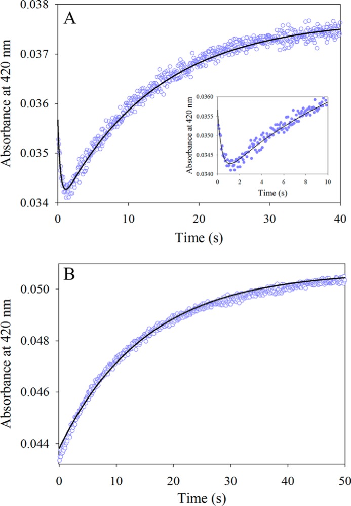
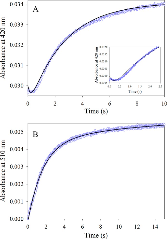
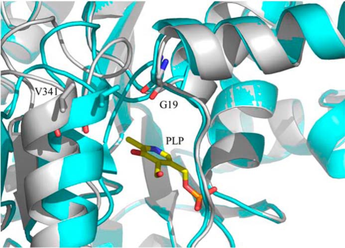
5-Aminolevulinate (ALA), an essential metabolite in all heme-synthesizing organisms, results from the pyridoxal 5'-phosphate (PLP)-dependent enzymatic condensation of glycine with succinyl-CoA in non-plant eukaryotes and α-proteobacteria. The predicted chemical mechanism of this ALA synthase (ALAS)-catalyzed reaction includes a short-lived glycine quinonoid intermediate and an unstable 2-amino-3-ketoadipate intermediate. Using liquid chromatography coupled with tandem mass spectrometry to analyze the products from the reaction of murine erythroid ALAS (mALAS2) with O-methylglycine and succinyl-CoA, we directly identified the chemical nature of the inherently unstable 2-amino-3-ketoadipate intermediate, which predicates the glycine quinonoid species as its precursor. With stopped-flow absorption spectroscopy, we detected and confirmed the formation of the quinonoid intermediate upon reacting glycine with ALAS. Significantly, in the absence of the succinyl-CoA substrate, the external aldimine predominates over the glycine quinonoid intermediate. When instead of glycine, L-serine was reacted with ALAS, a lag phase was observed in the progress curve for the L-serine external aldimine formation, indicating a hysteretic behavior in ALAS. Hysteresis was not detected in the T148A-catalyzed L-serine external aldimine formation. These results with T148A, a mALAS2 variant, which, in contrast to wild-type mALAS2, is active with L-serine, suggest that active site Thr-148 modulates ALAS strict amino acid substrate specificity. The rate of ALA release is also controlled by a hysteretic kinetic mechanism (observed as a lag in the ALA external aldimine formation progress curve), consistent with conformational changes governing the dissociation of ALA from ALAS.

Keywords: 5-Aminolevulinate Synthase; Enzyme Kinetics; Enzyme Mechanism; Heme; Hysteresis; Oxoamine Synthase; Porphyria; Porphyrin; Pyridoxal Phosphate; Sideroblastic Anemia.


Figures

SCHEME 1.
SCHEME 1.
Proposed chemical mechanism for the reaction catalyzed by ALAS. In the resting state, PLP binds covalently to an active site lysine as an internal aldimine (I) (PLP-K313 internal aldimine in mALAS2). The entry of glycine in the active site proceeds with the formation of an external aldimine (II). Removal of the pro-R proton of glycine results in formation of a first quinonoid intermediate (III), which facilitates the condensation with succinyl-CoA and subsequent release of the CoA moiety (IV). When the resulting 2-amino-3-ketoadipate intermediate (V) is decarboxylated (H207-assisted decarboxylation in the mALAS2 reaction), an enol intermediate is formed (VI), which is in rapid equilibrium with a second quinonoid intermediate (VII). The final reaction intermediate is the ALA external aldimine (VIII).
FIGURE 1.
FIGURE 1.
Analysis of the trapped intermediate in the ALAS-catalyzed reaction of O-methylglycine with succinyl-CoA by LC-MS/MS. A, UPLC profile of the reaction sample indicating that the retention time of the major species, which has a mass ion spectrum peak at 188.9 m/z, is 0.44 min. B, MS/MS scan spectrum (abundance versus m/z) of the 0.44 min-fraction derived from the reaction sample indicating that the predominant species, with a peak at 188.9 m/z, is consistent with the molecular mass of 189.17 for the predicted intermediate, 5-amino-6-methoxy-4,6-dioxohexanoic acid (inset in A).
FIGURE 2.
FIGURE 2.
Reaction between wild-type mALAS2 (30 μm) and glycine (100 mm). A, progress curve for the formation of the glycine external aldimine monitored at 420 nm. The data (open purple circles) were fit to a single-exponential equation with an observed rate constant (k1) of 0.080 ± 0.001 s−1 and amplitude (A1) of 4.00 × 10⁻³ ± 0.01 × 10⁻³. B, progress curve for the formation of the glycine quinonoid intermediate monitored at 510 nm. The data (open purple circles) were fit to a single-exponential equation with an observed rate constant (k1) of 0.070 ± 0.001 s−1 and amplitude (A1) of 0.80 × 10⁻³ ± 0.01 × 10⁻³.
FIGURE 3.
FIGURE 3.
Reaction of either wild-type mALAS2 or T148A variant (30 μm) with l-serine (100 mm). A, progress curve for the formation of the l-serine-mALAS2 external aldimine. The data points were fit to a two-exponential equation yielding the observed rate constants (k1 and k2) and amplitudes (A1 and A2): k1 = 2.5 ± 0.2 s−1and A1 = 1.70 × 10⁻³ ± 0.01 × 10⁻³ for the fast phase, and k2 = 0.070 ± 0.001 s−1 and A2 = 3.80 × 10⁻³ ± 0.01 × 10⁻³ for the slow phase. The inset shows the first 10 s of the reaction. B, progress curve for the formation of the l-serine-T148A external aldimine. The data were fit to a single-exponential equation yielding the observed rate constant (k1) of 0.070 ± 0.001 s−1 and the amplitude (A1) of 6.80 × 10⁻³ ± 0.01 × 10⁻³.
FIGURE 4.
FIGURE 4.
Reaction between wild-type mALAS2 (30 μm) and ALA (10 mm). A, progress curve for the formation of the ALA-mALAS2 external aldimine as monitored at 420 nm. The data points were fit to a two-exponential equation yielding the observed rate constants (k1 and k2) and amplitudes (A1 and A2): k1 = 4.7 ± 0.01 s−1 and A1 = 1.1 × 10⁻³ ± 0.07 × 10⁻³ for the fast phase, and k2 = 0.37 ± 0.003 s−1 and A2 = 5.2 × 10⁻³ ± 0.02 × 10⁻³ for the slow phase. The inset shows the first 2.5 s of the reaction. B, progress curve for the formation of the ALA quinonoid intermediate as monitored at 510 nm. The observed rate constants (k1 and k2) and amplitudes (A) were: k1 = 0.61 ± 0.01 s−1 and A1 = 4.50 × 10⁻³ ± 0.07 × 10⁻³ for the fast phase, and k2 = 0.08 ± 0.01 s−1 and A2 = 1.50 × 10⁻³ ± 0.04 × 10⁻³ for the slow phase.
FIGURE 5.
FIGURE 5.
Positioning of the glycine external aldimine in the active site of ALAS. Differences in the positioning of the α-carbon of glycine were detected upon superimposition of the crystal structures for monomers A and E from PDB file 2BWP. The ternary complex formed by the glycine external aldimine and succinyl-CoA was modeled in the active site of ALAS. The succinyl-CoA carbonyl group can strongly interact with the Schiff base nitrogen. The model was built by superimposing the structures with PDB coordinates 2BWP and 2BWO using PyMOL. Amino acid Lys-248 in R. capsulatus ALAS corresponds to Lys-313 in mALAS2.
FIGURE 6.
FIGURE 6.
Model depicting the positioning of the l-serine external aldimine in the active site of ALAS. The model was created by superimposing the l-serine external aldimine of S. paucimobilis SPT (PDB file 2W8J) with the glycine external aldimine of R. capsulatus ALAS (PDB file 2BWP). l-Serine is shown in green and glycine in cyan. Succinyl-CoA was subsequently modeled into the active site by superimposing the generated structure with the structure for the R. capsulatus·succinyl-CoA complex (PDB file 2BWO), using PyMOL. R. capsulatus ALAS amino acids Thr-83, His-142, Lys-248, and Arg-374 correspond to mALAS2 Thr-148, His-207, Lys-313, and Arg-439, respectively.
FIGURE 7.
FIGURE 7.
Alternative channel for the entry of substrates into the active site of R. capsulatus ALAS. A, alternative channel in the open and (B) closed conformations (PDB file 2BWN). This channel and the succinyl-CoA-specific channel are distinct. The N-terminal domain is shown in cyan, the catalytic domain in gray, and the C-terminal domain in magenta. The opposite monomer is shown in yellow. Succinyl-CoA is shown in stick representation.
FIGURE 8.
FIGURE 8.
Differences in the positioning of C-terminal α-helix Val-341 in the open and closed conformations. The open and closed conformations are shown in gray and cyan, respectively. Monomers A and B (PDB file 2BWN) were superimposed using PyMOL.

References

    1. Fratz E. J., Stojanovski B. M., Ferreira G. C. (2013) Toward Heme: 5-Aminolevulinate Synthase and Initiation of Porphyrin Synthesis in The Handbook of Porphyrin Science (Ferreira G. C., Kadish K. M., Smith K. M., Guilard R., ed), pp. 1–78, World Scientific Publishing Co., New Jersey
    1. Hunter G. A., Ferreira G. C. (2011) Molecular enzymology of 5-aminolevulinate synthase, the gatekeeper of heme biosynthesis. Biochim. Biophys. Acta 1814, 1467–1473 - PMC - PubMed
    1. Bottomley S. S. (2004) Sideroblastic Anemias. in Wintrobe's Clinical Hematology (Greer J., Foerster J., Lukens J. N., Rodgers G. M., Paraskevas R., Glader R. B., ed), pp. 1011–1033, Williams and Wilkins, Philadelphia, PA
    1. Whatley S. D., Ducamp S., Gouya L., Grandchamp B., Beaumont C., Badminton M. N., Elder G. H., Holme S. A., Anstey A. V., Parker M., Corrigall A. V., Meissner P. N., Hift R. J., Marsden J. T., Ma Y., Mieli-Vergani G., Deybach J. C., Puy H. (2008) C-terminal deletions in the ALAS2 gene lead to gain of function and cause X-linked dominant protoporphyria without anemia or iron overload. Am. J. Hum. Genet. 83, 408–414 - PMC - PubMed
    1. Mann S., Ploux O. (2011) Pyridoxal-5′-phosphate-dependent enzymes involved in biotin biosynthesis: structure, reaction mechanism and inhibition. Biochim. Biophys. Acta 1814, 1459–1466 - PubMed

Publication types

LinkOut - more resources