

The Drosophila egg chamber-a new spin on how tissues elongate
- PMID: 24920751
- PMCID: PMC4229889
- DOI: 10.1093/icb/icu067
The Drosophila egg chamber-a new spin on how tissues elongate
Abstract
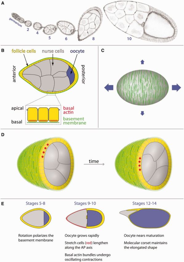
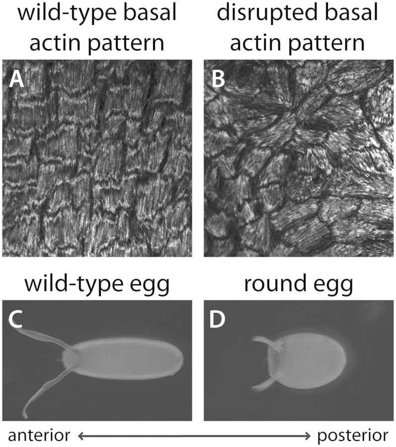
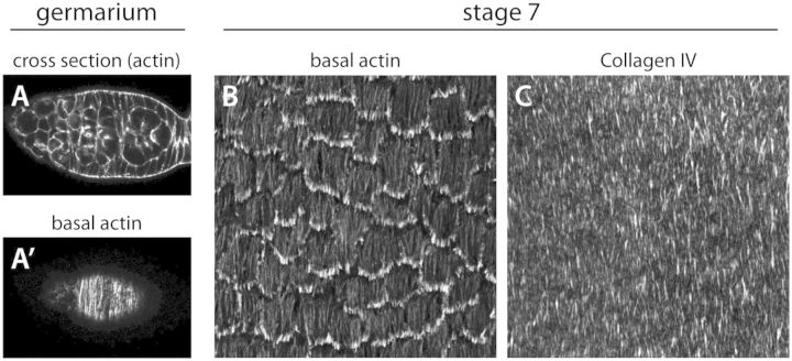
During development, tissues undergo complex cellular rearrangements and changes in shape that produce a diversity of body plans and the functional organs therein. The Drosophila egg chamber has emerged as an exciting and highly tractable model in which to investigate novel mechanisms driving the elongation of tissues. Egg chambers are multicellular assemblies within flies' ovaries that will each give rise to a single egg. Although initially spherical, these simple organ-like structures lengthen as they grow. This transformation depends on an unusual form of planar polarity in the egg chamber's outer epithelial layer, in which arrays of linear actin bundles and fibril-like structures in the basement membrane both align perpendicular to the axis of elongation. The resulting circumferential arrangement of structural molecules is then thought to act as a "molecular corset" that directionally biases growth of the egg chamber. I will explore four fundamental questions about this system: (1) How is the circumferential pattern generated in the follicular epithelium? (2) What is the physical nature of the corset? (3) How does a corset-type mechanism lead to the cellular rearrangements necessary for the elongation of tissues? and (4) To what extent are the cellular mechanisms controlling egg chamber elongation conserved in other systems? For each topic, I will present insights gleaned from the recent literature and highlight fertile areas for future investigation.
© The Author 2014. Published by Oxford University Press on behalf of the Society for Integrative and Comparative Biology. All rights reserved. For permissions please email: journals.permissions@oup.com.
Figures
References
-
- Aigouy B, Farhadifar R, Staple DB, Sagner A, Roper JC, Julicher F, Eaton S. Cell flow reorients the axis of planar polarity in the wing epithelium of Drosophila. Cell. 2010;142:773–86. - PubMed
-
- Bateman J, Reddy RS, Saito H, Van Vactor D. The receptor tyrosine phosphatase Dlar and integrins organize actin filaments in the Drosophila follicular epithelium. Curr Biol. 2001;11:1317–27. - PubMed
-
- Brangwynne C, Huang S, Parker KK, Ingber DE, Ostuni E. Symmetry breaking in cultured mammalian cells. In Vitro Cell Dev Biol Anim. 2000;36:563–5. - PubMed
Publication types
MeSH terms
Substances
Grants and funding
LinkOut - more resources
Full Text Sources
Other Literature Sources
Molecular Biology Databases
