. 2014 Aug 1;307(3):F337-45.
doi: 10.1152/ajprenal.00208.2014. Epub 2014 Jun 11.

Renal blood flow and oxygenation drive nephron progenitor differentiation

Affiliations

Renal blood flow and oxygenation drive nephron progenitor differentiation

Christopher Rymer et al. Am J Physiol Renal Physiol. .

Abstract

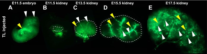
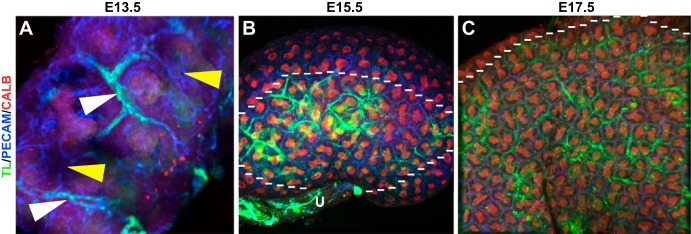
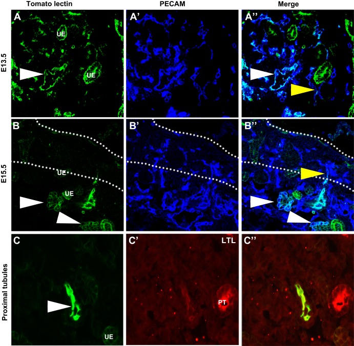
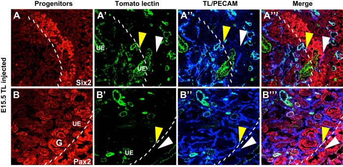
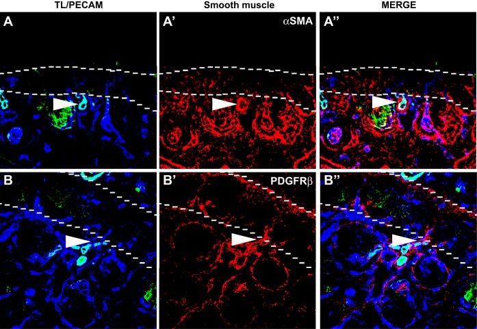
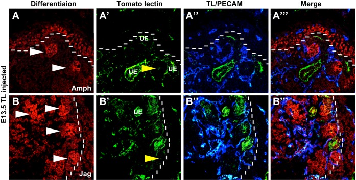
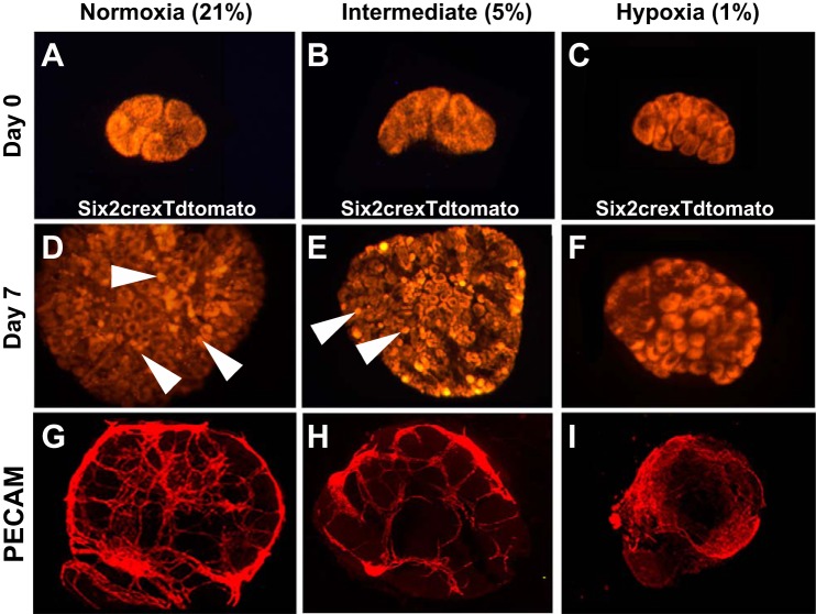
During kidney development, the vasculature develops via both angiogenesis (branching from major vessels) and vasculogenesis (de novo vessel formation). The formation and perfusion of renal blood vessels are vastly understudied. In the present study, we investigated the regulatory role of renal blood flow and O2 concentration on nephron progenitor differentiation during ontogeny. To elucidate the presence of blood flow, ultrasound-guided intracardiac microinjection was performed, and FITC-tagged tomato lectin was perfused through the embryo. Kidneys were costained for the vasculature, ureteric epithelium, nephron progenitors, and nephron structures. We also analyzed nephron differentiation in normoxia compared with hypoxia. At embryonic day 13.5 (E13.5), the major vascular branches were perfused; however, smaller-caliber peripheral vessels remained unperfused. By E15.5, peripheral vessels started to be perfused as well as glomeruli. While the interior kidney vessels were perfused, the peripheral vessels (nephrogenic zone) remained unperfused. Directly adjacent and internal to the nephrogenic zone, we found differentiated nephron structures surrounded and infiltrated by perfused vessels. Furthermore, we determined that at low O2 concentration, little nephron progenitor differentiation was observed; at higher O2 concentrations, more differentiation of the nephron progenitors was induced. The formation of the developing renal vessels occurs before the onset of blood flow. Furthermore, renal blood flow and oxygenation are critical for nephron progenitor differentiation.

Keywords: blood flow; endothelium; hypoxia; kidney development; vasculature.


Figures

Fig. 1.
Fig. 1.
Kidney blood flow occurs in a sequential spatiotemporal pattern. A–E: representative images of a tomato lectin (TL; green)-injected embryo (A) and kidneys (B–E) at various developmental stages. A: embryonic day 11.5 (E11.5) embryo showing perfused vessels throughout the head (white arrowheads) and body of the embryo, with the site of injection showing very bright staining (yellow arrowhead). B: dissected E11.5 kidney showing perfused vessels surrounding the developing kidney in a honeycomb arrangement (as marked by the dotted line). C: E13.5 kidney (as marked by the dotted line) showing perfusion of the major renal vessels (white arrowheads). Some lectin can also been seen sticking to the ureteric epithelium (yellow arrowhead). D: E15.5 kidney (as marked by the dotted line) showing perfusion of the smaller vascular branches (yellow arrowheads) as well as several perfused glomeruli (white arrowheads). E: E17.5 kidney showing significant perfusion throughout the developing kidney including numerous perfused glomeruli (white arrowheads) and smaller caliber vessels (yellow arrowheads).
Fig. 2.
Fig. 2.
Whole mount images suggesting that the formation of renal vessels precedes renal blood flow. A–C: whole mount images of blood flow in the developing kidney (green) at various developmental stages colabeled with vascular [platelet endothelial cell adhesion molecule (PECAM; blue)] and ureteric epithelial (calbindin, red) markers. A: representative E13.5 kidney showing the major vessels (white arrowhead) that are perfused and peripheral areas of the kidney that are vascularized but lacking blood flow (yellow arrowheads). B: E15.5 whole mount kidney showing that the more centralized vessels (inside the dotted line) that are closer to the ureter (U) and angiogenic vessels are perfused and interdigitating between the ureteric epithelium (red), but the peripheral vessels (outside the dotted line), likely of vasculogenic origin, are unperfused. C: representative E17.5 kidney showing significantly more perfusion throughout the kidney interdigitating between the branching ureteric epithelium (below the dotted line). However, in the presumptive nephrogenic zone, the vessels are unperfused (above the dotted line).
Fig. 3.
Fig. 3.
Confirmation that the renal vasculature develops before blood flow. A and B: sections of TL (green) perfused embryos colabeled with PECAM (blue). A–A″: representative E13.5 sections showing the major vessels as marked with PECAM perfused with TL (white arrowheads). In a small number of peripheral vessels, TL can also been seen in a subset of vessels that attached to unperfused vessels (yellow arrowhead). B–B″: E15.5 sections showing a peripheral vessel that showed positive lectin staining, indicating perfusion, until the outer cortex. Past this point, the vessel lacks perfusion (yellow arrowhead). Glomeruli are also apparent that are clearly perfused at this time point (white arrowheads). C–C″: E15.5 sections injected with TL and costained with the proximal tubule (PT) marker Lotus tetragonolobus lectin (LTL). C: TL injection site with a perfused vessel clearly demarcated (white arrowhead). Some staining was also observed in the ureteric epithelium (UE), whereas no staining is observed in the proximal tubule.
Fig. 4.
Fig. 4.
Blood flow does not extend into the undifferentiated nephron progenitors. A and B: representative images of E15.5 kidneys perfused with FITC-TL and colabeled for markers of nephron progenitors (Six2 and Pax2, red) and the vasculature (PECAM, blue). A–A″′: Six2 (red) is shown to mark nephron progenitors, which reside in the nephrogenic zone (marked by the dotted line). Perfused vessels (yellow arrowheads) stained with TL (green) and PECAM (blue) can be seen all the way to the nephrogenic zone, and unperfused vessels (white arrowheads) are then seen throughout the nephrogenic zone. B–B″′: a similar expression pattern is seen with Pax2 (red), although staining for Pax2 was also observed in the ureteric epithelium and developing glomeruli (G). Whereby perfused vessels (yellow arrowheads) marked by TL (green) abut the nephrogenic zone, however, the blood flow does not penetrate, leaving the vessels marked with PECAM (blue) within the nephrogenic zone unperfused (white arrowheads).
Fig. 5.
Fig. 5.
Smooth muscle cells and pericytes aggregate nephrogenic zone. A and B: representative E15.5 images showing smooth muscle/pericyte formation in relation to blood flow. A–A″: representative images showing blood flow in relation to smooth muscle formation. As shown, where there was cessation of blood flow into the nephrogenic zone (dotted line), there was an abundance of smooth muscle cells (white arrowheads). α-SMA, α-smooth muscle actin. B–B″: similarly, when we observed representative images of the pericyte marker platelet-derived growth factor receptor (PDGFR)-β, we found a similar clumping of PDGFR-β-positive cells (white arrowheads) at the border of blood flow and the nephrogenic zone (dotted line).
Fig. 6.
Fig. 6.
Renal blood flow is closely associated with nephron differentiation. A and B: representative images of E13.5 TL (green)-injected kidneys stained for nephron differentiation markers amphyphisin and jagged1 (Amph and Jag; red) and the vasculature (PECAM, blue). A–A″′: amphiphysin (red) staining was found in the early renal vesicles (white arrowheads) and also throughout the nephron progenitors in the neprogenic zone (dotted line). Perfused vessels were found encircling these differentiating structures and also contained within them (yellow arrowheads). B–B″′: similarly, jagged1-positive renal vesicles (white arrowheads) were observed surrounded by perfused vessels. The vessels were also located within the vesicles (yellow arrowheads).
Fig. 7.
Fig. 7.
Varying O2 concentrations mediate the amount of nephron progenitor differentiation. A–F: Six2cre mice bred with a tdTomato reporter mouse (red) were grown under varying O2 concentrations for 7 days. A–C: E12.5 kidneys at 0 days of culture showing the kidneys placed in normoxia (A), intermediate hypoxia (B), and hypoxia (C) at developmentally comparable stages. There was a presence of nephron progenitors but no differentiated structures. D–F: after 7 days of culture in normoxia (C), the kidney showed numerous differentiated nephron structures (white arrowheads), whereas a stepwise reduction in the number of glomerular structures was observed in the intermediate (E) and hypoxic (F) conditions. G–I: representative whole mount images of PECAM staining in explants at various O2 concentrations. Under the normal O2 concentration (G), significant differentiation and patterning of vessels were observed. There was still relatively well-defined differentiation at 3% O2 concentration (H), but a complete lack of vascular organization was observed in the 1% O2 concentration (I).

References

    1. Abrahamson DR, Robert B, Hyink DP, St John PL, Daniel TO. Origins and formation of microvasculature in the developing kidney. Kidney Int Suppl 67: S7–S11, 1998 - PubMed
    1. Andres AC, Munarini N, Djonov V, Bruneau S, Zuercher G, Loercher S, Rohrbach V, Ziemiecki A. EphB4 receptor tyrosine kinase transgenic mice develop glomerulopathies reminiscent of aglomerular vascular shunts. Mech Dev 120: 511–516, 2003 - PubMed
    1. Costantini F. Genetic controls and cellular behaviors in branching morphogenesis of the renal collecting system. Wiley Interdisip Rev Dev Biol 1: 693–713, 2012 - PMC - PubMed
    1. Costantini F, Kopan R. Patterning a complex organ: branching morphogenesis and nephron segmentation in kidney development. Dev Cell 18: 698–712, 2010 - PMC - PubMed
    1. Das A, Tanigawa S, Karner CM, Xin M, Lum L, Chen C, Olson EN, Perantoni AO, Carroll TJ. Stromal-epithelial crosstalk regulates kidney progenitor cell differentiation. Nat Cell Biol 15: 1035–1044, 2013 - PMC - PubMed

Publication types

MeSH terms

LinkOut - more resources