. 2014 Sep 1;88(17):9579-89.
doi: 10.1128/JVI.00975-14. Epub 2014 Jun 11.

CD40L-adjuvanted DNA/modified vaccinia virus Ankara simian immunodeficiency virus SIV239 vaccine enhances SIV-specific humoral and cellular immunity and improves protection against a heterologous SIVE660 mucosal challenge

Affiliations

CD40L-adjuvanted DNA/modified vaccinia virus Ankara simian immunodeficiency virus SIV239 vaccine enhances SIV-specific humoral and cellular immunity and improves protection against a heterologous SIVE660 mucosal challenge

Suefen Kwa et al. J Virol. .

Abstract

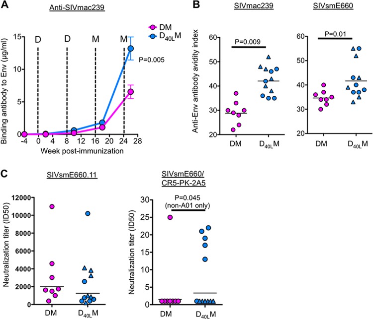
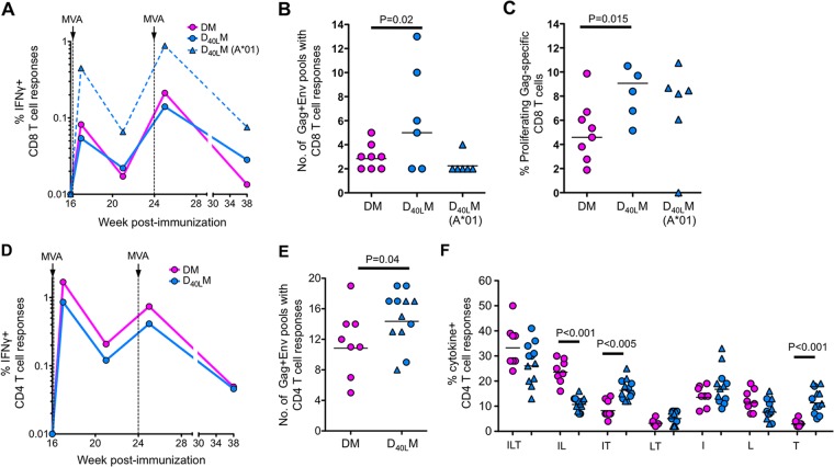
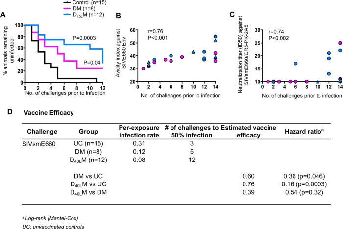
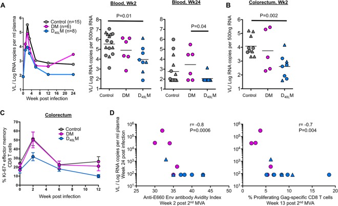
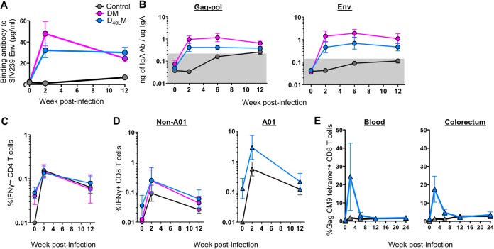
It remains a challenge to develop a successful human immunodeficiency virus (HIV) vaccine that is capable of preventing infection. Here, we utilized the benefits of CD40L, a costimulatory molecule that can stimulate both dendritic cells (DCs) and B cells, as an adjuvant for our simian immunodeficiency virus (SIV) DNA vaccine in rhesus macaques. We coexpressed the CD40L with our DNA/SIV vaccine such that the CD40L is anchored on the membrane of SIV virus-like particle (VLP). These CD40L containing SIV VLPs showed enhanced activation of DCs in vitro. We then tested the potential of DNA/SIV-CD40L vaccine to adjuvant the DNA prime of a DNA/modified vaccinia virus Ankara (MVA) vaccine in rhesus macaques. Our results demonstrated that the CD40L adjuvant enhanced the functional quality of anti-Env antibody response and breadth of anti-SIV CD8 and CD4 T cell responses, significantly delayed the acquisition of heterologous mucosal SIV infection, and improved viral control. Notably, the CD40L adjuvant enhanced the control of viral replication in the gut at the site of challenge that was associated with lower mucosal CD8 immune activation, one of the strong predictors of disease progression. Collectively, our results highlight the benefits of CD40L adjuvant for enhancing antiviral humoral and cellular immunity, leading to enhanced protection against a pathogenic SIV. A single adjuvant that enhances both humoral and cellular immunity is rare and thus underlines the importance and practicality of CD40L as an adjuvant for vaccines against infectious diseases, including HIV-1.

Importance: Despite many advances in the field of AIDS research, an effective AIDS vaccine that can prevent infection remains elusive. CD40L is a key stimulator of dendritic cells and B cells and can therefore enhance T cell and antibody responses, but its overly potent nature can lead to adverse effects unless used in small doses. In order to modulate local expression of CD40L at relatively lower levels, we expressed CD40L in a membrane-bound form, along with SIV antigens, in a nucleic acid (DNA) vector. We tested the immunogenicity and efficacy of the CD40L-adjuvanted vaccine in macaques using a heterologous mucosal SIV infection. The CD40L-adjuvanted vaccine enhanced the functional quality of anti-Env antibody response and breadth of anti-SIV T cell responses and improved protection. These results demonstrate that VLP-membrane-bound CD40L serves as a novel adjuvant for an HIV vaccine.


Figures

FIG 1
FIG 1
Design and expression of DNA vaccines. (A) Design of DNA vaccines without CD40L (DNA/SIV) and with CD40L (DNA/SIV-40L). The DNA/SIV immunogen expresses SIV239 Gag, Env, protease (PR), reverse transcriptase (RT), rev, and tat, using the cytomegalovirus (CMV) intermediate-early promoter with intron A and stabilized by a bovine growth hormone (BGH) polyadenylation sequence. The DNA/SIV-40L vaccine comprises the same structure as the DNA/SIV vaccine but includes the macaque CD40L sequence as a fusion to an internal ribosome entry site (IRES) inserted downstream of Env. X, safety mutations. (B) Expression of Gag, Env, or CD40L by flow cytometry in 293T cells transfected with DNA/SIV or DNA/SIV-40L plasmids. (C) Electron micrograph of virus-like particles (VLPs) budding from a transfected 293T cell. (D) Immunogold staining for CD40L on VLPs from cells transfected with DNA/SIV or DNA/SIV-40L plasmids. White arrows pointing to dark spots indicate immunogold staining for CD40L. (E) VLPs containing CD40L upregulate CD80 on myeloid DCs (mDC; HLA-DR+, CD11c+, CD123, CD3, CD20, and CD14). Tissue culture supernatants from 293T cells transfected with DNA/SIV or DNA/SIV-40L plasmids were used for stimulations and cells were stained after 12 h of stimulation. Longer periods (24 to 48 h) of stimulation resulted in significant spontaneous death of DCs, so data for 12 h is shown. RM1 to RM4 represent four rhesus macaques. Mock, supernatants from Lipofectamine-only cultures.
FIG 2
FIG 2
Anti-SIV antibody responses postvaccination. (A) Levels of binding antibody against vaccine immunogen SIVmac239 Env postvaccination. (B) Avidity index for full-length Env captured from Triton X-100-disrupted VLPs SIVmac239 or SIVE660 elicited Env-specific IgG at 2 weeks after a second MVA boost. (C) Neutralization titers to tier 1 (SIVE660.11) and tier 2 (SIVE660/CR5-PK-2A5) pseudoviruses. Titers were determined at 2 weeks after a second MVA boost. D, DNA vaccine; M, MVA vaccine.
FIG 3
FIG 3
Anti-SIV T cell responses postvaccination. (A) Magnitude of IFN-γ+ CD8 T cell responses. (B) Breadth of anti-SIV CD8 T cell responses. Breadth is measured using 13 Gag peptide pools and 11 Env peptide pools. These pools consist of a total of 125 Gag peptides and 225 Env peptides. (C) Percent proliferating Gag-specific CD8 T cells at 1 week after the second MVA, as measured by CFSE dilution. (D) Magnitude of IFN-γ+ CD4 T cell responses. (E) Breadth of anti-SIV CD4 T cell responses. (F) Boolean analysis of anti-SIV CD4 T cell responses at 1 week after the first MVA. Capital letters indicate the coexpression patterns for IFN-γ (I), IL-2 (L), and/or TNF-α (T).
FIG 4
FIG 4
Kaplan-Meier's survival curve analysis postchallenge. (A) Survival curve of DM, DCD40LM, and control groups. (B) Correlation between avidity index of vaccine-elicited SIVE660 Env-specific IgG at 2 weeks after the second MVA boost and the number of challenges to productive infection. (C) Correlation analysis between the neutralization titer against tier 2 SIVE660/CR5-PK-2A5 virus at 2 weeks after the second MVA boost and the number of challenges to productive infection. Uninfected animals are shown at challenge 14. (D) Vaccine efficacy against SIVE660 challenge.
FIG 5
FIG 5
Postinfection viral load, immune activation, and correlations. (A) Plasma viral load of non-A01+ and A01+ infected animals. (B) Viral RNA copies per 500 ng of total RNA in the colorectum at 2 weeks postinfection. (C) CD8 T cell immune activation in the gut (as measured by Ki67 expression on CD28 CD95+ effector memory CD8). (D) Correlation between viral load at week 24 postinfection and the avidity index against the SIVE660 at 2 weeks after the second MVA or the percentage of proliferating Gag-specific CD8 T cells at week 13 after the second MVA.
FIG 6
FIG 6
Anamnestic expansion of anti-SIV immunity postinfection in infected animals. (A) Titer of SIVmac239 Env-specific antibodies in serum. The mean is shown for each group. (B) Levels of Gag-Pol- or Env-specific IgA specific activity in rectal secretions. The geometric mean is shown for each group. (C) Levels of IFN-γ+ CD4 T cells in the blood. The geometric mean is shown for each group. (D) Levels of IFN-γ+ CD8 T cells in non-A01 and A01 animals. The geometric mean is shown for each group. (E) Frequency of Gag CM9 tetramer-positive CD8 T cells in the blood and colorectum. The mean is shown for each group. The legend for panels B to E is shown in panel A. The error bars indicate the standard errors.

References

    1. Grewal IS, Flavell RA. 1998. CD40 and CD154 in cell-mediated immunity. Annu. Rev. Immunol. 16:111–135. 10.1146/annurev.immunol.16.1.111 - DOI - PubMed
    1. Bennett SR, Carbone FR, Karamalis F, Flavell RA, Miller JF, Heath WR. 1998. Help for cytotoxic-T-cell responses is mediated by CD40 signaling. Nature 393:478–480. 10.1038/30996 - DOI - PubMed
    1. Schoenberger SP, Toes RE, van der Voort EI, Offringa R, Melief CJ. 1998. T-cell help for cytotoxic T lymphocytes is mediated by CD40-CD40L interactions. Nature 393:480–483. 10.1038/31002 - DOI - PubMed
    1. Ridge JP, Di Rosa F, Matzinger P. 1998. A conditioned dendritic cell can be a temporal bridge between a CD4+ T-helper and a T-killer cell. Nature 393:474–478. 10.1038/30989 - DOI - PubMed
    1. Elgueta R, Benson MJ, de Vries VC, Wasiuk A, Guo Y, Noelle RJ. 2009. Molecular mechanism and function of CD40/CD40L engagement in the immune system. Immunol. Rev. 229:152–172. 10.1111/j.1600-065X.2009.00782.x - DOI - PMC - PubMed

Publication types

MeSH terms