. 2014 Sep 1;88(17):9673-82.
doi: 10.1128/JVI.01147-14. Epub 2014 Jun 11.

Genomic flexibility of human endogenous retrovirus type K

Affiliations

Genomic flexibility of human endogenous retrovirus type K

Derek Dube et al. J Virol. .

Abstract

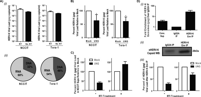
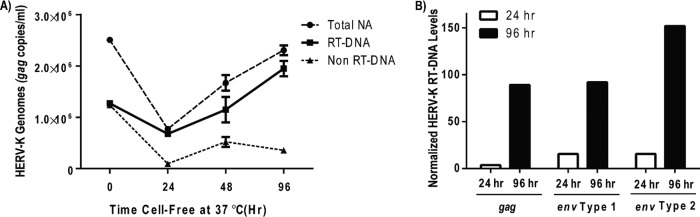
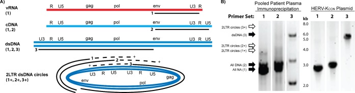
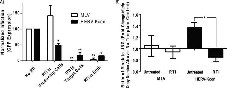
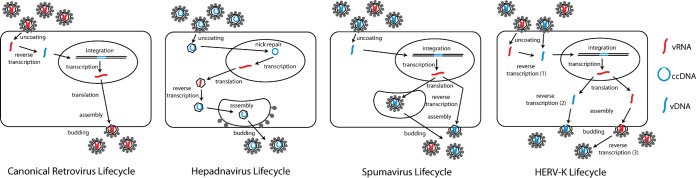
Human endogenous retrovirus type K (HERV-K) proviruses are scattered throughout the human genome, but as no infectious HERV-K virus has been detected to date, the mechanism by which these viruses replicated and populated the genome remains unresolved. Here, we provide evidence that, in addition to the RNA genomes that canonical retroviruses package, modern HERV-K viruses can contain reverse-transcribed DNA (RT-DNA) genomes. Indeed, reverse transcription of genomic HERV-K RNA into the DNA form is able to occur in three distinct times and locations: (i) in the virus-producing cell prior to viral release, yielding a DNA-containing extracellular virus particle similar to the spumaviruses; (ii) within the extracellular virus particle itself, transitioning from an RNA-containing particle to a DNA-containing particle; and (iii) after entry of the RNA-containing virus into the target cell, similar to canonical retroviruses, such as murine leukemia virus and HIV. Moreover, using a resuscitated HERV-K virus construct, we show that both viruses with RNA genomes and viruses with DNA genomes are capable of infecting target cells. This high level of genomic flexibility historically could have permitted these viruses to replicate in various host cell environments, potentially assisting in their many integration events and resulting in their high prevalence in the human genome. Moreover, the ability of modern HERV-K viruses to proceed through reverse transcription and package RT-DNA genomes suggests a higher level of replication competency than was previously understood, and it may be relevant in HERV-K-associated human diseases.

Importance: Retroviral elements comprise at least 8% of the human genome. Of all the endogenous retroviruses, HERV-K viruses are the most intact and biologically active. While a modern infectious HERV-K has yet to be found, HERV-K activation has been associated with cancers, autoimmune diseases, and HIV-1 infection. Thus, determining how this virus family became such a prevalent member of our genome and what it is capable of in its current form are of the utmost importance. Here, we provide evidence that HERV-K viruses currently found in the human genome are able to proceed through reverse transcription and historically utilized a life cycle with a surprising degree of genomic flexibility in which both RNA- and DNA-containing viruses were capable of mediating infection.


Figures

FIG 1
FIG 1
HERV-K particles containing RT-DNA are produced by teratocarcinoma cells in tissue culture. (A) Supernatants from NCCIT and Tera-1 cells were cleared of debris and contaminating DNA, concentrated, and subjected to qPCR or qRT-PCR for HERV-K gag. (i) HERV-K viral load in the supernatant from one representative experiment for each cell type on a log scale. (ii) Percentages of HERV-K vRNA and vDNA assuming 100% RT efficiency from the same experiments. (B) Supernatants from NCCIT or Tera-1 cells were prepared as described for panel A, either mock treated or UNG treated, and analyzed by qRT-PCR for HERV-K gag. The bars shown represent the percentages of HERV-K gag copies normalized to values for mock treatment of the respective cells from one representative experiment with Tera-1 cells and the averages from seven experiments with NCCIT cells. Statistical significance was determined by the Student t test comparing mock- and UNG-treated samples (*, P < 0.01). (C) NCCIT cells were cultured in the presence or absence of a reverse transcriptase inhibitor (3TC at 50 μM). The supernatant (Sup) was collected, cleared, concentrated, either mock or UNG treated, and then analyzed for HERV-K viral load as described above. The data shown display the effect of UNG relative to the level of HERV-K gag copies in each specific treatment. (D, i) Concentrated (Conc.) NCCIT cell supernatants were maintained or immunoprecipitated (IP) with a control IgG2A- or HERK-Env-specific capture antibody. The relative fold enrichment of HERV-K genomes as measured by qRT-PCR for HERV-K gag with respect to glyceraldehyde-3-phosphate dehydrogenase (GAPDH) was calculated using the ΔΔCT method. (ii) HERV-K particles from concentrated NCCIT supernatants were immunoprecipitated as described above and then probed by Western blotting (WB) for HERV-K capsid protein. Equivalent volumes were loaded into the lanes for the immunoprecipitated samples. (iii) NCCIT cells were cultured in the absence or presence of the reverse transcription inhibitor (RTI; 3TC at 50 μM), and HERV-K viral particles were immunoprecipitated as described above. Viral load was analyzed by qRT-PCR for HERV-K gag, and the effect of UNG was normalized to values for mock treatment for each sample independently.
FIG 2
FIG 2
Reverse transcription occurs within extracellular HERV-K virions. (A) Viral supernatants from NCCIT cells were collected, cleared, and DNase treated as described for Fig. 1. The resulting cell-free viral suspensions then were incubated at 37°C for various times prior to extracting the viral nucleic acid, mock or UNG treatment, and viral load analysis by qRT-PCR for HERV-K gag. The three lines represent the mock-treated sample (Total NA), the UNG-treated sample (Non RT-DNA), and the difference between the two (RT-DNA). (B) In a similar but separate experiment, the HERV-K RT-DNA levels were examined over time with three distinct primer sets (the gag product is from the 5′ end of the HERV-K coding region, and the two env primer sets amplify products in the 3′ end of the HERV-K coding region). For each primer set, time zero is set at 100. Of note, at later time points (>168 h) the overall viral load was significantly diminished, as would be expected with long-term exposure at 37°C due to general virus instability. A single representative experiment (of eight in total) is shown for both panels A and B.
FIG 3
FIG 3
HERV-K vDNA is present in the plasma of patients with lymphoma. (A) Plasma from a single representative patient was treated with DNase and then assayed for HERV-K genomes with gag-specific primers with or without reverse transcriptase (RT) as described for Fig. 2. (B) Multiple patients were assayed as described for panel A, and the percentage of HERV-K vDNA genomes in the population was calculated by dividing the no-RT viral load by the RT viral load. (C) Lymphoma patient plasma (distinct from those shown in panels A and B) was treated with DNase prior to the extraction of viral nucleic acids and then treated with DNase again after viral lysis prior to assay by qRT-PCR. The data from one representative experiment are shown. (D) A subset of plasma from patients with lymphoma was analyzed for their sensitivity to UNG treatment as described in the legend to Fig. 1. The percentage of HERV-K nucleic acids susceptible to UNG (RT-DNA genomes) is shown for each patient. (E) Plasma from four patients with diffuse large B cell lymphoma (DLBCL) were pooled, and then HERV-K particles were immunoprecipitated as described for Fig. 1. HERV-K nucleic acids were extracted and subjected to mock or UNG treatment prior to qRT-PCR for HERV-K gag. The bar shown represents the percentage of HERV-K genomes susceptible to UNG (RT-DNA genomes) of total HERV-K genomes from a single representative experiment.
FIG 4
FIG 4
HERV-K particles from pooled lymphoma patient plasma contain dsDNA genomes. (A) Three different primer sets were employed to take advantage of differences in the viral genomes before, during, and after reverse transcription. Primer set 1 (env forward and R reverse) will amplify a 2.9-kb product in all forms of HERV-K genomes and an additional 3.9-kb product in 2LTR dsDNA circles (1+). Primer set 2 (env forward and U5 reverse) will amplify a 3-kb product in all species except vRNA and an additional 4-kb product in 2LTR circles (2+). Primer set 3 (U3 forward and env reverse) will amplify a 5.9-kb product in dsDNA and 2LTR circles and an additional 6.9-kb product in 2LTR circles (3+; depicted on the 2LTR circular DNA by the dashed line). (B) RT-PCR (primer set 1) or PCR (primer sets 2 and 3) was done with both pooled immunoprecipitated patient plasma (as described for Fig. 1D) and a control HERV-KCON plasmid (which should yield all amplification products except for the 2LTR circular DNA form). The lanes shown are labeled with the primer set used, are from the same experiment, and were run on the same gel. Solid arrows indicate a DNA band at the expected size in the indicated lane, while empty arrows indicate no band of the predicted size, suggesting the absence of 2LTR circles in this experiment. Bands of interest were confirmed as HERV-K by sequence verification. Note that the doublets seen in the patient serum samples with primer sets 1 and 2 represent HERV-K type 1 (smaller) and 2 (larger) genomes.
FIG 5
FIG 5
RT-DNA- and RNA-containing HERV-KCON particles both yield productive infection. HERV-KCON particles bearing VSV-G and encoding a GFP reporter were produced in 293FT cells as described in reference . Recombinant MLV particles bearing VSV-G and encoding a GFP reporter were produced in parallel. (A) Analysis of the effects of RTI on infection levels of HERV-K and MLV. Viruses were produced in cells in the presence or absence of a reverse transcriptase inhibitor (AZT at 50 μM) for 48 h, and the supernatant was collected, cleared for debris by centrifugation, and filtered through a 0.45-μm Whatman filter. 293FT cells in the presence or absence of reverse transcriptase inhibitor (AZT at 50 μM) were spinfected with the resulting cleared viral supernatants for 1 h at 800 × g and incubated at 37°C for an additional 36 h to allow infection to occur. The cells then were prepared for flow cytometry, and infection was assessed by GFP reporter expression. Overall infection levels for HERV-KCON were low (100 to 1,000 IU/ml) but reproducible. The bars shown are the normalized averages from three experiments. Statistical significance was determined by a Student t test comparing the various RTI treatments to no RTI (*, P < 0.01; **, P < 0.001). (B) Analysis of the genomic constituents of infectious virus particles. Viral preparations used for panel A were treated with DNase prior to extraction of the viral nucleic acids and subjected to mock or UNG treatment prior to qRT-PCR with primers specific to the gfp reporter gene found in both recombinant viruses. The ΔΔCT method was used to determine the fold change of the gfp gene copy number above the no-template control for the mock- and UNG-treated samples (representing the relative levels of the viral genomes). The bars shown represent the fold change from mock-treated samples divided by that from UNG-treated samples. A value of 1 suggests that no RT-DNA-containing particles were present in the viral population (the quantity of viral genomes was the same in mock- and UNG-treated samples), and a value significantly above 1 suggests the presence of RT-DNA genomes in the population (UNG treatment reduced the number of viral genomes amplified). Significance was determined by the Student t test comparing the fold expression of the untreated and RTI-treated virus-producing cells within each virus type (*, P < 0.001).
FIG 6
FIG 6
Model for the viral life cycle of HERV-K compared to those of the canonical Retroviridae, Hepadnaviridae, and Spumaviridae. The three distinct replication pathways used by retroviruses, hepadnaviruses, and spumaviruses are depicted. Reverse transcription of the vRNA (red) genome occurs early in the infection process for canonical retroviruses and late during infection in hepadnaviruses and spumaviruses, so that the latter two virus families produce progeny virions containing vDNA (blue) genomes. Of note, while the spumavirus particles with vDNA genomes are much more important for downstream infection, vRNA genomes also can be packaged into viral particles. Alternatively, putative infectious HERV-K viruses can undergo reverse transcription either early or late in the infection process (and potentially in extracellular virions).

References

    1. de Parseval N, Heidmann T. 2005. Human endogenous retroviruses: from infectious elements to human genes. Cytogenet. Genome Res. 110:318–332. 10.1159/000084964 - DOI - PubMed
    1. Bannert N, Kurth R. 2004. Retroelements and the human genome: new perspectives on an old relation. Proc. Natl. Acad. Sci. U. S. A. 101(Suppl 2):S14572–S14579. 10.1073/pnas.0404838101 - DOI - PMC - PubMed
    1. Lower R, Lower J, Kurth R. 1996. The viruses in all of us: characteristics and biological significance of human endogenous retrovirus sequences. Proc. Natl. Acad. Sci. U. S. A. 93:5177–5184. 10.1073/pnas.93.11.5177 - DOI - PMC - PubMed
    1. Kitamura M, Maruyama N, Shirasawa T, Nagasawa R, Watanabe K, Tateno M, Yoshiki T. 1994. Expression of an endogenous retroviral gene product in human placenta. Int. J. Cancer 58:836–840. 10.1002/ijc.2910580615 - DOI - PubMed
    1. Mi S, Lee X, Li X, Veldman GM, Finnerty H, Racie L, LaVallie E, Tang XY, Edouard P, Howes S, Keith JC, Jr, McCoy JM. 2000. Syncytin is a captive retroviral envelope protein involved in human placental morphogenesis. Nature 403:785–789. 10.1038/35001608 - DOI - PubMed

Publication types

LinkOut - more resources