

Microinfarct disruption of white matter structure: a longitudinal diffusion tensor analysis
- PMID: 24920857
- PMCID: PMC4117171
- DOI: 10.1212/WNL.0000000000000579
Microinfarct disruption of white matter structure: a longitudinal diffusion tensor analysis
Abstract
Objective: To evaluate the local effect of small asymptomatic infarctions detected by diffusion-weighted imaging (DWI) on white matter microstructure using longitudinal structural and diffusion tensor imaging (DTI).
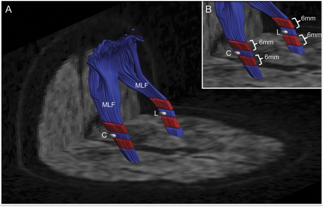
Methods: Nine acute to subacute DWI lesions were identified in 6 subjects with probable cerebral amyloid angiopathy who had undergone high-resolution MRI both before and after DWI lesion detection. Regions of interest (ROIs) corresponding to the site of the DWI lesion (lesion ROI) and corresponding site in the nonlesioned contralateral hemisphere (control ROI) were coregistered to the pre- and postlesional scans. DTI tractography was additionally performed to reconstruct the white matter tracts containing the ROIs. DTI parameters (fractional anisotropy [FA], mean diffusivity [MD]) were quantified within each ROI, the 6-mm lesion-containing tract segments, and the entire lesion-containing tract bundle. Lesion/control FA and MD ratios were compared across time points.
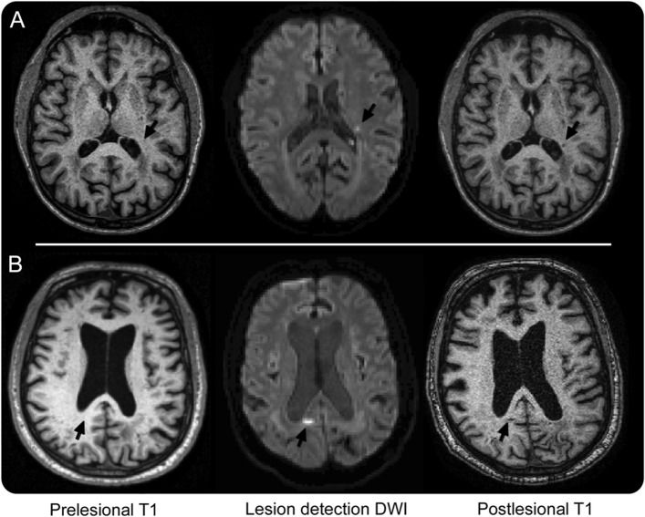
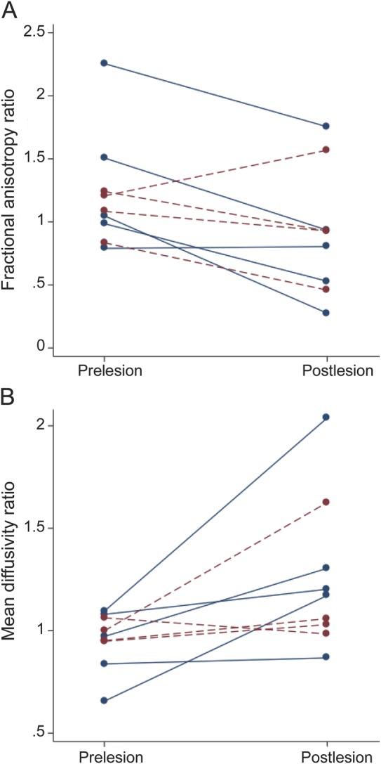
Results: The postlesional scans (performed a mean 7.1 ± 4.7 months after DWI lesion detection) demonstrated a decrease in median FA lesion/control ROI ratio (1.08 to 0.93, p = 0.038) and increase in median MD lesion/control ROI ratio (0.97 to 1.17, p = 0.015) relative to the prelesional scans. There were no visible changes on postlesional high-resolution T1-weighted and fluid-attenuated inversion recovery images in 4 of 9 lesion ROIs and small (2-5 mm) T1 hypointensities in the remaining 5. No postlesional changes in FA or MD ratios were detected in the 6-mm lesion-containing tract segments or full tract bundles.
Conclusions: Asymptomatic DWI lesions produce chronic local microstructural injury. The cumulative effects of these widely distributed lesions may directly contribute to small-vessel-related vascular cognitive impairment.
© 2014 American Academy of Neurology.
Figures
References
-
- Neuropathology Group. Medical Research Council Cognitive Function and Ageing Study. Pathological correlates of late-onset dementia in a multicentre, community-based population in England and Wales. Neuropathology Group of the Medical Research Council Cognitive Function and Ageing Study (MRC CFAS). Lancet 2001;357:169–175 - PubMed
Publication types
MeSH terms
Grants and funding
LinkOut - more resources
Full Text Sources
Other Literature Sources