. 2014 May 23:9:2581-95.
doi: 10.2147/IJN.S63472. eCollection 2014.

Manganese-containing Prussian blue nanoparticles for imaging of pediatric brain tumors

Affiliations

Manganese-containing Prussian blue nanoparticles for imaging of pediatric brain tumors

Matthieu F Dumont et al. Int J Nanomedicine. .

Abstract

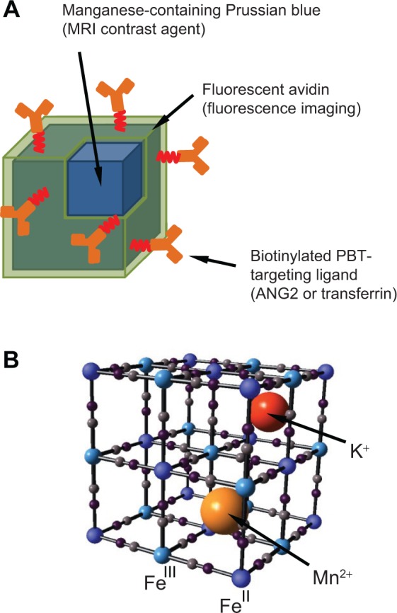
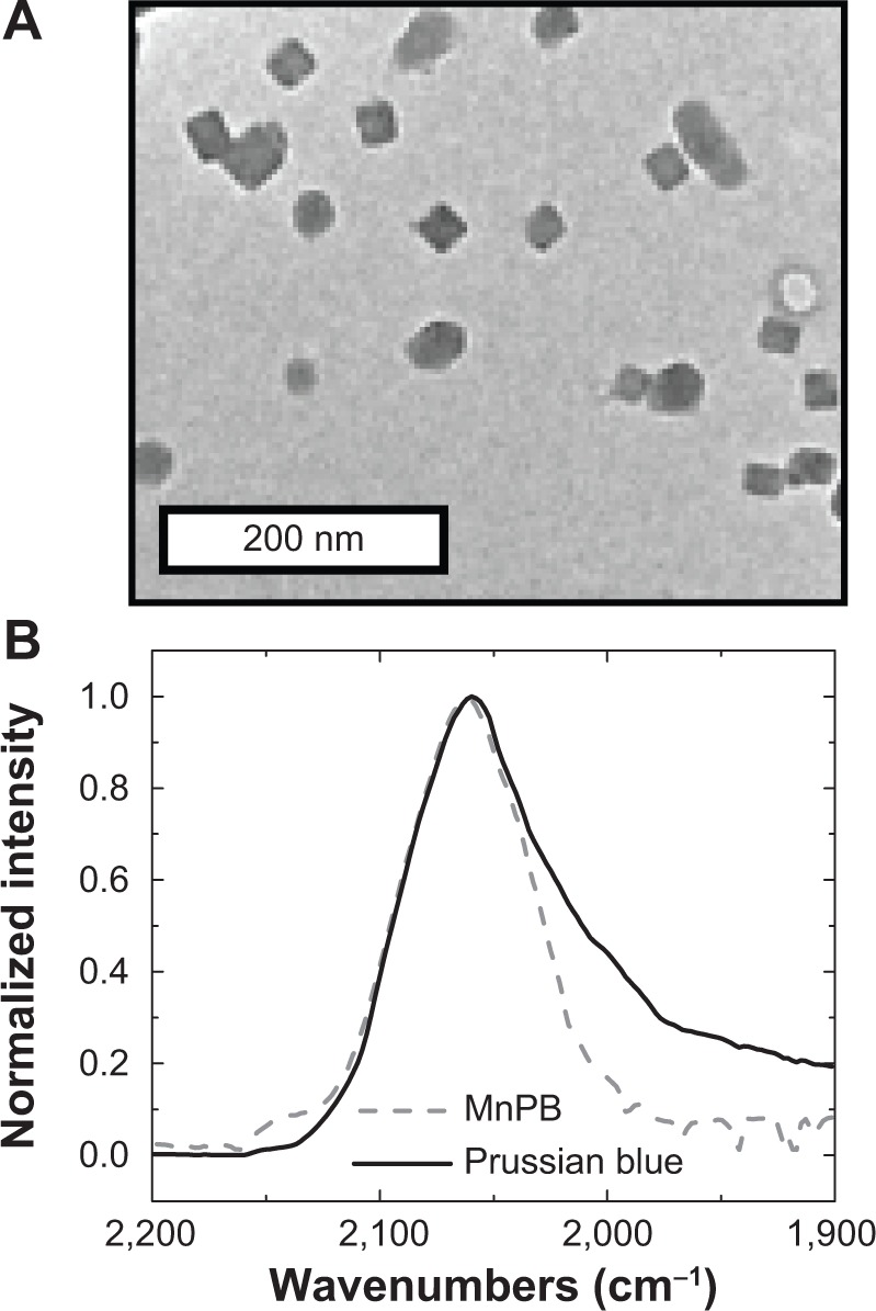
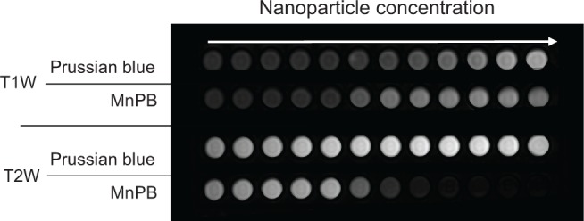
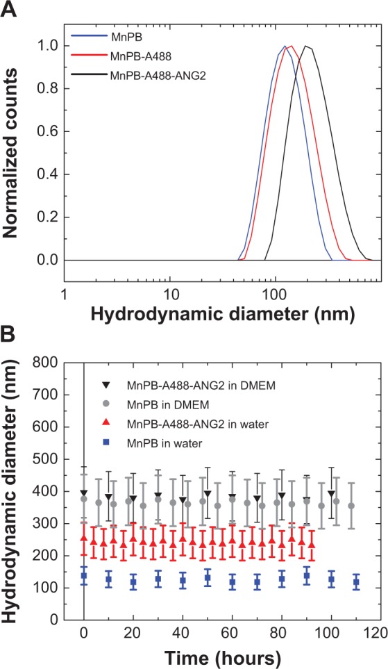
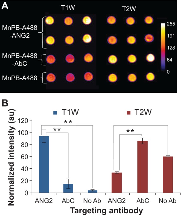
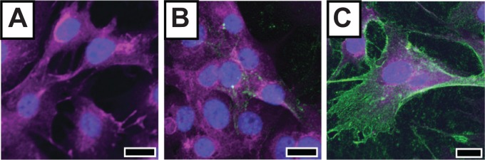
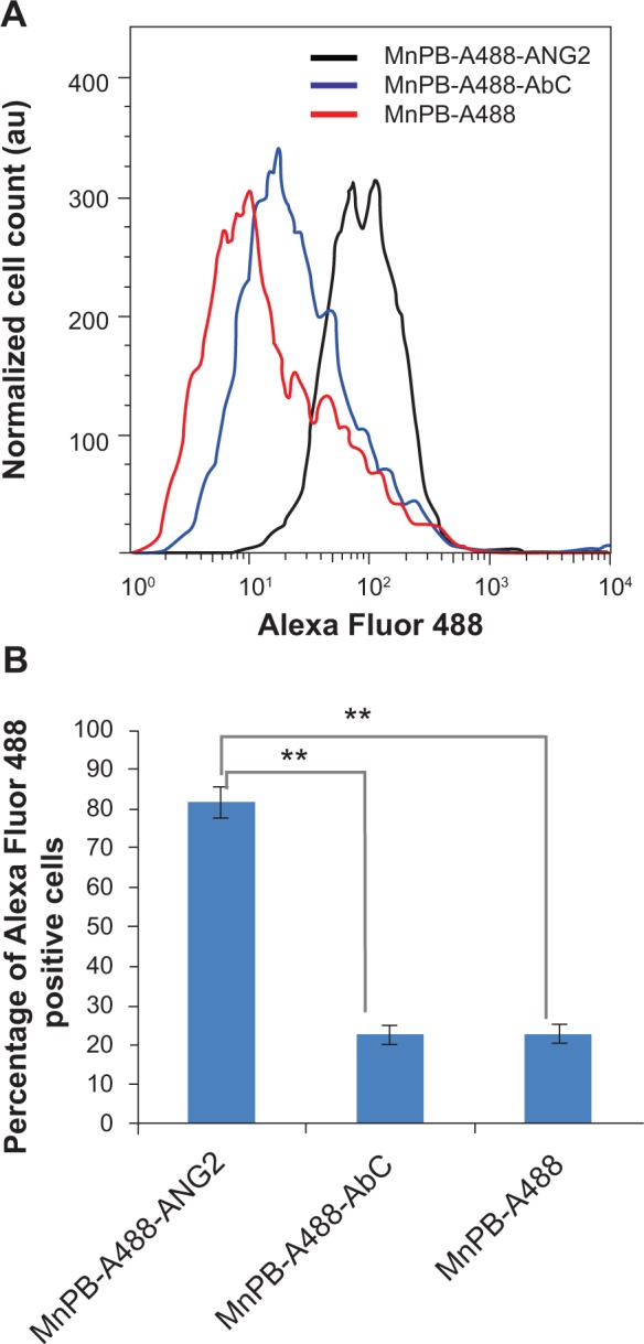
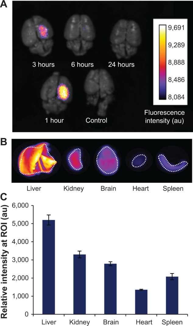
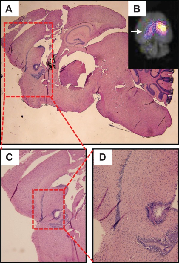
Pediatric brain tumors (PBTs) are a leading cause of death in children. For an improved prognosis in patients with PBTs, there is a critical need to develop molecularly-specific imaging agents to monitor disease progression and response to treatment. In this paper, we describe manganese-containing Prussian blue nanoparticles as agents for molecular magnetic resonance imaging (MRI) and fluorescence-based imaging of PBTs. Our core-shell nanoparticles consist of a core lattice structure that incorporates and retains paramagnetic Mn(2+) ions, and generates MRI contrast (both negative and positive). The biofunctionalized shell is comprised of fluorescent avidin, which serves the dual purpose of enabling fluorescence imaging and functioning as a platform for the attachment of biotinylated ligands that target PBTs. The surfaces of our nanoparticles are modified with biotinylated antibodies targeting neuron-glial antigen 2 or biotinylated transferrin. Both neuron-glial antigen 2 and the transferrin receptor are protein markers overexpressed in PBTs. We describe the synthesis, biofunctionalization, and characterization of these multimodal nanoparticles. Further, we demonstrate the MRI and fluorescence imaging capabilities of manganese-containing Prussian blue nanoparticles in vitro. Finally, we demonstrate the potential of these nanoparticles as PBT imaging agents by measuring their organ and brain biodistribution in an orthotopic mouse model of PBTs using ex vivo fluorescence imaging.

Keywords: Prussian blue; fluorescence; imaging; magnetic resonance imaging; manganese; multimodal; nanoparticles; pediatric brain tumors.


Figures

Figure 1
Figure 1
Manganese-containing Prussian blue nanoparticles for imaging of PBTs. Notes: (A) Schematic representation of manganese-containing Prussian blue nanoparticles comprised of an inorganic core that provides MRI contrast and a biofunctionalized shell consisting of fluorescently-labeled avidin and biotinylated PBT-targeting ligands. (B) Schematic representation of the Prussian blue lattice (ball and stick) containing interstitial Mn2+ and K+ ions. Abbreviations: ANG2, anti-neuron-glial antigen 2; MRI, magnetic resonance imaging; PBT, pediatric brain tumor.
Figure 2
Figure 2
Properties of the MnPB nanoparticles. Notes: (A) Representative transmission electron microscopy image of MnPB nanoparticles. (B) Fourier transform infrared spectra of Prussian blue without interstitial manganese (solid line) and MnPB (dashed line) in the cyanide stretching region (1,900 cm−1 to 2,200 cm−1). Abbreviation: MnPB, manganese-containing Prussian blue.
Figure 3
Figure 3
T1W and T2W magnetic resonance images of Prussian blue and MnPB at 3 T. Abbreviations: T1W, T1-weighted; T2W, T2-weighted; MnPB, manganese-containing Prussian blue.
Figure 4
Figure 4
Size and stability of biofunctionalized MnPB nanoparticles. Notes: (A) Size distribution of MnPB-A488-ANG2 after each functionalization step. Hydrodynamic size distributions of the MnPB nanoparticles, MnPB nanoparticles coated with avidin-Alexa Fluor 488 (MnPB-A488), and MnPB-A488 modified with biotinylated anti-neuron-glial antigen 2 (MnPB-A488-ANG2), respectively. (B) Temporal stability of MnPB nanoparticles and MnPB-A488-ANG2 in Milli-Q water and Dulbecco’s Modified Eagle’s Medium for up to 4 days post synthesis. Abbreviations: MnPB, manganese-containing Prussian blue; A488, avidin-Alexa Fluor 488; ANG2, anti-neuron-glial antigen 2 antibody; DMEM, Dulbecco’s Modified Eagle’s Medium.
Figure 5
Figure 5
Molecular MRI-based detection of PBT cells using biofunctionalized Prussian blue nanoparticles. (A) T1-weighted and T2-weighted contrast enhancement in phantoms comprised of a fixed number of BSG D10 treated with MnPB-A488-ANG2 (n=6), MnPB-A488-AbC (n=3), or MnPB-A488 (no antibody, triplicate). (B) Normalized fluorescence signal intensity (au) for BSG D10 treated with ANG2, AbC, and no antibody-modified MnPB-A488. **P<0.05. Abbreviations: MnPB, manganese-containing Prussian blue; A488, avidin-Alexa Fluor 488; ANG2, anti-neuron-glial antigen 2; AbC, eotaxin antibody; BSG, brainstem glioma; T1W, T1-weighted; T2W, T2-weighted; PBT, pediatric brain tumor; au, arbitrary units; Ab, antibody; MRI, magnetic resonance imaging.
Figure 6
Figure 6
Fluorescence-based detection of PBT cells using biofunctionalized Prussian blue nanoparticles. Notes: Fluorescent image of BSG D10 treated with control nanoparticles, ie, (A) MnPB-A488 and (B) MnPB-A488-AbC, and experimental nanoparticles, ie, (C) MnPB-A488-ANG2. The green fluorescence comes from A488 on the constructs. Scale bars represent 20 μm. Abbreviations: BSG, brainstem glioma; MnPB, manganese-containing Prussian blue; A488, avidin-Alexa Fluor 488; ANG2, anti-neuron-glial antigen 2; AbC, eotaxin antibody; PBT, pediatric brain tumor.
Figure 7
Figure 7
Flow cytometric analysis of the specificity of biofunctionalized MnPB nanoparticles for PBT cells. Notes: (A) Representative histograms of cell count plotted against Alexa Fluor 488 detection levels for BSG D10 cells treated with MnPB-A488 (no antibody, red line), MnPB-A488-AbC (blue line), and MnPB-A488-ANG2 (black line), and stained with 7-AAD. (B) Percentage Alexa Fluor 488-positive cells (fluorescence intensity cutoff 50) cells for BSG D10 treated with MnPB-A488-ANG2, MnPB-A488-AbC, or MnPB-A488. **P<0.05. Abbreviations: BSG, brainstem glioma; 7-AAD, 7-aminoactinomycin D; MnPB, manganese-containing Prussian blue; A488, avidin-Alexa Fluor 488; ANG2, anti-neuron-glial antigen 2; PBT, pediatric brain tumor; au, arbitrary units.
Figure 8
Figure 8
Ex vivo fluorescence imaging of biodistribution of the nanoparticles in an orthotopic mouse model of PBT. Notes: (A) Fluorescent signal observed in the brains of four separate mice with PBT (1–24 hours) injected intravenously (via tail vein) with transferrin-coated Prussian blue nanoparticles (MnPB-ATxRd-Tf). A control mouse with PBT was not injected with nanoparticles. (B) Representative ex vivo fluorescence imaging of organ biodistribution of the nanoparticles at 3 hours post-injection. (C) Histograms quantifying the observed fluorescence biodistribution of the nanoparticles 3 hours post-injection. ROIs for intensity measurements are indicated by white dashed lines in (B). Abbreviations: PBT, pediatric brain tumor; MnPB, manganese-containing Prussian blue; ATxRd, Texas Red-labeled avidin; Tf, transferrin; ROI, region of interest; au, arbitrary units.
Figure 9
Figure 9
Histological analysis of fluorescence-positive regions of mice brains with PBTs. Notes: (A) Sagittal slice of mouse brain stained with hematoxylin and eosin. (B) Inset showing a fluorescence image of the brain, with arrow indicating the viewing direction of the sagittal section in (A). (C, D) Progressive zooms of the hematoxylin and eosin-stained sagittal brain sections containing a hypercellular ventricular and periventricular region within the fluorescence-positive region. Abbreviation: PBT, pediatric brain tumor.

References

    1. Mostoufi-Moab S, Grimberg A. Pediatric brain tumor treatment: growth consequences and their management. Pediatr Endocrinol Rev. 2010;8(1):6–17. - PMC - PubMed
    1. Pollack I. Multidisciplinary management of childhood brain tumors: a review of outcomes, recent advances, and challenges. J Neurosurg Pediatr. 2011;8(2):135–148. - PubMed
    1. Jansen MH, Kaspers GJ. A new era for children with diffuse intrinsic pontine glioma: hope for cure? Expert Rev Anticancer Ther. 2012;12(9):1109–1112. - PubMed
    1. Jansen MH, van Vuurden DG, Vandertop WP, Kaspers GJ. Diffuse intrinsic pontine gliomas: a systematic update on clinical trials and biology. Cancer Treat Rev. 2012;38(1):27–35. - PubMed
    1. Schwartzentruber J, Korshunov A, Liu X-Y, et al. Driver mutations in histone H3.3 and chromatin remodelling genes in paediatric glioblastoma. Nature. 2012;482(7384):226–231. - PubMed

Publication types