. 2014 May 27:9:2677-87.
doi: 10.2147/IJN.S61940. eCollection 2014.

Increased antiparkinson efficacy of the combined administration of VEGF- and GDNF-loaded nanospheres in a partial lesion model of Parkinson's disease

Affiliations

Increased antiparkinson efficacy of the combined administration of VEGF- and GDNF-loaded nanospheres in a partial lesion model of Parkinson's disease

Enara Herrán et al. Int J Nanomedicine. .

Abstract

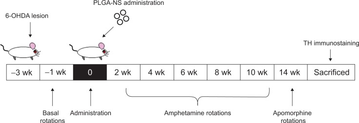
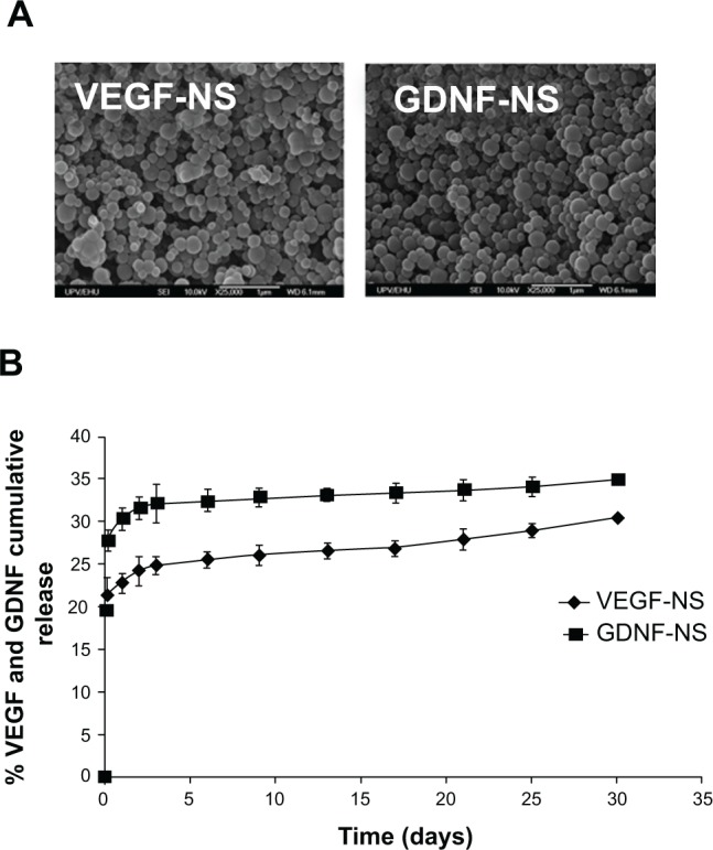
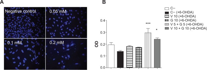
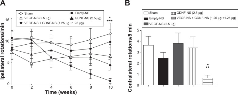
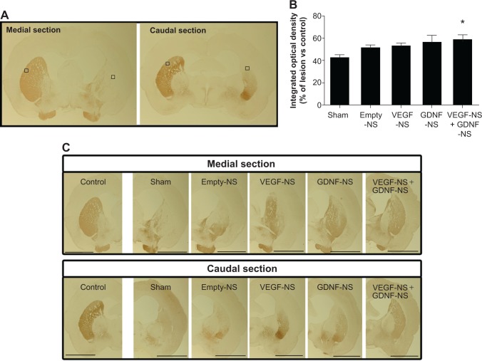
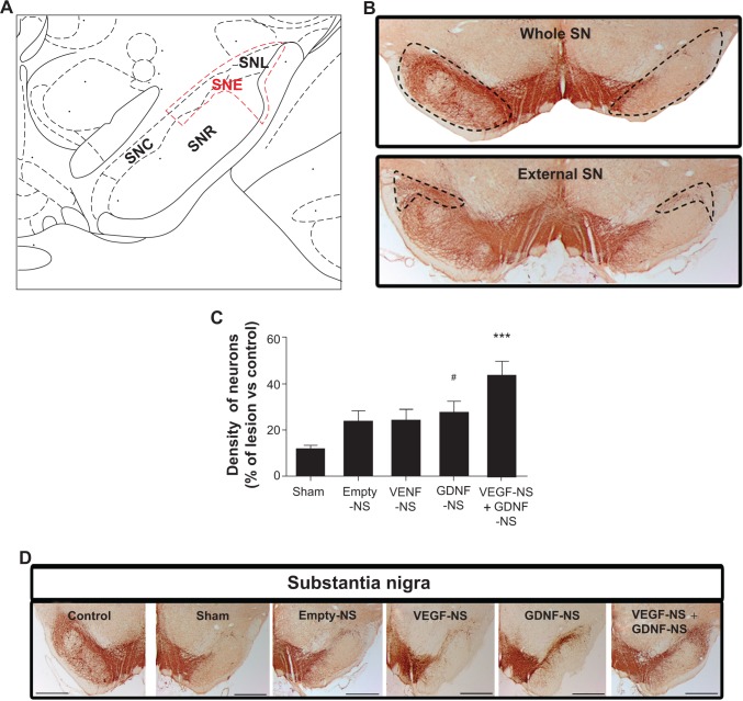
Current research efforts are focused on the application of growth factors, such as glial cell line-derived neurotrophic factor (GDNF) and vascular endothelial growth factor (VEGF), as neuroregenerative approaches that will prevent the neurodegenerative process in Parkinson's disease. Continuing a previous work published by our research group, and with the aim to overcome different limitations related to growth factor administration, VEGF and GDNF were encapsulated in poly(lactic-co-glycolic acid) nanospheres (NS). This strategy facilitates the combined administration of the VEGF and GDNF into the brain of 6-hydroxydopamine (6-OHDA) partially lesioned rats, resulting in a continuous and simultaneous drug release. The NS particle size was about 200 nm and the simultaneous addition of VEGF NS and GDNF NS resulted in significant protection of the PC-12 cell line against 6-OHDA in vitro. Once the poly(lactic-co-glycolic acid) NS were implanted into the striatum of 6-OHDA partially lesioned rats, the amphetamine rotation behavior test was carried out over 10 weeks, in order to check for in vivo efficacy. The results showed that VEGF NS and GDNF NS significantly decreased the number of amphetamine-induced rotations at the end of the study. In addition, tyrosine hydroxylase immunohistochemical analysis in the striatum and the external substantia nigra confirmed a significant enhancement of neurons in the VEGF NS and GDNF NS treatment group. The synergistic effect of VEGF NS and GDNF NS allows for a reduction of the dose by half, and may be a valuable neurogenerative/neuroreparative approach for treating Parkinson's disease.

Keywords: 6-OHDA; PLGA; nanoparticles; neuroregeneration; neurotrophic factors; tyrosine hydroxylase.


Figures

Figure 1
Figure 1
Experimental design for the NS treatments in 6-OHDA lesioned rats. Abbreviations: 6-OHDA, 6-hydroxydopamine; NS, nanosphere; PLGA, poly(lactic-co-glycolic acid); TH, tyrosine hydroxylase; wk, weeks.
Figure 2
Figure 2
GDNF NS and VEGF NS characterization and in vitro toxicity study. Notes: (A) Scanning electron microscope (SEM) photomicrographs of GDNF NS and VEGF NS. (B) The in vitro VEGF NS and GDNF NS release profiles at 37°C in phosphate-buffered saline (pH 7.4). The results are represented as the mean ± standard deviation (n=3). Abbreviations: GDNF, glial cell line-derived neurotrophic factor; NS, nanospheres; VEGF, vascular endothelial growth factor.
Figure 3
Figure 3
In vitro neurotoxicity assay. Notes: (A) DAPI immunostaining after different 6-OHDA concentration treatments to establish the neurotoxic cell model. (B) PC-12 cell viability evaluation after 6-OHDA induced toxicity in vitro. *P<0.05, V 10 + G 10 (+6-OHDA) versus C−, and C− (+6-OHDA) groups. ***P<0.001, V 5 + G 5 (+6-OHDA) versus C−, and C− (+6-OHDA) groups. Abbreviations: 6-OHDA, 6-hydroxydopamine; C−, negative control; DAPI, 4′,6-diamidino-2-phenylindole; G 5, GDNF 5 ng/mL; G 10, GDNF 10 ng/mL; GDNF, glial cell line-derived neurotrophic factor; OD, optical density; V 5, VEGF 5 ng/mL; V 10, VEGF 10 ng/mL; VEGF, vascular endothelial growth factor.
Figure 4
Figure 4
Behavioral study: amphetamine and apomorphine rotational tests. Notes: (A) The results obtained from the amphetamine rotational behavior test after NS administration. GDNF NS group versus sham group show statistically significant differences (*P<0.05). The VEGF NS and GDNF NS group versus sham and empty NS groups showed statistically significant differences (***P<0.001). (B) Data obtained from the apomorphine rotational behavior test 14 weeks after NS implantation. The VEGF NS and GDNF NS-treated group exhibited the best behavioral recovery (*P<0.05, VEGF NS and GDNF NS versus empty NS; **P<0.01, VEGF NS and GDNF NS versus sham). The data are shown as the mean ± standard deviation (n=6–8). Abbreviations: GDNF, glial cell line-derived neurotrophic factor; NS, nanospheres; VEGF, vascular endothelial growth factor.
Figure 5
Figure 5
Histological evaluation of the treatments in the striatum. Notes: (A) The images give an illustrative overview of the medial and caudal sections of the caudoputamen complex for TH immunohistochemistry. Squares placed on lesioned and non-lesioned (control) hemispheres delimit the surfaces where the integrated optical density was measured using a computerized image analysis system. Scale bar =2 mm. (B) The graphic shows the percentage of integrated optical density of the lesioned hemisphere with respect to the non-lesioned hemisphere (control) for each experimental group. Data are shown as the mean ± standard error of the mean (n=6–8) (*P<0.05 VEGF NS and GDNF NS group versus sham group). (C) Photomicrographs of striata immunostained for TH from a representative intact hemisphere (control) and 6-OHDA lesioned hemispheres from the different experimental groups. Abbreviations: 6-OHDA, 6-hydroxydopamine; GDNF, glial cell line-derived neurotrophic factor; NS, nanospheres; TH, tyrosine hydroxylase; VEGF, vascular endothelial growth factor.
Figure 6
Figure 6
Histological evaluation of the treatments in the SN. Notes: (A) Schematic illustration of the SN with the “external SN” delimited. This area is topologically related to the lesioned area of striatum and includes SNL, a part of the SNR, and half of the SNC. (B) Picture of whole SN and delimited “external SN”. Scale bar =1 mm. (C) Density of dopaminergic neurons in “external SN”. The results are expressed as a percentage of lesioned hemisphere compared to the non-lesioned one (control). Data are shown as the mean ± standard error of the mean (n=6–8) (#P<0.05 GDNF NS group versus sham group; ***P<0.001 VEGF NS and GDNF NS group versus sham and empty NS groups). (D) Photomicrographs of SN immunostained for tyrosine hydroxylase from a representative intact hemisphere (control) and 6-OHDA lesioned hemispheres from the different experimental groups. Scale bar =1 mm. Abbreviations: 6-OHDA, 6-hydroxydopamine; GDNF, glial cell line-derived neurotrophic factor; NS, nanospheres; SN, substantia nigra; SNC, SN pars compacta; SNL, SN lateral; SNR, SN pars reticulata; SNE, SN externa; VEGF, vascular endothelial growth factor.

Similar articles

Cited by

References

    1. Linazasoro G. A global view of Parkinson’s disease pathogenesis: implications for natural history and neuroprotection. Parkinsonism Relat Disord. 2009;15(6):401–405. - PubMed
    1. Obeso JA, Rodriguez-Oroz MC, Goetz CG, et al. Missing pieces in the Parkinson’s disease puzzle. Nat Med. 2010;16(6):653–661. - PubMed
    1. Deierborg T, Soulet D, Roybon L, Hall V, Brundin P. Emerging restorative treatments for Parkinson’s disease. Prog Neurobiol. 2008;85(4):407–432. - PubMed
    1. Sullivan AM, Toulouse A. Neurotrophic factors for the treatment of Parkinson’s disease. Cytokine Growth Factor Rev. 2011;22(3):157–165. - PubMed
    1. Ruozi B, Belletti D, Bondioli L, et al. Neurotrophic factors and neurodegenerative diseases: a delivery issue. Int Rev Neurobiol. 2012;102:207–247. - PubMed

Publication types

MeSH terms