. 2014 Jun 12;9(6):e99116.
doi: 10.1371/journal.pone.0099116. eCollection 2014.

CellFIT: a cellular force-inference toolkit using curvilinear cell boundaries

Affiliations

CellFIT: a cellular force-inference toolkit using curvilinear cell boundaries

G Wayne Brodland et al. PLoS One. .

Abstract

Mechanical forces play a key role in a wide range of biological processes, from embryogenesis to cancer metastasis, and there is considerable interest in the intuitive question, "Can cellular forces be inferred from cell shapes?" Although several groups have posited affirmative answers to this stimulating question, nagging issues remained regarding equation structure, solution uniqueness and noise sensitivity. Here we show that the mechanical and mathematical factors behind these issues can be resolved by using curved cell edges rather than straight ones. We present a new package of force-inference equations and assessment tools and denote this new package CellFIT, the Cellular Force Inference Toolkit. In this approach, cells in an image are segmented and equilibrium equations are constructed for each triple junction based solely on edge tensions and the limiting angles at which edges approach each junction. The resulting system of tension equations is generally overdetermined. As a result, solutions can be obtained even when a modest number of edges need to be removed from the analysis due to short length, poor definition, image clarity or other factors. Solving these equations yields a set of relative edge tensions whose scaling must be determined from data external to the image. In cases where intracellular pressures are also of interest, Laplace equations are constructed to relate the edge tensions, curvatures and cellular pressure differences. That system is also generally overdetermined and its solution yields a set of pressures whose offset requires reference to the surrounding medium, an open wound, or information external to the image. We show that condition numbers, residual analyses and standard errors can provide confidence information about the inferred forces and pressures. Application of CellFIT to several live and fixed biological tissues reveals considerable force variability within a cell population, significant differences between populations and elevated tensions along heterotypic boundaries.


Conflict of interest statement

Competing Interests: The authors have declared that no competing interests exist.

Figures

Figure 1
Figure 1. A synthetic planar aggregate and its analysis by CellFIT.
(A) shows a representative region consisting of 50 complete cells, 177 complete edges, and 30 partial cells and corresponding incomplete edges taken from a larger aggregate. Its cells were assigned to one of three types, as indicated by coloured shading and a finite element model was used to determine its annealed state, as shown. The edge tensions were assigned the following values according to their type 5∶6∶7∶10∶11∶12. (B) shows the topological and geometric information provided to the CellFIT algorithms. The spectra shown in (C) provide legends for the tension and pressure colours used in the remaining parts of the figure. (D) shows the ground truth Standard Tensions and Pressures as determined by the FE annealing process, and extracted using Equation 16 and its associated text. (E) shows the values that the standard Polyarc version of CellFIT calculates based solely on the data in (B). (F) shows the results obtained by calculating the angles at triple junctions using straight edges only. (G) and (H) show CellFIT results when noise levels of 2 or 5 are respectively introduced into the CellFIT input data. A noise level of x corresponds to x degrees of angle error and x% curvature error. (I) shows CellFIT results obtained after the original mesh was converted into an image and redigitized before analysis.
Figure 2
Figure 2. Equilibrium considerations.
(A) shows a curved cell edge and the forces acting on it, while (B) shows how edge tension, curvature and pressure are related. Specifically, the pressure difference Δp generates a force formula image 
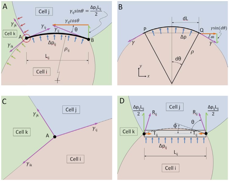
in the y-direction. This force must be just balanced by the vertical components of the tension γ. Thus we have that formula image which, when simplified, gives formula image, the Laplace equation. (C) shows the forces that act at a typical triple junction, while (D) shows the forces that act on an edge that is constrained to remain straight by beam action, as described in the text.
Figure 3
Figure 3. The tension equations are generally overdetermined.
Geometric considerations show that the tension equations are, within a scale factor (see text), adequately determined (A, and B). When a full cell is enclosed (C) and as more cells become fully enclosed, they generally become increasingly overdetermined (D and E), though scaling information external to the image is still needed.
Figure 4
Figure 4. The pressure equations are generally overdetermined.
As the number of cells increases and cells acquire multiple neighbours, the Pressure Equations become increasingly well determined (A to E). Even when the system is seemingly overdetermined (D and E), external data still is needed to ascertain the pressure offset (see text).
Figure 5
Figure 5. CellFIT analysis of cells near the amnioserosa/lateral epidermis boundary during early dorsal closure in a living Drosophila embryo, as imaged in (A) and with inferred Standard Tensions and Pressures illustrated in (B) according to the color bars.
The amnioserosa is visible in a wide band from the lower left-hand corner towards the upper right, while the lateral epidermis, identifiable by its smaller cells, is confined to a large triangle in the upper left corner. The boundary between these two tissues is indicated by the black arrows. The blue and green arrows point to features discussed in the text. Overall, the CellFIT equations were very well conditioned – having tension and pressure condition numbers of 30.3 and 15.6, respectively. The tension and pressure residuals are shown in (B) as thin lines emanating from each triple junction and bisecting each cell edge, respectively. These residuals are scaled so that a residual equal to the mean tension has a length equal to the mean cell radius. Even at this scaling, the residuals are generally quite small and many are barely discernable. Finally, confidence limits are shown for individual tensions and pressures, (C) and (D) respectively, by boundary type. The points and bars indicate best estimate +/− one standard error (based on the covariance matrix), respectively. These confidence limits are a significant, but modest, fraction of the inferred tensions and pressures. Prior investigations have suggested the existence of a uniform high-tension purse-string along the edge of the amnioserosa, but CellFIT reveals a more complex and interesting scenario. See text for details.
Figure 6
Figure 6. CellFIT analysis of a wing imaginal disc from a living Drosophila larva.
(A) shows the inferred Standard Tensions based on Fig. 4D of Reference . The tension equations are well conditioned (condition number of 11.1), the confidence limits are acceptable and the force residuals are a bit larger than for the previous case. The black arrows in (A) indicate the boundary between compartments within the imaginal disc. (B) shows that the forces along that boundary tend to be higher than those elsewhere (see also text). Pressures were not calculated as boundary curvatures could not be obtained from the source image.
Figure 7
Figure 7. CellFIT analysis of an image of a dragonfly wing, published as Fig. 162 in Thompson's On Growth and Form .
(A) shows the inferred Standard Tensions and Pressures. The CellFIT equations were well conditioned (tension and pressure condition numbers of 16.7 and 13.9 respectively), the residuals are quite small, and the confidence limits for the tensions and pressures are acceptable (B and C). As noted in the text, the tensions along the veins (indicated with arrows) are significantly higher than those along the other cell edges. This example shows that CellFIT can extract useful information from historical images or from fixed or otherwise non-living tissues.

References

    1. Brodland GW, Chen X, Lee P, Marsden M (2010) From genes to neural tube defects (NTDs): Insights from multiscale computational modeling. HFSP J 4: 142–152. - PMC - PubMed
    1. Varner VD, Voronov DA, Taber LA (2010) Mechanics of head fold formation: Investigating tissue-level forces during early development. Development 137: 3801–3811. - PMC - PubMed
    1. Martin AC (2010) Pulsation and stabilization: Contractile forces that underlie morphogenesis. Dev Biol 341: 114–125. - PubMed
    1. Dahmann C, Oates AC, Brand M (2011) Boundary formation and maintenance in tissue development. Nat Rev Genet 12: 43–55. - PubMed
    1. Fernandez-Gonzalez R, Simoes Sde M, Roper JC, Eaton S, Zallen JA (2009) Myosin II dynamics are regulated by tension in intercalating cells. Dev Cell 17: 736–743. - PMC - PubMed

Publication types