. 2014 Jun 12;9(6):e99091.
doi: 10.1371/journal.pone.0099091. eCollection 2014.

Establishment of highly tumorigenic human colorectal cancer cell line (CR4) with properties of putative cancer stem cells

Affiliations

Establishment of highly tumorigenic human colorectal cancer cell line (CR4) with properties of putative cancer stem cells

Rebecca A Rowehl et al. PLoS One. .

Abstract

Background: Colorectal cancer (CRC) has the third highest mortality rates among the US population. According to the most recent concept of carcinogenesis, human tumors are organized hierarchically, and the top of it is occupied by malignant stem cells (cancer stem cells, CSCs, or cancer-initiating cells, CICs), which possess unlimited self-renewal and tumor-initiating capacities and high resistance to conventional therapies. To reflect the complexity and diversity of human tumors and to provide clinically and physiologically relevant cancer models, large banks of characterized patient-derived low-passage cell lines, and especially CIC-enriched cell lines, are urgently needed.

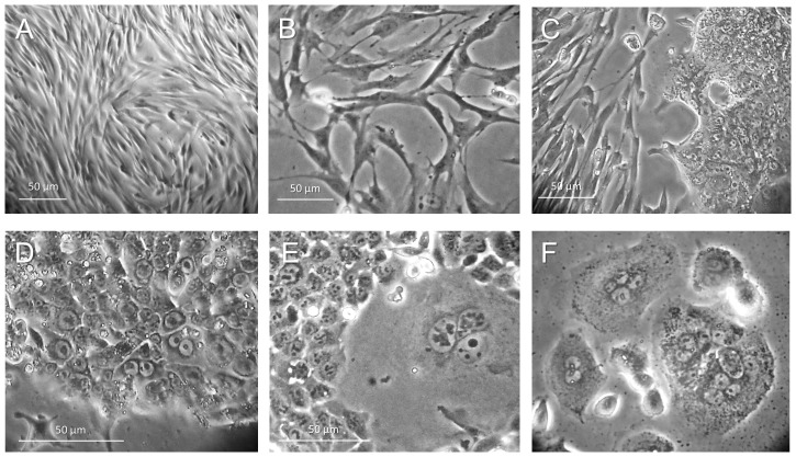
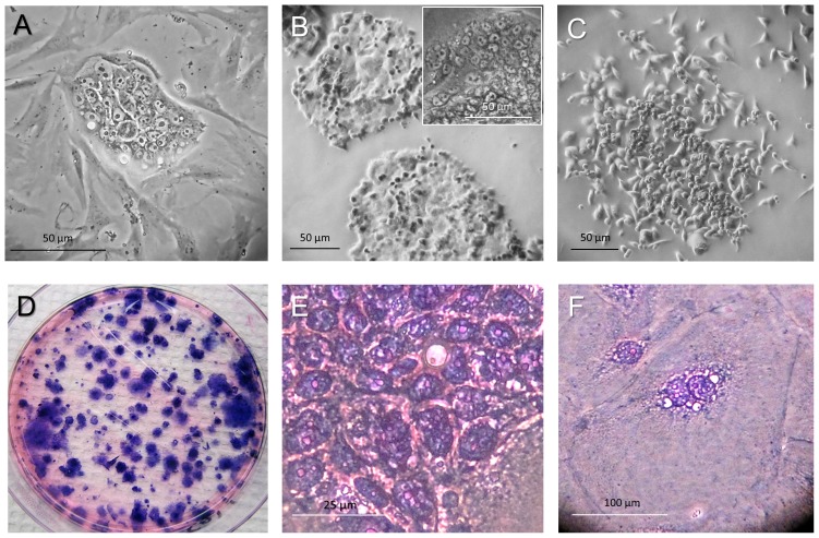
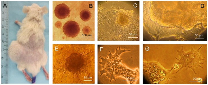
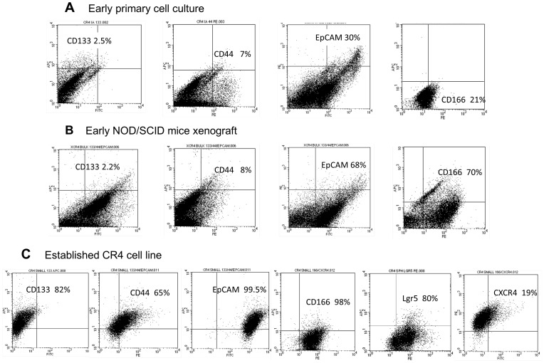
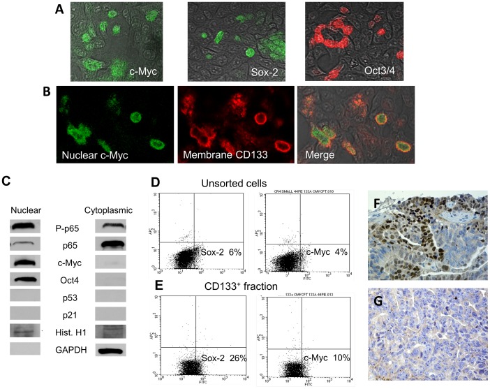
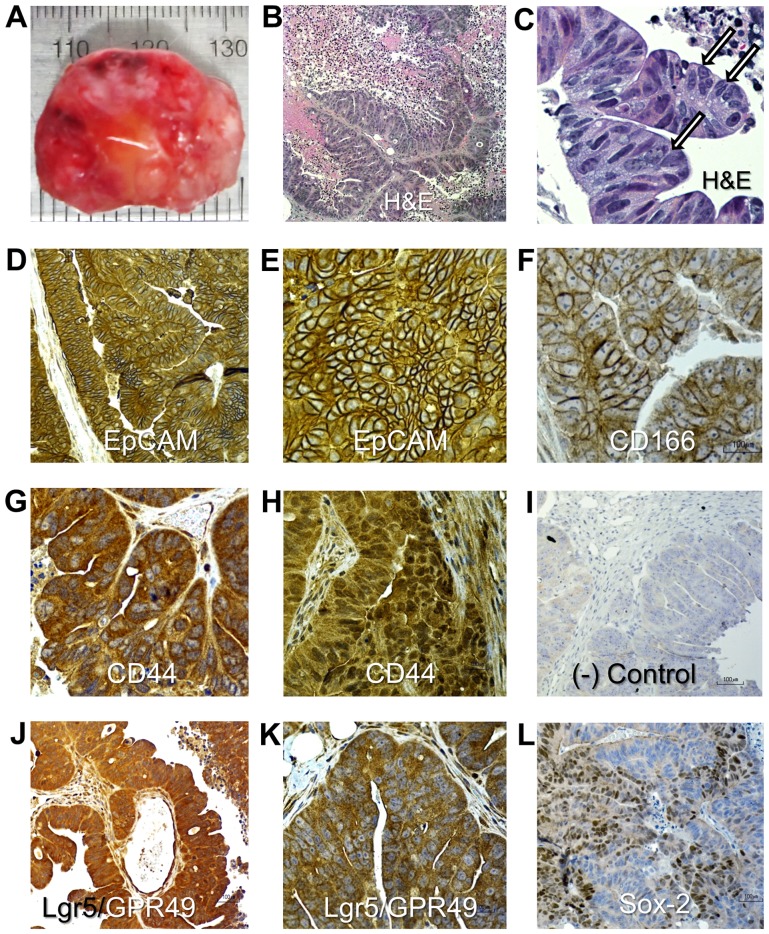
Principal findings: Here we report the establishment of a novel CIC-enriched, highly tumorigenic and clonogenic colon cancer cell line, CR4, derived from liver metastasis. This stable cell line was established by combining 3D culturing and 2D culturing in stem cell media, subcloning of cells with particular morphology, co-culture with carcinoma associated fibroblasts (CAFs) and serial transplantation to NOD/SCID mice. Using RNA-Seq complete transcriptome profiling of the tumorigenic fraction of the CR4 cells in comparison to the bulk tumor cells, we have identified about 360 differentially expressed transcripts, many of which represent stemness, pluripotency and resistance to treatment. Majority of the established CR4 cells express common markers of stemness, including CD133, CD44, CD166, EpCAM, CD24 and Lgr5. Using immunocytochemical, FACS and western blot analyses, we have shown that a significant ratio of the CR4 cells express key markers of pluripotency markers, including Sox-2, Oct3/4 and c-Myc. Constitutive overactivation of ABC transporters and NF-kB and absence of tumor suppressors p53 and p21 may partially explain exceptional drug resistance of the CR4 cells.

Conclusions: The highly tumorigenic and clonogenic CIC-enriched CR4 cell line may provide an important new tool to support the discovery of novel diagnostic and/or prognostic biomarkers as well as the development of more effective therapeutic strategies.


Conflict of interest statement

Competing Interests: The authors have declared that no competing interests exist.

Figures

Figure 1
Figure 1. Cellular heterogeneity of the CR4 tumor cells.
(A) Initially dominating population of the densely packed elongated fibroblast-like cells. (B) Sparser elongated tumor cells with processes. (C) Appearance of small clusters of very small cells (∼7 µm) adjacent to fibroblast-like cells. (D) Typical holoclone induced by small CR4 cells. (E) Giant multinucleated cell within CR4 holoclone of small cells. (F) Colony of multinucleated cells.
Figure 2
Figure 2. Subcloning of the small CR4 cells.
(A) Clone of small CR4 cells surrounded by long fibroblast-like cells with dychotomized processes. (B) After subcloning, small CR4 cells seeded at low density in serum-free SPCM on type I collagen produced typical densely packed holoclones characteristic for stem cells of different origin. (C) Paraclone of elongated larger cells adjacent to small CR4 cells. (D) Low number of the purified small cells produced predominantly round-edged holoclones and rare paraclones adherent to type I collagen with high efficiency. (E) Higher magnification of the holoclone cells with large nuclei and thin rim of cytoplasm. (F) Large MNCs (≥200 µm; hematoxylin and eosin staining) are often located at the periphery of the holoclones.
Figure 3
Figure 3. Functional characterization of the CR4 cells.
(A) Large subcutaneous tumor induced by transplantation of the CR4 cells (1×10³) in NOD/SCID mouse (passage 2). (B) Dense floating spheroids induced by serial passaging of the CR4 cells in 3D culture on the ultra-low-adherent plates. (C) Three-dimensional spheroid induced on the surface of the adherent CR4 holoclone. (D) Formation of a large 3D organoid on the surface of the adherent CR4 holoclone. (E) Appearance of long processes in spheroid cells induced by CR4 cells in 3D culture containing 15% of collagen gel, which indicates their high invasive potential . (F) Long dychotomized processes developed by adherent small CR4 cells. (G) Long processes of adherent MNCs.
Figure 4
Figure 4. Molecular characterization of the CR4 cell line: expression of cell surface markers.
(A) Representative FACS analyses of the different cell surface markers expression in early primary culture of the fast-adherent CR4 cells grown on type I collagen in MSCB medium. (B) FACS analysis of the first passage of FA tumor cells isolated from NOD/SCID mice tumor xenografts induced by early-passage CR4 cells. (C) Dramatic increase in the expression of the common markers of stemness, including CD133, CD44, CD166, Lgr5 and EpCAM in the late-passage (p13) CR4 cells. Note that 19% of cells also expressed marker of CICs with metastatic activity, CXCR4, which was identified in multiple human cancers.
Figure 5
Figure 5. Expression of key pluripotency markers in CR4 cells.
(A) Nuclear localization of key pluripotency markers, c-Myc, Sox-2 and Oct3/4. (B) Co­localization of nuclear c-Myc with high membrane expression of CD133. (A, B: Immunocytochemical analysis of CR4 cells grown on type I collagen-coated chambered slides). (C) Western blot analysis confirms expression of c-Myc and Oct-3/4 in nuclear fractions of the CR4 cells. In contrast, they are negative for p53 and p21. Nuclear fraction also expressed higher levels of the phosphorilated p65 (which indicate constitutive activation of NF-kB), whereas unphosphorilated p65 was located predominantly in a cytoplasm. (D, E) Representative FACS analyses show higher expression of Sox-2 and c-Myc in CD133-positive cells compared to unsorted cells. (F) Immunohistochemical analysis shows high nuclear expression of Sox-2 in the CR4-induced NOD/SCID mice tumor xenograft. (G) Negative control (tissue section without incubation with primary anti-Sox-2 Abs).
Figure 6
Figure 6. CR4 cells are highly resistant to treatment with cytotoxic drugs (comparative cytotoxicity of paclitaxel and new-generation taxoids against CR4 cells).
Commonly used Paclitaxel (Taxol) in doses lower than 10 µM is not effective against CR4 cells and often increases their proliferation. In contrast, several new-generation taxoids induce dose-dependent inhibition of proliferation of these potent tumor-initiating cells. (MTT assay after drug treatment for 48 hr). The obtained p values for all the drugs and all the drug concentrations were much smaller that 0.05. The largest p value was obtained for SBT-1214 at 10 nM concentration (p = 0.0131); in particular, at 10 µM concentration of SBT-1214 p = 0.00032.
Figure 7
Figure 7. Histopathological and immunohistocheminal analyses of the CR4-induced mice tumor xenograft tissues.
(A) Large tumor induced by transplantation of 1×10³ CR4 cells. (B) Hematoxylin ­and eosin-stained tissue section shows classic histologic features of human metastatic colon cancer. (C) High power magnification of region shown in (B); arrows show giant multinucleated cells within the papillary structures. (D, E) Strong cell surface expression of the epithelial marker, EpCAM. (F) Strong cell surface expression of CD166. (G) Cytoplasmic expression of CD44. (H) Microscopic focus with strong nuclear and weaker cytoplasmic expression of CD44. (I) Negative control (primary Abs were omitted). (J, K) Strong and moderate expression, respectively, of the colon stem cell marker, Lgr5/GPR49. (L) Strong nuclear expression of the pluripotency marker, Sox-2 in large areas of the tumor.

References

    1. Siegel R, Naishadham D, Jemal A (2012) Cancer statistics, 2012. CA Cancer J Clin 62: 10–29. - PubMed
    1. Hutchinson L, Kirk R (2011) High drug attrition rates—where are we going wrong? Nat Rev Clin Oncol 8: 189–90. - PubMed
    1. Gillet JP, Calcagno AM, Varma S, Marino M, Green LJ, et al. (2011) Redefining the relevance of established cancer cell lines to the study of mechanisms of clinical anti-cancer drug resistance. Proc Natl Acad Sci USA 108: 18708–13. - PMC - PubMed
    1. Reya T, Morrison SJ, Clarke MF, Weissman IL (2001) Stem cells, cancer, and cancer stem cells. Nature 414: 105–11. - PubMed
    1. Clarke MF, Fuller M (2006) Stem cells and cancer: Two faces of eve. Cell 124: 1111–5. - PubMed

Publication types

Substances