. 2014 Jun 12;8(6):e2924.
doi: 10.1371/journal.pntd.0002924. eCollection 2014 Jun.

Protein kinase C and extracellular signal-regulated kinase regulate movement, attachment, pairing and egg release in Schistosoma mansoni

Affiliations

Protein kinase C and extracellular signal-regulated kinase regulate movement, attachment, pairing and egg release in Schistosoma mansoni

Margarida Ressurreição et al. PLoS Negl Trop Dis. .

Abstract

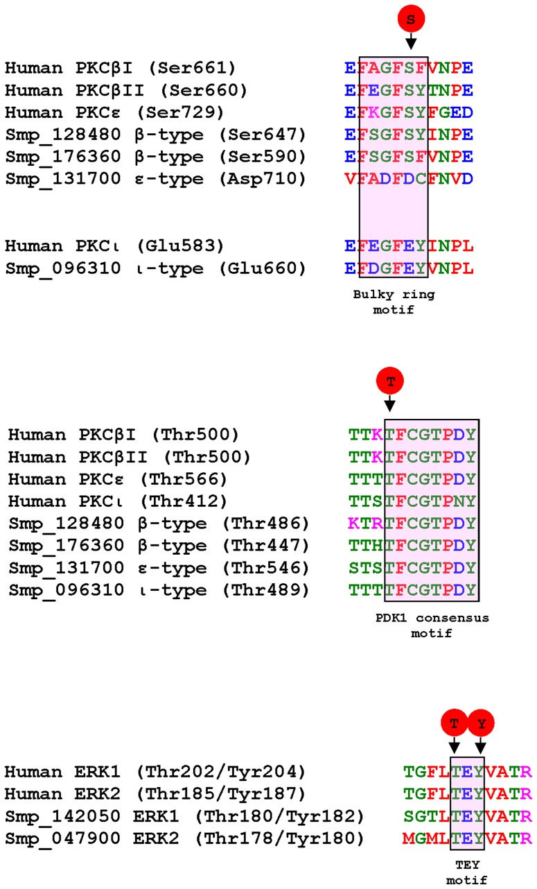
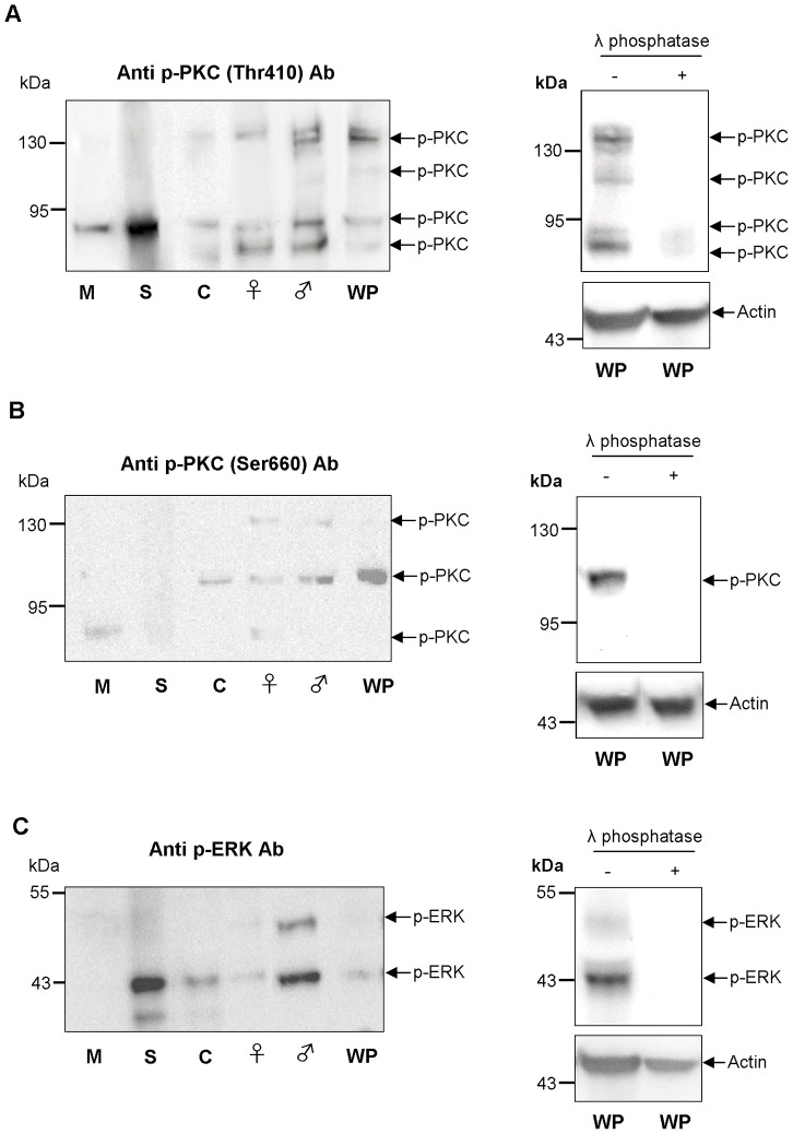
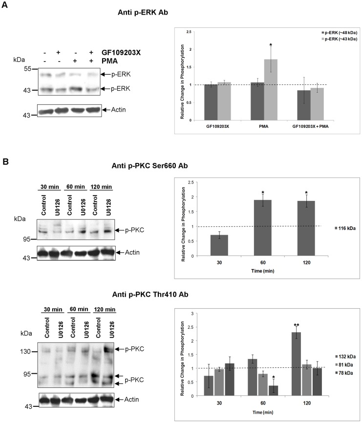
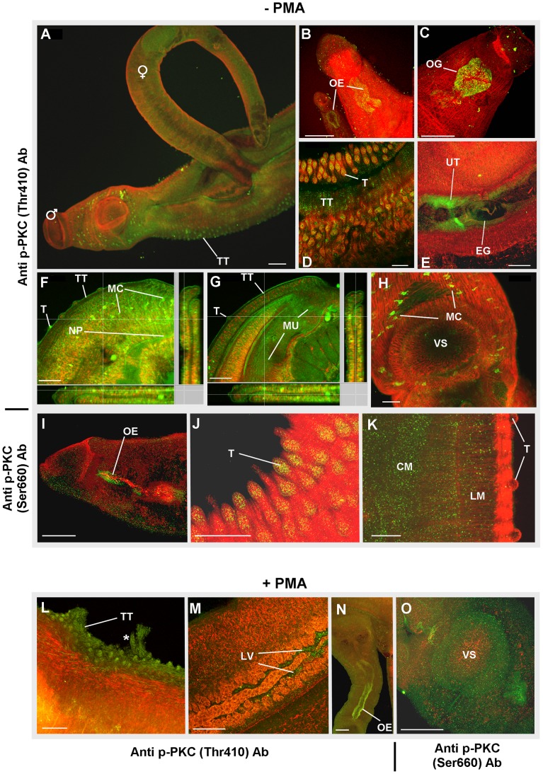
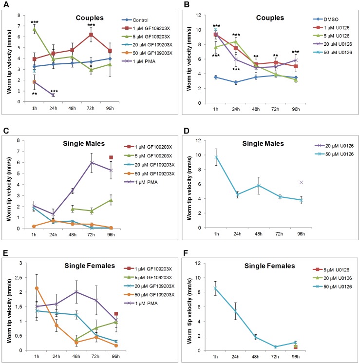
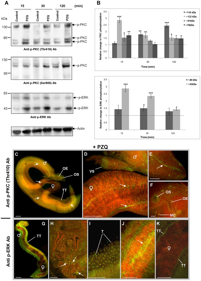
Protein kinases C (PKCs) and extracellular signal-regulated kinases (ERKs) are evolutionary conserved cell signalling enzymes that coordinate cell function. Here we have employed biochemical approaches using 'smart' antibodies and functional screening to unravel the importance of these enzymes to Schistosoma mansoni physiology. Various PKC and ERK isotypes were detected, and were differentially phosphorylated (activated) throughout the various S. mansoni life stages, suggesting isotype-specific roles and differences in signalling complexity during parasite development. Functional kinase mapping in adult worms revealed that activated PKC and ERK were particularly associated with the adult male tegument, musculature and oesophagus and occasionally with the oesophageal gland; other structures possessing detectable activated PKC and/or ERK included the Mehlis' gland, ootype, lumen of the vitellaria, seminal receptacle and excretory ducts. Pharmacological modulation of PKC and ERK activity in adult worms using GF109203X, U0126, or PMA, resulted in significant physiological disturbance commensurate with these proteins occupying a central position in signalling pathways associated with schistosome muscular activity, neuromuscular coordination, reproductive function, attachment and pairing. Increased activation of ERK and PKC was also detected in worms following praziquantel treatment, with increased signalling associated with the tegument and excretory system and activated ERK localizing to previously unseen structures, including the cephalic ganglia. These findings support roles for PKC and ERK in S. mansoni homeostasis, and identify these kinase groups as potential targets for chemotherapeutic treatments against human schistosomiasis, a neglected tropical disease of enormous public health significance.


Conflict of interest statement

The authors have declared that no competing interests exist.

Figures

Figure 1
Figure 1. Alignment of antibody recognition sites.
Comparison of the amino acid recognition sequences, together with the phosphorylated site, for the three anti-phospho antibodies used between S. mansoni PKC and ERK predicted protein sequences obtained from the S. mansoni genome database and relevant human protein sequences. The anti-phospho PKC (pan) (βII Ser660) antibodies recognize multiple human PKCs including PKCβI, βII and ε; based on sequence similarity these antibodies are predicted to react with Smp_128480 and Smp_176360 (both β-type PKCs) only when phosphorylated on Ser647 and Ser590, respectively within the bulky ring motif. The S. mansoni ε-type PKC (Smp_131700) lacks the conserved Ser phosphorylation site, as does ι-type PKC (Smp_096310) in common with human PKCι. The anti-phospho PKC (pan) (ζ Thr410) antibodies also recognize multiple human PKCs including PKCβI, βII, ε and ι; based on sequence similarity these antibodies are predicted to react with all S. mansoni PKCs only when phosphorylated on the conserved Thr residue within the PDK1 consensus motif. The anti-phospho p44/42 MAPK (Thr202/Tyr204) antibodies recognize human ERK1 and ERK2 only when phosphorylated on Thr and Tyr within the conserved TEY motif; based on sequence similarity, these antibodies are predicted to detect S. mansoni ERK1 and ERK 2 (Smp_142050 and Smp_047900, respectively) when phosphorylated.
Figure 2
Figure 2. Phosphorylated (activated) PKC- and ERK-like proteins exist in four different life stages of S. mansoni.
Western blot showing immunoreactive bands detected with (A) anti-phospho PKC (pan) (ζ Thr410), (B) anti-phospho PKC (pan) (βII Ser660), and (C) anti-phospho p44/42 MAPK (ERK1/2) (Thr202/Tyr204) antibodies (Ab) in miracidia (M), mother sporocysts (S), cercariae (C), adult female (♀) and male (♂) worms, and coupled worm pairs (WP) (∼12 µg for each sample). Results are representative of three independent experiments. Lambda phosphatase was also employed to confirm that the antibodies reacted only with the phosphorylated form of each protein; actin was used a loading control.
Figure 3
Figure 3. S. mansoni phosphorylated (activated) PKC-like proteins possess characteristic PKC activity.
(A) Immunodetection of phosphorylated (activated) S. mansoni PKC-like proteins after exposure of live adult worm pairs to GF109203X (20 µM; 2 h), GF109203X (20 µM; 2 h) followed by PMA (1 µM; 30 min), or PMA (1 µM; 30 min) only. Protein homogenates (12 µg) were processed for Western blotting and blots probed with anti-phospho PKC (pan) (ζ Thr410) or anti-phospho PKC (pan) (βII Ser660) antibodies (Ab); anti-actin antibodies were used to assess protein loading between samples. Immunoreactive bands from three independent experiments, each with two replicates, were analyzed with GeneTools and the mean relative change (± SEM; see graphs) in phosphorylation for each protein calculated relative to the phosphorylation levels of untreated (DMSO or RPMI 1640) controls that were assigned a value of 1 (shown as the dotted line). * p≤0.05, ** p≤0.01, and *** p≤0.001 (ANOVA). (B) Adult S. mansoni were either left untreated (upper graph) or were exposed to PMA (1 µM) for 30 min or DMSO (vehicle) (lower graph) prior to homogenization; equal amounts of protein were then used for immunoprecipitation with anti-phospho PKC (pan) (ζ Thr410) or (βII Ser 660) antibodies, or for the negative (beads only) control. The PKC activities of immunocomplexes, negative control, supernatant, and positive control (recombinant human PKC) were then determined using the Onmia PKC assay kit with Ser/Thr 8 substrate peptide. Florescence was recorded every 5 min for up to 200 min. Results are representative of two independent experiments.
Figure 4
Figure 4. S. mansoni ERK-like proteins are phosphorylated (activated) by MEK and possess characteristic ERK activity.
(A) Live adult worm pairs were exposed to 1 µM U0126 or 0.1% DMSO (control) for 30, 60 or 120 min. Protein homogenates (12 µg) were processed for Western blotting and blots probed with anti-phospho p44/42 MAPK (ERK1/2) (Thr202/Tyr204) antibodies (Ab). Immunoreactive bands from three independent experiments were analyzed with GeneTools and the mean relative change (± SEM; see graph) in phosphorylation of each protein calculated relative to the phosphorylation levels of controls that were assigned a value of 1 (shown here as the dotted line). **p≤0.01 and ***p≤0.001 (ANOVA). (B) Phosphorylation of Elk-1 fusion protein by immunoprecipitated ERK. Adult worm pairs were homogenized and equal amounts of protein used for immunoprecipitation with immobilized anti-phospho p44/42 MAPK (ERK1/2) (Thr202/Tyr204) antibodies or with beads only (negative control). Immunocomplexes were then incubated with Elk-1 fusion protein as the ERK substrate and phosphorylated Elk-1 was detected on Western blots using anti-phospho Elk-1 antibodies. Results are representative of two independent experiments.
Figure 5
Figure 5. Connectivity between ERK and PKC signalling in S. mansoni.
Adult S. mansoni were treated with (A) GF109203X (20vM; 120 min), PMA (1 µM; 30 min), GF109203X (20 µM; 120 min) followed by PMA (1 µM; 30 min), or DMSO (vehicle control), or with (B) U0126 (1 µM; 30, 60, and 120 min) or DMSO (vehicle control). Proteins (12 µg) were then processed for Western blotting using (A) anti-phospho p44/p42 MAPK (ERK1/2) (Thr202/Tyr204), or (B) anti-phospho PKC (pan) (ζ Thr410) or anti-phospho PKC (pan) (βII Ser660) antibodies (Ab). Anti-actin antibodies were used to assess protein loading. Immunoreactive bands from three independent experiments were analysed and quantified using GeneTools and the mean relative change (± SEM; see graphs) in phosphorylation for each protein calculated relative to the phosphorylation levels of untreated (DMSO or RPMI-1640) controls that were assigned a value of 1 (shown as the dotted line). *p≤0.05, **p≤0.01 (ANOVA).
Figure 6
Figure 6. Immunolocalization of phosphorylated (activated) PKC in intact S. mansoni.
Adult worm pairs were either (A–K) fixed immediately after perfusion, or (L–O) treated with PMA (1 µM; 15 min) before fixing. Worm pairs were then incubated with anti-phospho PKC (pan) (ζ Thr410) or anti-phospho PKC (pan) (βII Ser660) primary antibodies (Ab) and Alexa Fluor 488 secondary antibodies (green); specimens were additionally stained with rhodamine phalloidin to reveal actin filaments (red). Activated PKC was found associated with: (B, C, I, N) oesophagus (OE) and oesophageal gland (OG); (A, D, F, G, J–L) tubercles (T) of the tegument (TT); (E, M) reproductive structures including the uterus (UT) surrounding an egg (EG) during expulsion and lumen of the vitellaria (LV); (K) longitudinal (LM) and circular muscle (CM) layers; (F,G) myocytons (MC) including those close to the (H) ventral sucker (VS); (O) the VS; and (F,G) the neural plexus (NP) innervating the musculature (MU). (L) The asterisk (*) identifies an area of the tegument possibly disrupted by PMA. All images are of z-axis projections displayed in maximum pixel brightness mode. Bar: A = 100 µM, B–O = 50 µm.
Figure 7
Figure 7. Immunolocalization of phosphorylated (activated) ERK in intact S. mansoni.
Adult worm pairs were either (A–H) fixed immediately after perfusion, or (I–M) treated with PMA (1 µM; 15 min) before fixing. Worm pairs were then incubated in anti-phospho p44/42 MAPK (ERK1/2) (Thr202/Tyr204) antibodies and Alexa Fluor 488 secondary antibodies (green); specimens were additionally stained with rhodamine phalloidin to reveal actin filaments (red). Under normal conditions activated ERK was found associated with: (A, B) flame cells (FC) and excretory tubules (ET); (C) oesophagus (OE); (D) tubercles (T) of the tegument (TT); (E) seminal receptacle (SR) adjacent to the ovary (OV); (F) the uterus (UT) surrounding an egg (EG) during expulsion; (G, H) the region of the Mehlis' gland (MG) adjacent to the ootype (OT) containing the egg. Following exposure to PMA increased ERK activation was evident over (I) the worm surface including at the (J) ventral sucker (VS), and (K) the tubercles (T) of the tegument (TT). Activated ERK was also associated with (L) the testicular lobes (TL) and (M) the ovary (OV). All images are of z-axis projections displayed in maximum pixel brightness mode. Bar:  = 50 µm.
Figure 8
Figure 8. Modulation of PKC and ERK activity in adult S. mansoni induces worm uncoupling, suppresses egg output, and causes male worms to detach.
Adult worm pairs were incubated in increasing concentrations of U0126, GF109203X, or 1 µM PMA and movies captured at various time points over 96 h and imported into Image J. The effects of these compounds on (A, B) pairing, (C, D) egg release by paired worms (uncoupled worms were not included), and (E, F) male adult worm detachment from the culture plate were then determined against vehicle controls (which in A–D were assigned a value of 100% and are shown as the dotted line). Mean values (± SEM) shown represent those from four independent experiments, each of which contained a minimum of six adult worm pairs. *p≤0.05, **p≤0.01, and ***p≤0.001 (ANOVA).
Figure 9
Figure 9. Modulation of PKC and ERK activity in adult S. mansoni affects worm movement.
Adult worm pairs were incubated with increasing concentrations of U0126, GF109203X, or 1 µM PMA and movies captured at various time points over 96 h and imported into Image J. Movies were then analyzed to determine the speed of movement (velocity, mm/s) that the posterior tip of each worm travelled in 10 s when (A, B) coupled. In some cases worms uncoupled as a consequence of treatment (as shown in Figure 8A, B). When this was the case, the speed of movement of the uncoupled (C, D) single male or (E, F) female worms was determined for their respective treatments for the remainder of the assay; note that no vehicle control exists when worms had separated because the control worms remained paired. Mean values (± SEM) shown represent those from four independent experiments each of which contained a minimum of six adult worm pairs; the actual numbers of worms in each analysis is, however, dependent upon the uncoupling effect of the individual treatment (shown in Figures 8A, B). *p≤0.05, **p≤0.01, and ***p≤0.001 (ANOVA).
Figure 10
Figure 10. Modulation of PKC activity in adult S. mansoni induces sustained coiling.
Adult worm pairs were incubated with increasing concentrations of GF109203X, or 1 µM PMA and movies captured at (B) 1 h, (C) 24 h, (D) 48 h, (E) 72 h, and (F) 96 h and imported into Image J. Movies were then analyzed to determine the number of coils (shown in A) present per worm that were sustained for at least 10 s. Values shown represent the mean (± SEM) percentage of worms displaying a given number of coils at each time point for each treatment. *p≤0.05, **p≤0.01, and ***p≤0.001 (ANOVA).
Figure 11
Figure 11. Praziquantel (PZQ) induces the phosphorylation (activation) of ERK and PKC in adult S. mansoni.
(A) Live adult worms were exposed to 0.02 mg/ml PZQ or 0.2% DMSO (vehicle control) for 15, 30 and 120 min. Proteins (12 µg) were processed for Western blotting and blots probed with anti-phospho PKC (pan) (ζ Thr410), anti-phospho PKC (pan) (βII Ser660), or anti-phospho p44/42 MAPK (ERK1/2) (Thr202/Tyr204) antibodies (Ab); anti-actin antibodies were used to assess protein loading differences between samples. (B) Immunoreactive bands from three independent experiments, each with two replicates, were analyzed and mean relative change (± SEM) in phosphorylation for each protein calculated relative to the phosphorylation of controls that were assigned a value of 1 (shown as the dotted line). *p≤0.05, **p≤0.01, and ***p≤0.001 (ANOVA). (C–K) Adult worm pairs were treated with 0.02 mg/ml PZQ for 15 min prior to fixing. Worm pairs were then incubated in either (C–F) anti-phospho PKC (pan) (ζ Thr410) or (G–K) anti-phospho p44/42 MAPK (ERK1/2) (Thr202/Tyr204) primary antibodies, followed by Alexa Fluor 488 secondary antibodies (green); specimens were also stained with rhodamine phalloidin to show actin filaments (red). Activated PKC was found particularly associated with: (C) the tegument (TT) and oesophagus (OE); (C, F) opening of the oral sucker (OS) and myocytons (MC); (D) the musculature of the ventral sucker (VS); and (C–E) collecting ducts of the excretory system (arrowed). Activated ERK was found particularly associated with (G, I) tubercles (T) of the tegument (TT); (H) cephalic ganglia (arrowed); (J) musculature (arrow); and (K) surface of the female worm tegument (TT) adjacent to the male gynaecophoric canal. All microscopy images are of z-axis projections displayed in maximum pixel brightness mode Bar: C, D, G, K = 100 µm; E, F, H, I = 25 µm.

References

    1. Reyland ME (2009) Protein kinase C isoforms: multi-functional regulators of cell life and death. Front Biosci 14: 2386–2399. - PMC - PubMed
    1. Krishna M, Narang H (2008) The complexity of mitogen-activated protein kinases (MAPKs) made simple. Cell Mol Life Sci 65: 3525–3544. - PMC - PubMed
    1. Berriman M, Haas BJ, LoVerde PT, Wilson RA, Dillon GP, et al. (2009) The genome of the blood fluke Schistosoma mansoni . Nature 460: 352–358. - PMC - PubMed
    1. Andrade LF, Nahum LA, Avelar LGA, Silva LL, Zerlotini A, et al. (2011) Eukaryotic protein kinases (ePKs) of the helminth parasite Schistosoma mansoni . BMC Genomics 12: 215. - PMC - PubMed
    1. The Schistosoma japonicum Genome Sequencing and Functional Analysis Consortium (2009) The Schistosoma japonicum genome reveals features of host-parasite interplay. Nature 460: 345–351. - PMC - PubMed

Publication types

MeSH terms

LinkOut - more resources